Cervical Myelopathy: Difference between revisions
Rachael Lowe (talk | contribs) mNo edit summary |
Rachael Lowe (talk | contribs) mNo edit summary |
||
| Line 6: | Line 6: | ||
== Definition/Description == | == Definition/Description == | ||
Cervical myelopathy refers to compression on the cervical spinal cord | Cervical myelopathy refers to compression on the cervical spinal cord. Any space occupying lesion within the cervical spine with the potential to compress the spinal cord can cause cervical myelopathy.<ref name="Richard">Richard K. Root Clinical Infectious Diseases: A Practical Approach, 1999</ref><ref name="Kong">Kong LD, Meng LC, Wang LF, Shen Y, Wang P and Shang ZK. Evaluation of conservative treatment and timing of surgical intervention for mild forms of cervical spondylotic myelopathy. Exp Ther Med. 2013 Sep;6(3):852-856.</ref> Cervical myelopathy is predominantly due to pressure on the anterior spinal cord with ischaemia as a result of deformation of the cord by anterior herniated discs, spondylitic spurs, an ossified posterior longitudinal ligament or [[Cervical Stenosis|spinal stenosis]]<ref name="Dai">Dai L, Ni B, Yuan W and Jia L. Radiculopathy after laminectomy for cervical compression myelopathy. J Bone Joint Surg Br. 1998 Sep;80(5):846-9.</ref> | ||
The spontaneous course of myelopathy is characterised either by long periods of stable disability followed by episodes of deterioration or a linear progressive course. The presentation of a cervical myelopathy varies in accordance to the severity of the spinal cord compression as well as its location.<ref name="Boos">Boos N and Aebi M (Eds). Spinal disorders: Fundamentals of Diagnosis and Treatment. Springer-Verlag Berlin Heidelberg. 2008.</ref> | |||
== Clinically Relevant Anatomy == | == Clinically Relevant Anatomy == | ||
There are seven cervical vertebrae and eight cervical nerve roots.<ref name="Kong" /><ref name="Cook">Cook C, Brown C, Isaacs R, Roman M, David S and Richardson W. Clustered clinical findings for diagnosis of cervical spine myelopathy. J Man Manip Ther. 2010 Dec; 18(4): 175–180.</ref> The spinal cord is the extension of the central nervous system outside the cranium. It is encased by the vertebral column and begins at the foramen magnum.<ref name="Cramer">Cramer GD and Darby SA. Basic and Clinical Anatomy of the Spine, Spinal Cord, and ANS. 2nd Edition. Elsevier 2008.</ref> The spinal cord is an extremely vital part of the central nervous system, and even a small injury can lead to severe disability.<ref name="Selzer">Selzer ME and Dobkin BH. Spinal Cord Injury (American Academy of Neurology Quality of Life Series). Demos Medical Publishing (New York). 2008</ref> | There are seven cervical vertebrae and eight cervical nerve roots.<ref name="Kong" /><ref name="Cook">Cook C, Brown C, Isaacs R, Roman M, David S and Richardson W. Clustered clinical findings for diagnosis of cervical spine myelopathy. J Man Manip Ther. 2010 Dec; 18(4): 175–180.</ref> The spinal cord is the extension of the central nervous system outside the cranium. It is encased by the vertebral column and begins at the foramen magnum.<ref name="Cramer">Cramer GD and Darby SA. Basic and Clinical Anatomy of the Spine, Spinal Cord, and ANS. 2nd Edition. Elsevier 2008.</ref> The spinal cord is an extremely vital part of the central nervous system, and even a small injury can lead to severe disability.<ref name="Selzer">Selzer ME and Dobkin BH. Spinal Cord Injury (American Academy of Neurology Quality of Life Series). Demos Medical Publishing (New York). 2008</ref> | ||
A complex system of ligaments serves to | A complex system of ligaments serves to stabilise and protect the cervical spine. For example, ligamentum flavum extends from the anterior surface of the cephalic vertebra to the posterior surface of the caudal vertebra and connects to the ventral aspect of the facet joint capsules. A ligament that is often involved in this condition is the posterior longitudinal ligament. It is situated within the vertebral canal, originating from the body of the axis, where it is continuous with the membrana tectoria, and extends along the posterior surfaces of the bodies of the vertebrae until inserting into the sacrum.<ref name="Selzer" /> | ||
{| border="0" cellpadding="2" | {| border="0" cellpadding="2" | ||
| Line 23: | Line 22: | ||
Chronic cervical degeneration is the most common cause of progressive spinal cord and nerve root compression. Spondylotic changes can result in stenosis of the spinal canal, lateral recess, and foramina. Spinal canal stenosis can lead to myelopathy, whereas the latter two can lead to radiculopathy. | Chronic cervical degeneration is the most common cause of progressive spinal cord and nerve root compression. Spondylotic changes can result in stenosis of the spinal canal, lateral recess, and foramina. Spinal canal stenosis can lead to myelopathy, whereas the latter two can lead to radiculopathy. | ||
== Epidemiology == | |||
Cervical spondylotic myelopathy is the most common disorder of the spinal cord in persons older than 55 years of age.<ref name="Cook" /><ref name="Amenta">Amenta PS, Ghobrial GM, Krespan K, Nguyen P, Ali M, Harrop JS. Cervical spondylotic myelopathy in the young adult: a review of the literature and clinical diagnostic criteria in an uncommon demographic. Clin Neurol Neurosurg. 2014. 120:68-72.</ref><ref name="Kadanka">Kadanka Z, Bednarík J, Vohánka S, Vlach O, Stejskal L, Chaloupka R et al. Conservative treatment versus surgery in spondylotic cervical myelopathy: a prospective randomised study. Eur Spine J. 2000;9(6):538-44.</ref> Radiologic spondylotic changes increase with patient age - 90% of asymptomatic persons older than 70 years have some form of degenerative change in the cervical spine. Cervical spine myelopathy resulting from sagittal narrowing of the spinal canal and compression of the spinal cord is present in 90% of individuals by the seventh decade of life.<ref name="cook">Cook C, Roman M, Stewart KM, Leithe LG, Isaacs R. [http://www.pt.armstrong.edu/mincer/journalclub/cspinemylepathy.pdf Reliability and diagnostic accuracy of clinical special tests for myelopathy in patients seen for cervical dysfunction]. J Orthop Sports Phys Ther. 2009 Mar;39(3):172-8. doi: 10.2519/jospt.2009.2938.</ref> Both sexes are affected equally. Cervical spondylosis usually starts earlier in men (50 years) than in women (60 years). It causes hospitalization at a rate of 4.04 per 100,000 person-years.<ref name="Cook" /><ref name="Koakutsu">Koakutsu T,Nakajo J, Morozumi N, Hoshikawa T, Ogawa S, and Ishii Y. Cervical myelopathy due to degenerative spondylolisthesis. Ups J Med Sci. 2011; 116(2): 129–132.</ref> | |||
== Epidemiology | == Pathological Process == | ||
Cervical spondylotic myelopathy is the most common disorder of the spinal cord in persons older than 55 years of age.<ref name="Cook" /><ref name="Amenta">Amenta PS, Ghobrial GM, Krespan K, Nguyen P, Ali M, Harrop JS. Cervical spondylotic myelopathy in the young adult: a review of the literature and clinical diagnostic criteria in an uncommon demographic. Clin Neurol Neurosurg. 2014. 120:68-72.</ref><ref name="Kadanka">Kadanka Z, Bednarík J, Vohánka S, Vlach O, Stejskal L, Chaloupka R et al. Conservative treatment versus surgery in spondylotic cervical myelopathy: a prospective randomised study. Eur Spine J. 2000;9(6):538-44.</ref> Radiologic spondylotic changes increase with patient age - 90% of asymptomatic persons older than 70 years have some form of degenerative change in the cervical spine. Both sexes are affected equally. Cervical spondylosis usually starts earlier in men (50 years) than in women (60 years). It causes hospitalization at a rate of 4.04 per 100,000 person-years.<ref name="Cook" /><ref name="Koakutsu">Koakutsu T,Nakajo J, Morozumi N, Hoshikawa T, Ogawa S, and Ishii Y. Cervical myelopathy due to degenerative spondylolisthesis. Ups J Med Sci. 2011; 116(2): 129–132.</ref> | |||
The causes of cervical myelopathy can be divided into different categories: | The causes of cervical myelopathy can be divided into different categories: | ||
*'''Static factors''': A narrowing of the spinal canal size | *'''Static factors''': A narrowing of the spinal canal size commonly results from degenerative changes in the cervical spine anatomy such as disc degeneration, spondylosis, stenosis, osteophyte formation at the level of facet joints, segmental ossification of the posterior longitudinal ligament and yellow ligament hypertrophy, calcification or ossification. Patients with a congenitally narrow spinal canal (<13mm) have a higher risk for the development of symptomatic cervical myelopathy.<ref name="Boos" /><ref name="cook" /><ref name="Koakutsu" /><ref name="Yonenobu">Yonenobu K. Cervical radiculopathy and myelopathy: when and what can surgery contribute to treatment? Eur Spine J. 2000; 9(1): 1-7.</ref> | ||
*'''Dynamic factors''': Due to mechanical abnormalities of the cervical spine or instability.<ref name="Boos" /> | *'''Dynamic factors''': Due to mechanical abnormalities of the cervical spine or instability.<ref name="Boos" /> | ||
*'''Vascular and cellular factors''': Spinal cord ischemia affects oligodendrocytes, which results in demyelination exhibiting features of chronic degenerative disorders. Also glutamatergic toxicity, cell injury and apoptosis may occur.<ref name="Boos" /> | *'''Vascular and cellular factors''': Spinal cord ischemia affects oligodendrocytes, which results in demyelination exhibiting features of chronic degenerative disorders. Also glutamatergic toxicity, cell injury and apoptosis may occur.<ref name="Boos" /> | ||
Cord compression is thought to be a combination of static compression and intermittent dynamic compression from cervical motion (flexion/extension). | |||
== | == Characteristics and Clinical Presentation == | ||
Cervical myelopathy can cause a variety of signs and symptoms. Symptoms are divided into two groups: long-tract and segmental symptoms. Onset is insidious, typically in persons aged 50-60 years. | |||
Early symptoms of this condition are ‘numb, clumsy, painful hands’ and disturbance of fine motor skills.<ref name="Boos" /> Weakness and numbness occur in a non-specific/non-dermatomal pattern. As spinal cord degeneration progresses, lower motor neuron findings in the upper extremities, such as loss of strength, atrophy of the interosseous muscles and difficulty in fine finger movements, may present. | |||
Additional clinical findings may include: neck pain and stiffness (decreased ROM, especially extension), shoulder and scapular pain, paresthesia in one or both arms or hands, signs of radiculopathy, Babinski and Hoffman's sign, ataxia and dexterity loss.<ref name="Cook" /><ref name="Harrop">Harrop JS, Naroji S, Maltenfort M, Anderson DG, Albert T, Ratliff JK et al. Cervical myelopathy: a clinical and radiographic evaluation and correlation to cervical spondylotic myelopathy. Spine (Phila Pa 1976). 2010 Mar 15;35(6):620-624.</ref><ref name="Park">Park SJ, Kim SB, Kim MK, Lee SH and Oh IH. Clinical features and surgical results of cervical myelopathy caused by soft disc herniation. Korean J Spine. 2013;10(3):138-143.</ref> Typical neurological signs of long-tract involvement are exaggerated tendon reflexes (patellar and achilles), presence of pathological reflexes (e.g. clonus, Babinski and Hoffman's sign), spastic quadriplegia, sensory loss and bladder-bowel disturbance.<ref name="Yonenobu" /> | |||
Cervical spondylotic myelopathy | |||
Once the disorder is diagnosed, complete remission to normality never occurs and spontaneous temporary remission is uncommon. In 75% of the patients, episodic worsening with neurological deterioration occurs, 20% have slow steady progression, and 5% experience rapid onset and progression.<ref name="Boos" /><br> | |||
'''Common Symptoms''' | '''Common Symptoms''' | ||
| Line 68: | Line 52: | ||
*Radiculopathic signs | *Radiculopathic signs | ||
== | == Diagnostic Procedures == | ||
A detailed and thorough neurological examination plus MRI is the current standard to diagnose the presence of cervical myelopathy. | |||
Plain radiographs alone are of little use as an initial diagnostic procedure. A magnetic resonance image (MRI) is considered the best imaging method for confirming the presence of spinal canal stenosis, cord compression, or myelomalacia, elements germane to cervical spine myelopathy. MRI of the cervical spine can also rule out spinal cord tumours. | |||
An MRI is most useful because it expresses the amount of compression placed on the spinal cord and demonstrates relatively high levels of sensitivity and specificity<ref name="Cook" /><ref name="Harrop" />. Anterior-posterior width reduction, cross-sectional evidence of cord compression, obliteration of the subarachnoid space and signal intensity changes to the cord found on MR imaging are considered the most appropriate parameters for confirmation of a spinal cord compression myelopathy<ref name="Cook" />. More than half of patients with cervical spine myelopathy show intramedullary high signal intensity on T2-weighted imaging, mainly in the spinal gray matter<ref name="Sato">Sato T, Horikoshi T, Watanabe A, Uchida M, Ishigame K, Araki T et al. Evaluation of cervical myelopathy using apparent diffusion coefficient measured by diffusion-weighted imaging. AJNR Am J Neuroradiol. 2012; 33(2):388-392.</ref>. Radiographic cervical spinal cord compression and hyperintense T2 intraparenchymal signal abnormalities (MRI) correlate well with the presence of myelopathic findings on physical examination<ref>Harrop, James S; Naroji, Swetha; Maltenfort, Mitchell; Anderson, D. Greg; Albert, Todd; Ratliff, John K; Ponnappan, Ravi K; Rihn, Jeffery A; Smith, Harvey E; Hilibrand, Alan; Sharan, Ashwini D; Vaccaro, Alexander. [http://journals.lww.com/spinejournal/Abstract/publishahead/Cervical_Myelopathy__A_Clinical_and_Radiographic.99733.aspx Cervical Myelopathy: A Clinical and Radiographic Evaluation and Correlation to Cervical Spondylotic Myelopathy]. Spine 10 February 2010 [epub ahead of print]</ref>. | |||
== | {| border="0" cellpadding="2" | ||
|- | |||
! scope="col" width="400" | [[Image:Cervical Osteophytes.jpg|center|400px|Osteophytes with Cord Compression]] | |||
! scope="col" width="400" | [[Image:Cervical Spondylosis.jpg|center|400px|Cervical Spondylosis]] | |||
|} | |||
== Clinical Examination == | == Clinical Examination == | ||
The diagnosis of CSM is primarily based on the clinical signs found on physical examination and is supported by imaging findings | The diagnosis of CSM is primarily based on the clinical signs found on physical examination and is supported by imaging findings.<ref name="Amenta" /> According to Cook et al,<ref name="cook 2">Chad Cook, Christopher Brown, Robert Isaacs, Matthew Roman, Samuel Davis, and William Richardson. Clustered clinical findings for diagnosis of cervical spine myelopathy. J Man Manip Ther. 2010 December; 18(4): 175–180.</ref> selected combinations of the following clinical findings are effective in ruling out and ruling in cervical spine myelopathy. Combinations of three of five or four of five of these tests enable post-test probability of the condition to 94–99%: | ||
#gait deviation | #gait deviation | ||
| Line 112: | Line 86: | ||
*[[Romberg Test|Romberg test]] | *[[Romberg Test|Romberg test]] | ||
Although these tests exhibit moderate to substantial reliability among skilled clinicians, they demonstrate low sensitivity and are not appropriate for ruling out myelopathy. One method used to improve the diagnostic accuracy of clinical testing is combining tests into clusters. These often overcome the inherent weakness of stand alone tests.<ref name="Cook" /><ref name="cook 2" /> | Although these tests exhibit moderate to substantial reliability among skilled clinicians, they demonstrate low sensitivity and are not appropriate for ruling out myelopathy. One method used to improve the diagnostic accuracy of clinical testing is combining tests into clusters. These often overcome the inherent weakness of stand alone tests.<ref name="Cook" /><ref name="cook 2" /> | ||
==== '''Outcome Measures''' ==== | |||
• [[Neck Disability Index|Neck disability index]]<br>• [[Neck Pain and Disability Scale|Neck pain and disability scale]] <br>• Nurick-score <ref name="Vitzthum">Vitzthum H and Dalitz K. Analysis of five specific scores for cervical spondylogenic myelopathy. Eur Spine J. 2007; 16(12): 2096–2103.</ref> <br>• Japanese-orthopaedic-association-score (JOA-Score) <ref name="Vitzthum" /> <ref name="Tanaka">Tanaka N, Konno S, Takeshita K, Fukui M, Takahashi K, Chiba K et al. An outcome measure for patients with cervical myelopathy: the Japanese Orthopaedic Association Cervical Myelopathy Evaluation Questionnaire (JOACMEQ): an average score of healthy volunteers. J Orthop Sci. 2014 Jan;19(1):33-48.</ref> <br>• Cooper-myelopathy-scale (CMS) <ref name="Vitzthum" /> <br>• European-myelopathy-score (EMS) | |||
== Differential Diagnosis == | |||
*[[Adhesive Capsulitis|Adhesive Capsulitis]] | |||
*Brown-Sequard Syndrome | |||
*[[Carpal Tunnel Syndrome|Carpal Tunnel Syndrome]] | |||
'''Outcome Measures''' | *Central Cord Syndrome | ||
*Cervical Disc Disease | |||
• [[Neck Disability Index|Neck disability index]]<br>• [[Neck Pain and Disability Scale|Neck pain and disability scale]] <br>• Nurick-score <ref name="Vitzthum">Vitzthum H and Dalitz K. Analysis of five specific scores for cervical spondylogenic myelopathy. Eur Spine J. 2007; 16(12): 2096–2103.</ref> <br>• Japanese-orthopaedic-association-score (JOA-Score) <ref name="Vitzthum" /> <ref name="Tanaka">Tanaka N, Konno S, Takeshita K, Fukui M, Takahashi K, Chiba K et al. An outcome measure for patients with cervical myelopathy: the Japanese Orthopaedic Association Cervical Myelopathy Evaluation Questionnaire (JOACMEQ): an average score of healthy volunteers. J Orthop Sci. 2014 Jan;19(1):33-48.</ref> <br>• Cooper-myelopathy-scale (CMS) <ref name="Vitzthum" /> <br>• European-myelopathy-score (EMS) | *Cervical Myofascial Pain | ||
*Cervical Sprain and Strain | |||
*Chronic Pain Syndrome | |||
*Diabetic Neuropathy | |||
*[[MS Multiple Sclerosis|Multiple Sclerosis]] | |||
*Myofascial Pain | |||
*Neoplastic Brachial Plexopathy | |||
*[[Osteoporosis|Osteoporosis]] | |||
*Spinal Cord Injury | |||
*Radiation-Induced Brachial Plexopathy | |||
*[[Rheumatoid Arthritis|Rheumatoid Arthritis]] | |||
*Traumatic Brachial Plexopathy | |||
*Tumors | |||
[[ | |||
* | |||
* | |||
* | |||
* | |||
* | |||
* | |||
* | |||
* | |||
* | |||
* | |||
* | |||
* | |||
== Management == | == Management == | ||
There is no consensus about the treatment of mild and moderate forms of cervical myelopathy. Surgical treatment has no better results than conservative treatment over two years of follow-up | There is no consensus about the treatment of mild and moderate forms of cervical myelopathy. Surgical treatment has no better results than conservative treatment over two years of follow-up<ref>Kadanka Z, Bednarík J, Vohánka S, Vlach O, Stejskal L, Chaloupka R, Filipovicová D, Surelová D, Adamová B, Novotný O, Nemec M, Smrcka V, Urbánek I. Conservative treatment versus surgery in spondylotic cervical myelopathy: a prospective randomised study. Eur Spine J (2000) 9 :538–544</ref>. Patients with cervical myelopathy that are treated with a conservative approach (anti-inflammatory medication and physical therapy) may have some short term benefit in relief of painful symptoms. Because the condition is degenerative and progressive, slow and continued progressive neurologic deterioration will occur. | ||
=== Medical Management === | === Medical Management === | ||
People who have progressive neurologic changes (such as weakness, numbness or falling) with signs of severe spinal cord compression or spinal cord swelling are candidates for surgery. Patients with severe or disabling pain may also be helped with surgery.<ref name="Almeida">Almeida GP, Carneiro KK and Marques AP. Manual therapy and therapeutic exercise in patient with symptomatic cervical spondylotic myelopathy: a case report. J Bodyw Mov Ther. 2013 Oct;17(4):504-9.</ref> When myelopathy is caused by factors of a progressive nature, such as spinal cord tumors, surgical treatment is likewise indicated | People who have progressive neurologic changes (such as weakness, numbness or falling) with signs of severe spinal cord compression or spinal cord swelling are candidates for surgery. Patients with severe or disabling pain may also be helped with surgery.<ref name="Almeida">Almeida GP, Carneiro KK and Marques AP. Manual therapy and therapeutic exercise in patient with symptomatic cervical spondylotic myelopathy: a case report. J Bodyw Mov Ther. 2013 Oct;17(4):504-9.</ref> When myelopathy is caused by factors of a progressive nature, such as spinal cord tumors, surgical treatment is likewise indicated<ref name="1 Massimo Leonardi, Norbert Boos , Degenerative disorders of the cervical spine">Massimo Leonardi, Norbert Boos , Degenerative disorders of the cervical spine</ref><ref>7 Law MD Jr, Bernhardt M, White AA 3rd.Cervical spondylotic myelopathy: a review of surgical indications and decision making. Yale J Biol Med. 1993 May-Jun;66(3):165-77.</ref>. | ||
People who experience better surgical outcomes often have these characteristics: | People who experience better surgical outcomes often have these characteristics: | ||
* The symptom of an electrical sensation that runs down the back and into the limbs | |||
* Younger age | |||
* Shorter duration of symptoms | |||
* Single rather than multiple areas of involvement | |||
* Larger areas available for the cord | |||
The principal aim of surgery for cervical myelopathy is decompression of the spinal cord. The surgical techniques include multilevel discectomies or corpectomies with or without instrumented fusion, laminectomy with or without instrumented fusion or laminoplasty<ref name="Boos" />. Surgical decompression is generally considered if the symptoms affect daily life but early surgical intervention is thought to be more effective. Therefore, early detection may be the key to minimize postoperative sequelae<ref name="Sato" />. | |||
Final outcomes from the surgery vary. Typically, one-third of patients improve, one-third stay the same, and one-third continue to worsen over time, with respect to their pre-surgical symptoms<ref name="Kadanka" /><ref name="Almeida" /> | |||
Final outcomes from the surgery vary. Typically, one-third of patients improve, one-third stay the same, and one-third continue to worsen over time, with respect to their pre-surgical symptoms | |||
=== Physical therapy management === | === Physical therapy management === | ||
Patients | Patients can be treated conservatively. Kadaňka et al. found no difference in long term outcome (2 years after the intervention) between patient who received a conservative or surgical treatment<ref>Kadaňka, Z., et al. "Conservative treatment versus surgery in spondylotic cervical myelopathy: a prospective randomised study." ''European Spine Journal'' 9.6 (2000): 538-544. </ref>. Even after 10 years, there were no differences found between the surgery and conservative group<ref>Kadaňka, Zdeněk, et al. "Cervical spondylotic myelopathy: conservative versus surgical treatment after 10 years." ''European Spine Journal'' 20.9 (2011): 1533-1538.</ref>. Also in the Cochrane review no differences between surgical or conservative treatment were found<ref name=":0">Fouyas, Ioannis P., Patrick FX Statham, and Peter AG Sandercock. "Cochrane review on the role of surgery in cervical spondylotic radiculomyelopathy." ''Spine'' 27.7 (2002): 736-747. </ref>. The only prognostic factor in which surgery can be recommended is the circumferential spinal cord compression in the maximum compression segment on axial MRI<ref>Shimomura, Takatoshi, et al. "Prognostic factors for deterioration of patients with cervical spondylotic myelopathy after nonsurgical treatment." ''Spine'' 32.22 (2007): 2474-2479. </ref> | ||
Rhee JM et al. describes myelopathy as a typically progressive disorder and that there is little of evidence that conservative treatment halts or reverses its progression. So, they recommend not routinely prescribing nonoperative treatment as the primary modality in patients with moderate to severe myelopathy.<ref>Rhee, John M., et al. "Nonoperative management of cervical myelopathy: a systematic review." ''Spine'' 38.22S (2013): S55-S67. </ref> | Rhee JM et al. describes myelopathy as a typically progressive disorder and that there is little of evidence that conservative treatment halts or reverses its progression. So, they recommend not routinely prescribing nonoperative treatment as the primary modality in patients with moderate to severe myelopathy.<ref>Rhee, John M., et al. "Nonoperative management of cervical myelopathy: a systematic review." ''Spine'' 38.22S (2013): S55-S67. </ref> | ||
In general, the goals of | In general, the goals of physiotherapy treatment are: <ref name="Boos" /> | ||
* relieve pain | |||
* improve function | |||
* prevent neurological deterioration | |||
* reverse or improve neurological deficits | |||
Cervical myelopathy can be treated symptomatically. Possible therapies include: | Cervical myelopathy can be treated symptomatically. Possible therapies include: | ||
*Cervical traction and manipulation of the thoracic spine: useful for the reduction of pain scores and level of disability in patients with mild cervical myelopathy. Other signs and symptoms, such as weakness, headache, dizziness, and hypoesthesia, are positively affected | *'''Cervical traction and manipulation of the thoracic spine''': useful for the reduction of pain scores and level of disability in patients with mild cervical myelopathy. Other signs and symptoms, such as weakness, headache, dizziness, and hypoesthesia, are positively affected<ref name="Browder">Browder DA, Erhard RE and Piva SR. Intermittent cervical traction and thoracic manipulation for management of mild cervical compressive myelopathy attributed to cervical herniated disc: a case series. J Orthop Sports Phys Ther. 2004;34(11):701-712.</ref>. Cervical traction can be combined with other treatments like electrotherapy and exercises. Joghataei et al. reported a significant increase in grip strength after 10 weeks of this combined treatment<ref>Joghataei, Mohammad Taghi, Amir Massoud Arab, and Hossein Khaksar. "The effect of cervical traction combined with conventional therapy on grip strength on patients with cervical radiculopathy." ''Clinical rehabilitation'' 18.8 (2004): 879-887.</ref> | ||
*[[Manual Therapy and Exercise for Neck Pain: Clinical Treatment Tool-kit|Manual therapy techniques]]: employed to reduce the neck pain with natural apophyseal glides and sustained natural apophyseal glides for cervical extension and rotation | *[[Manual Therapy and Exercise for Neck Pain: Clinical Treatment Tool-kit|'''Manual therapy techniques''']]: employed to reduce the neck pain with natural apophyseal glides and sustained natural apophyseal glides for cervical extension and rotation<ref name="Almeida" /> Manipulation and mobilizations can be effective when they are combined with exercise therapy. When you use them without exercises, there is only poor evidence that it could be effective<ref>Binder, Allan I. "Cervical spondylosis and neck pain." ''BMJ: British Medical Journal'' 334.7592 (2007): 527-531. </ref><ref>Kay TM, Gross A, Goldsmith C, Santaguida PL, Hoving J, Brontfort G, et al, Cervical Overview Group. Exercises for mechanical neck disorders. Cochrane Database Syst Rev 2005</ref>. | ||
*Exercises: The | *'''Exercises''': The effects of exercise therapy specifically on cervical myelopathy have not been studied but there is evidence for exercises for mechanical neck pain. For example: stretching, strengthening exercises, active range of motion exercises, home exercise programs<ref name=":0" /> | ||
*[[Deep Neck Flexor Stabilisation Protocol|Cervical | *[[Deep Neck Flexor Stabilisation Protocol|'''Cervical stabilisation exercises''']]: when there is anteroposterior instability of the vertebral bodies of a degenerative nature, vertebral segment stabilisation of the cervical spine can be performed with a pressure biofeedback unit (PBU), performing 10 repetitions sustained for 10s, beginning with 22mmHg with the intention to progress to 30mmHg<ref name="Almeida" />. | ||
*Dynamic upper and lower limb exercises (flexion and extension) with the use of the PBU on the neck.<ref name="Almeida" /> | *'''Dynamic upper and lower limb exercises''' (flexion and extension) with the use of the PBU on the neck.<ref name="Almeida" /> | ||
*Proprioceptive neuromuscular facilitation: for the upper and lower limbs.<ref name="Almeida" /> | *'''Proprioceptive neuromuscular facilitation''': for the upper and lower limbs.<ref name="Almeida" /> | ||
*Improve posture | *'''Improve posture''' | ||
*Motor training programmes | *'''Motor training programmes''' may improve arm and hand functioning at function and/or activity level in cervical spinal cord injured patients<ref>Annemie I. F. Spooren, Yvonne J. M. Janssen-Potten, Eric Kerckhofs and Henk A. M. Seelen. Outcome of motor training programmes on arm and hand functioning in patients with cervical spinal cord injury according to different levels of the icf: a systematic review. J Rehabil Med 2009; 41: 497–505</ref>. | ||
*Mobility and proprioception exercises | *'''Mobility and proprioception exercises''' | ||
*Aerobic exercises such as the treadmill <ref name="Almeida" /> | *'''Aerobic exercises''' such as the treadmill <ref name="Almeida" /> | ||
*Balance training e.g. standing on one leg with eyes open and evolving to eyes closed; standing on a stable platform and evolving to an unstable platform with a rocker board <ref name="Almeida" /> | *'''Balance training''' e.g. standing on one leg with eyes open and evolving to eyes closed; standing on a stable platform and evolving to an unstable platform with a rocker board <ref name="Almeida" /> | ||
*Core stability | *'''Core stability exercises''' <ref name="Almeida" /><br> | ||
{| width="40%" cellspacing="1" cellpadding="1" border="0" align="centre" class="FCK__ShowTableBorders" | {| width="40%" cellspacing="1" cellpadding="1" border="0" align="centre" class="FCK__ShowTableBorders" | ||
|- | |- | ||
Revision as of 10:24, 10 September 2017
Original Editors - Bridgit A Finley and Stefanie Van De Vijver
Top Contributors - Laura Ritchie, Rachael Lowe, Bridgit A Finley, Stefanie Van De Vijver, Kim Jackson, Admin, Evan Thomas, Vidya Acharya, Loes Verspecht, Lise Buelens, Dale Boren, Manisha Shrestha, Bram Van Laer, Lucinda hampton, Simisola Ajeyalemi, WikiSysop, Karen Wilson, Timothy Assi, Jess Bell, Olajumoke Ogunleye, Aminat Abolade, Naomi O'Reilly, Habibu Salisu Badamasi, Ilke Joukes, Kai A. Sigel and Scott Cornish
Definition/Description[edit | edit source]
Cervical myelopathy refers to compression on the cervical spinal cord. Any space occupying lesion within the cervical spine with the potential to compress the spinal cord can cause cervical myelopathy.[1][2] Cervical myelopathy is predominantly due to pressure on the anterior spinal cord with ischaemia as a result of deformation of the cord by anterior herniated discs, spondylitic spurs, an ossified posterior longitudinal ligament or spinal stenosis[3]
The spontaneous course of myelopathy is characterised either by long periods of stable disability followed by episodes of deterioration or a linear progressive course. The presentation of a cervical myelopathy varies in accordance to the severity of the spinal cord compression as well as its location.[4]
Clinically Relevant Anatomy[edit | edit source]
There are seven cervical vertebrae and eight cervical nerve roots.[2][5] The spinal cord is the extension of the central nervous system outside the cranium. It is encased by the vertebral column and begins at the foramen magnum.[6] The spinal cord is an extremely vital part of the central nervous system, and even a small injury can lead to severe disability.[7]
A complex system of ligaments serves to stabilise and protect the cervical spine. For example, ligamentum flavum extends from the anterior surface of the cephalic vertebra to the posterior surface of the caudal vertebra and connects to the ventral aspect of the facet joint capsules. A ligament that is often involved in this condition is the posterior longitudinal ligament. It is situated within the vertebral canal, originating from the body of the axis, where it is continuous with the membrana tectoria, and extends along the posterior surfaces of the bodies of the vertebrae until inserting into the sacrum.[7]
 |
 |
|---|
Chronic cervical degeneration is the most common cause of progressive spinal cord and nerve root compression. Spondylotic changes can result in stenosis of the spinal canal, lateral recess, and foramina. Spinal canal stenosis can lead to myelopathy, whereas the latter two can lead to radiculopathy.
Epidemiology[edit | edit source]
Cervical spondylotic myelopathy is the most common disorder of the spinal cord in persons older than 55 years of age.[5][8][9] Radiologic spondylotic changes increase with patient age - 90% of asymptomatic persons older than 70 years have some form of degenerative change in the cervical spine. Cervical spine myelopathy resulting from sagittal narrowing of the spinal canal and compression of the spinal cord is present in 90% of individuals by the seventh decade of life.[10] Both sexes are affected equally. Cervical spondylosis usually starts earlier in men (50 years) than in women (60 years). It causes hospitalization at a rate of 4.04 per 100,000 person-years.[5][11]
Pathological Process[edit | edit source]
The causes of cervical myelopathy can be divided into different categories:
- Static factors: A narrowing of the spinal canal size commonly results from degenerative changes in the cervical spine anatomy such as disc degeneration, spondylosis, stenosis, osteophyte formation at the level of facet joints, segmental ossification of the posterior longitudinal ligament and yellow ligament hypertrophy, calcification or ossification. Patients with a congenitally narrow spinal canal (<13mm) have a higher risk for the development of symptomatic cervical myelopathy.[4][10][11][12]
- Dynamic factors: Due to mechanical abnormalities of the cervical spine or instability.[4]
- Vascular and cellular factors: Spinal cord ischemia affects oligodendrocytes, which results in demyelination exhibiting features of chronic degenerative disorders. Also glutamatergic toxicity, cell injury and apoptosis may occur.[4]
Cord compression is thought to be a combination of static compression and intermittent dynamic compression from cervical motion (flexion/extension).
Characteristics and Clinical Presentation[edit | edit source]
Cervical myelopathy can cause a variety of signs and symptoms. Symptoms are divided into two groups: long-tract and segmental symptoms. Onset is insidious, typically in persons aged 50-60 years.
Early symptoms of this condition are ‘numb, clumsy, painful hands’ and disturbance of fine motor skills.[4] Weakness and numbness occur in a non-specific/non-dermatomal pattern. As spinal cord degeneration progresses, lower motor neuron findings in the upper extremities, such as loss of strength, atrophy of the interosseous muscles and difficulty in fine finger movements, may present.
Additional clinical findings may include: neck pain and stiffness (decreased ROM, especially extension), shoulder and scapular pain, paresthesia in one or both arms or hands, signs of radiculopathy, Babinski and Hoffman's sign, ataxia and dexterity loss.[5][13][14] Typical neurological signs of long-tract involvement are exaggerated tendon reflexes (patellar and achilles), presence of pathological reflexes (e.g. clonus, Babinski and Hoffman's sign), spastic quadriplegia, sensory loss and bladder-bowel disturbance.[12]
Once the disorder is diagnosed, complete remission to normality never occurs and spontaneous temporary remission is uncommon. In 75% of the patients, episodic worsening with neurological deterioration occurs, 20% have slow steady progression, and 5% experience rapid onset and progression.[4]
Common Symptoms
- Distal weakness
- Decreased ROM in the cervical spine, especially extension.
- Clumsy or weak hands
- Pain in shoulder or arms
- Unsteady or clumsy gait
- Increased reflexes in the lower extremities and in the upper extremities below the level of the lesion.
- Numbness and parasthesia in one or both hands
- Radiculopathic signs
Diagnostic Procedures[edit | edit source]
A detailed and thorough neurological examination plus MRI is the current standard to diagnose the presence of cervical myelopathy.
Plain radiographs alone are of little use as an initial diagnostic procedure. A magnetic resonance image (MRI) is considered the best imaging method for confirming the presence of spinal canal stenosis, cord compression, or myelomalacia, elements germane to cervical spine myelopathy. MRI of the cervical spine can also rule out spinal cord tumours.
An MRI is most useful because it expresses the amount of compression placed on the spinal cord and demonstrates relatively high levels of sensitivity and specificity[5][13]. Anterior-posterior width reduction, cross-sectional evidence of cord compression, obliteration of the subarachnoid space and signal intensity changes to the cord found on MR imaging are considered the most appropriate parameters for confirmation of a spinal cord compression myelopathy[5]. More than half of patients with cervical spine myelopathy show intramedullary high signal intensity on T2-weighted imaging, mainly in the spinal gray matter[15]. Radiographic cervical spinal cord compression and hyperintense T2 intraparenchymal signal abnormalities (MRI) correlate well with the presence of myelopathic findings on physical examination[16].
 |
|---|
Clinical Examination[edit | edit source]
The diagnosis of CSM is primarily based on the clinical signs found on physical examination and is supported by imaging findings.[8] According to Cook et al,[17] selected combinations of the following clinical findings are effective in ruling out and ruling in cervical spine myelopathy. Combinations of three of five or four of five of these tests enable post-test probability of the condition to 94–99%:
- gait deviation
- +ve Hoffmann’s test
- inverted supinator sign
- +ve Babinski test
- age 45 years or older
Other clinical examination tests often used for myelopathy include:[5][8]
- Spurling’s test
- Distraction test
- +ve clonus/Babinski/Hoffman's
- Hyperreflexic biceps
- Hyperreflexic quadriceps
- Hyperreflexic achilles
- Pain constancy
- L’hermitte’s sign
- Romberg test
Although these tests exhibit moderate to substantial reliability among skilled clinicians, they demonstrate low sensitivity and are not appropriate for ruling out myelopathy. One method used to improve the diagnostic accuracy of clinical testing is combining tests into clusters. These often overcome the inherent weakness of stand alone tests.[5][17]
Outcome Measures[edit | edit source]
• Neck disability index
• Neck pain and disability scale
• Nurick-score [18]
• Japanese-orthopaedic-association-score (JOA-Score) [18] [19]
• Cooper-myelopathy-scale (CMS) [18]
• European-myelopathy-score (EMS)
Differential Diagnosis[edit | edit source]
- Adhesive Capsulitis
- Brown-Sequard Syndrome
- Carpal Tunnel Syndrome
- Central Cord Syndrome
- Cervical Disc Disease
- Cervical Myofascial Pain
- Cervical Sprain and Strain
- Chronic Pain Syndrome
- Diabetic Neuropathy
- Multiple Sclerosis
- Myofascial Pain
- Neoplastic Brachial Plexopathy
- Osteoporosis
- Spinal Cord Injury
- Radiation-Induced Brachial Plexopathy
- Rheumatoid Arthritis
- Traumatic Brachial Plexopathy
- Tumors
Management[edit | edit source]
There is no consensus about the treatment of mild and moderate forms of cervical myelopathy. Surgical treatment has no better results than conservative treatment over two years of follow-up[20]. Patients with cervical myelopathy that are treated with a conservative approach (anti-inflammatory medication and physical therapy) may have some short term benefit in relief of painful symptoms. Because the condition is degenerative and progressive, slow and continued progressive neurologic deterioration will occur.
Medical Management[edit | edit source]
People who have progressive neurologic changes (such as weakness, numbness or falling) with signs of severe spinal cord compression or spinal cord swelling are candidates for surgery. Patients with severe or disabling pain may also be helped with surgery.[21] When myelopathy is caused by factors of a progressive nature, such as spinal cord tumors, surgical treatment is likewise indicated[22][23].
People who experience better surgical outcomes often have these characteristics:
- The symptom of an electrical sensation that runs down the back and into the limbs
- Younger age
- Shorter duration of symptoms
- Single rather than multiple areas of involvement
- Larger areas available for the cord
The principal aim of surgery for cervical myelopathy is decompression of the spinal cord. The surgical techniques include multilevel discectomies or corpectomies with or without instrumented fusion, laminectomy with or without instrumented fusion or laminoplasty[4]. Surgical decompression is generally considered if the symptoms affect daily life but early surgical intervention is thought to be more effective. Therefore, early detection may be the key to minimize postoperative sequelae[15].
Final outcomes from the surgery vary. Typically, one-third of patients improve, one-third stay the same, and one-third continue to worsen over time, with respect to their pre-surgical symptoms[9][21]
Physical therapy management[edit | edit source]
Patients can be treated conservatively. Kadaňka et al. found no difference in long term outcome (2 years after the intervention) between patient who received a conservative or surgical treatment[24]. Even after 10 years, there were no differences found between the surgery and conservative group[25]. Also in the Cochrane review no differences between surgical or conservative treatment were found[26]. The only prognostic factor in which surgery can be recommended is the circumferential spinal cord compression in the maximum compression segment on axial MRI[27]
Rhee JM et al. describes myelopathy as a typically progressive disorder and that there is little of evidence that conservative treatment halts or reverses its progression. So, they recommend not routinely prescribing nonoperative treatment as the primary modality in patients with moderate to severe myelopathy.[28]
In general, the goals of physiotherapy treatment are: [4]
- relieve pain
- improve function
- prevent neurological deterioration
- reverse or improve neurological deficits
Cervical myelopathy can be treated symptomatically. Possible therapies include:
- Cervical traction and manipulation of the thoracic spine: useful for the reduction of pain scores and level of disability in patients with mild cervical myelopathy. Other signs and symptoms, such as weakness, headache, dizziness, and hypoesthesia, are positively affected[29]. Cervical traction can be combined with other treatments like electrotherapy and exercises. Joghataei et al. reported a significant increase in grip strength after 10 weeks of this combined treatment[30]
- Manual therapy techniques: employed to reduce the neck pain with natural apophyseal glides and sustained natural apophyseal glides for cervical extension and rotation[21] Manipulation and mobilizations can be effective when they are combined with exercise therapy. When you use them without exercises, there is only poor evidence that it could be effective[31][32].
- Exercises: The effects of exercise therapy specifically on cervical myelopathy have not been studied but there is evidence for exercises for mechanical neck pain. For example: stretching, strengthening exercises, active range of motion exercises, home exercise programs[26]
- Cervical stabilisation exercises: when there is anteroposterior instability of the vertebral bodies of a degenerative nature, vertebral segment stabilisation of the cervical spine can be performed with a pressure biofeedback unit (PBU), performing 10 repetitions sustained for 10s, beginning with 22mmHg with the intention to progress to 30mmHg[21].
- Dynamic upper and lower limb exercises (flexion and extension) with the use of the PBU on the neck.[21]
- Proprioceptive neuromuscular facilitation: for the upper and lower limbs.[21]
- Improve posture
- Motor training programmes may improve arm and hand functioning at function and/or activity level in cervical spinal cord injured patients[33].
- Mobility and proprioception exercises
- Aerobic exercises such as the treadmill [21]
- Balance training e.g. standing on one leg with eyes open and evolving to eyes closed; standing on a stable platform and evolving to an unstable platform with a rocker board [21]
- Core stability exercises [21]
| [34] | [35] |
In surgical cases, the physiotherapist still has an important role, both before and after the surgery. In the pre-operative phase, the physiotherapist needs to become thoroughly familiar with the patient's history and about his/her activities of daily living that they are aiming to return to. The physiotherapist will inform the patient about the treatment program and the expectations after the surgery. There are different tests to develop a thorough picture of the patient's baseline pre-operative status such as walking tolerance, Neck Pain and Disability Scale, Neck Disability Index and lung function. Nomura et. al found that the maximum voluntary ventilation should significantly increase after the surgery.[36]
Exercises to improve mobility and proprioception will be given to the patient. The patient starts unencumbered stabilisation exercises and then progresses to more active mobilisation exercises. During the day the patient is encouraged to perform ADLs. On the second day, the intensity of the exercises is increased and are then progressed to include standing and walking exercises. Assuming typical progress with rehabilitation, the patient can go home after the ninth day. At home, physiotherapy continues with active exercises. The physiotherapist has to make sure that the patient can continue his/her ADLs and increases the intensity of it daily. After a straightforward rehabilitation, there are no limitations to the ADLs for the patient. Also important in the rehabilitation is to improve the posture[37]. The main goal is to make the patient able to participate in society again without permanent restrictions due to the surgery.This is in line with the findings of Ogawa et al. who found that dynamic stress after the surgery may improve the functional recovery.[38]
Resources[edit | edit source]
In this video, Dr. Jeffrey Wang, a professor in the Department of Orthopaedic Surgery of UCLA, reviews Spinal Stenosis
| [39] | [40] |
Case Studies[edit | edit source]
- 57 Year-old male diagnosed with Cervical Myelopathy
- Cervical Spondylotic Myelopathy in a Patient Presenting With Low Back Pain
Clinical Bottom Line[edit | edit source]
Cervical myelopathy is the result of spinal cord compression in the cervical spine and is the most common disorder of the spinal cord in persons older than 55 years of age. Cervical compression in myelopathy is predominantly due to pressure on the anterior spinal cord with ischaemia and to deformation of the cord by anterior herniated discs, spondylitic spurs or an ossified posterior longitudinal ligament. Early symptoms of this condition are ‘numb, clumsy, painful hands’ and disturbance of fine motor skill. The diagnosis of CSM is primarily based on the clinical signs found on physical examination and is supported by imaging findings of cervical spondylosis with cord compression. Once the disorder is diagnosed, complete remission to normality never occurs, and spontaneous remission to normal normality is uncommon. Exercises and techniques that may help relieve symptoms of cervical myelopathy include: cervical traction, manual therapy techniques, proprioceptive neuromuscular facilitation, cervical stabilization exercises and dynamic upper and lower limb exercises.
Presentations[edit | edit source]
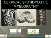 |
Cervical Arthritis, Cervical Spondylotic Myelopathy
This presentation was created by Pablo Pazmino MD. Cervical Arthritis, Cervical Spondylotic Myelopathy/ View the presentation |
References[edit | edit source]
- ↑ Richard K. Root Clinical Infectious Diseases: A Practical Approach, 1999
- ↑ 2.0 2.1 Kong LD, Meng LC, Wang LF, Shen Y, Wang P and Shang ZK. Evaluation of conservative treatment and timing of surgical intervention for mild forms of cervical spondylotic myelopathy. Exp Ther Med. 2013 Sep;6(3):852-856.
- ↑ Dai L, Ni B, Yuan W and Jia L. Radiculopathy after laminectomy for cervical compression myelopathy. J Bone Joint Surg Br. 1998 Sep;80(5):846-9.
- ↑ 4.0 4.1 4.2 4.3 4.4 4.5 4.6 4.7 Boos N and Aebi M (Eds). Spinal disorders: Fundamentals of Diagnosis and Treatment. Springer-Verlag Berlin Heidelberg. 2008.
- ↑ 5.0 5.1 5.2 5.3 5.4 5.5 5.6 5.7 Cook C, Brown C, Isaacs R, Roman M, David S and Richardson W. Clustered clinical findings for diagnosis of cervical spine myelopathy. J Man Manip Ther. 2010 Dec; 18(4): 175–180.
- ↑ Cramer GD and Darby SA. Basic and Clinical Anatomy of the Spine, Spinal Cord, and ANS. 2nd Edition. Elsevier 2008.
- ↑ 7.0 7.1 Selzer ME and Dobkin BH. Spinal Cord Injury (American Academy of Neurology Quality of Life Series). Demos Medical Publishing (New York). 2008
- ↑ 8.0 8.1 8.2 Amenta PS, Ghobrial GM, Krespan K, Nguyen P, Ali M, Harrop JS. Cervical spondylotic myelopathy in the young adult: a review of the literature and clinical diagnostic criteria in an uncommon demographic. Clin Neurol Neurosurg. 2014. 120:68-72.
- ↑ 9.0 9.1 Kadanka Z, Bednarík J, Vohánka S, Vlach O, Stejskal L, Chaloupka R et al. Conservative treatment versus surgery in spondylotic cervical myelopathy: a prospective randomised study. Eur Spine J. 2000;9(6):538-44.
- ↑ 10.0 10.1 Cook C, Roman M, Stewart KM, Leithe LG, Isaacs R. Reliability and diagnostic accuracy of clinical special tests for myelopathy in patients seen for cervical dysfunction. J Orthop Sports Phys Ther. 2009 Mar;39(3):172-8. doi: 10.2519/jospt.2009.2938.
- ↑ 11.0 11.1 Koakutsu T,Nakajo J, Morozumi N, Hoshikawa T, Ogawa S, and Ishii Y. Cervical myelopathy due to degenerative spondylolisthesis. Ups J Med Sci. 2011; 116(2): 129–132.
- ↑ 12.0 12.1 Yonenobu K. Cervical radiculopathy and myelopathy: when and what can surgery contribute to treatment? Eur Spine J. 2000; 9(1): 1-7.
- ↑ 13.0 13.1 Harrop JS, Naroji S, Maltenfort M, Anderson DG, Albert T, Ratliff JK et al. Cervical myelopathy: a clinical and radiographic evaluation and correlation to cervical spondylotic myelopathy. Spine (Phila Pa 1976). 2010 Mar 15;35(6):620-624.
- ↑ Park SJ, Kim SB, Kim MK, Lee SH and Oh IH. Clinical features and surgical results of cervical myelopathy caused by soft disc herniation. Korean J Spine. 2013;10(3):138-143.
- ↑ 15.0 15.1 Sato T, Horikoshi T, Watanabe A, Uchida M, Ishigame K, Araki T et al. Evaluation of cervical myelopathy using apparent diffusion coefficient measured by diffusion-weighted imaging. AJNR Am J Neuroradiol. 2012; 33(2):388-392.
- ↑ Harrop, James S; Naroji, Swetha; Maltenfort, Mitchell; Anderson, D. Greg; Albert, Todd; Ratliff, John K; Ponnappan, Ravi K; Rihn, Jeffery A; Smith, Harvey E; Hilibrand, Alan; Sharan, Ashwini D; Vaccaro, Alexander. Cervical Myelopathy: A Clinical and Radiographic Evaluation and Correlation to Cervical Spondylotic Myelopathy. Spine 10 February 2010 [epub ahead of print]
- ↑ 17.0 17.1 Chad Cook, Christopher Brown, Robert Isaacs, Matthew Roman, Samuel Davis, and William Richardson. Clustered clinical findings for diagnosis of cervical spine myelopathy. J Man Manip Ther. 2010 December; 18(4): 175–180.
- ↑ 18.0 18.1 18.2 Vitzthum H and Dalitz K. Analysis of five specific scores for cervical spondylogenic myelopathy. Eur Spine J. 2007; 16(12): 2096–2103.
- ↑ Tanaka N, Konno S, Takeshita K, Fukui M, Takahashi K, Chiba K et al. An outcome measure for patients with cervical myelopathy: the Japanese Orthopaedic Association Cervical Myelopathy Evaluation Questionnaire (JOACMEQ): an average score of healthy volunteers. J Orthop Sci. 2014 Jan;19(1):33-48.
- ↑ Kadanka Z, Bednarík J, Vohánka S, Vlach O, Stejskal L, Chaloupka R, Filipovicová D, Surelová D, Adamová B, Novotný O, Nemec M, Smrcka V, Urbánek I. Conservative treatment versus surgery in spondylotic cervical myelopathy: a prospective randomised study. Eur Spine J (2000) 9 :538–544
- ↑ 21.0 21.1 21.2 21.3 21.4 21.5 21.6 21.7 21.8 Almeida GP, Carneiro KK and Marques AP. Manual therapy and therapeutic exercise in patient with symptomatic cervical spondylotic myelopathy: a case report. J Bodyw Mov Ther. 2013 Oct;17(4):504-9.
- ↑ Massimo Leonardi, Norbert Boos , Degenerative disorders of the cervical spine
- ↑ 7 Law MD Jr, Bernhardt M, White AA 3rd.Cervical spondylotic myelopathy: a review of surgical indications and decision making. Yale J Biol Med. 1993 May-Jun;66(3):165-77.
- ↑ Kadaňka, Z., et al. "Conservative treatment versus surgery in spondylotic cervical myelopathy: a prospective randomised study." European Spine Journal 9.6 (2000): 538-544.
- ↑ Kadaňka, Zdeněk, et al. "Cervical spondylotic myelopathy: conservative versus surgical treatment after 10 years." European Spine Journal 20.9 (2011): 1533-1538.
- ↑ 26.0 26.1 Fouyas, Ioannis P., Patrick FX Statham, and Peter AG Sandercock. "Cochrane review on the role of surgery in cervical spondylotic radiculomyelopathy." Spine 27.7 (2002): 736-747.
- ↑ Shimomura, Takatoshi, et al. "Prognostic factors for deterioration of patients with cervical spondylotic myelopathy after nonsurgical treatment." Spine 32.22 (2007): 2474-2479.
- ↑ Rhee, John M., et al. "Nonoperative management of cervical myelopathy: a systematic review." Spine 38.22S (2013): S55-S67.
- ↑ Browder DA, Erhard RE and Piva SR. Intermittent cervical traction and thoracic manipulation for management of mild cervical compressive myelopathy attributed to cervical herniated disc: a case series. J Orthop Sports Phys Ther. 2004;34(11):701-712.
- ↑ Joghataei, Mohammad Taghi, Amir Massoud Arab, and Hossein Khaksar. "The effect of cervical traction combined with conventional therapy on grip strength on patients with cervical radiculopathy." Clinical rehabilitation 18.8 (2004): 879-887.
- ↑ Binder, Allan I. "Cervical spondylosis and neck pain." BMJ: British Medical Journal 334.7592 (2007): 527-531.
- ↑ Kay TM, Gross A, Goldsmith C, Santaguida PL, Hoving J, Brontfort G, et al, Cervical Overview Group. Exercises for mechanical neck disorders. Cochrane Database Syst Rev 2005
- ↑ Annemie I. F. Spooren, Yvonne J. M. Janssen-Potten, Eric Kerckhofs and Henk A. M. Seelen. Outcome of motor training programmes on arm and hand functioning in patients with cervical spinal cord injury according to different levels of the icf: a systematic review. J Rehabil Med 2009; 41: 497–505
- ↑ Rach Rauw. Cervical Traction Techniques. Available from: http://www.youtube.com/watch?v=ouoQd6PPJcI[last accessed 19/02/16]
- ↑ ReligiosoPT. Supine Thoracic Thrust. Available from: http://www.youtube.com/watch?v=Rj4Y5JGNPZs[last accessed 19/02/16]|}
- ↑ Nomura, Takuo, et al. "A subclinical impairment of ventilatory function in cervical spondylotic myelopathy." Archives of physical medicine and rehabilitation 85.7 (2004): 1210-1211.
- ↑ G. Aufdemkampe, J.B. Den Dekker, I. Van Ham, B.C.M. Smits-Engelsman, P. Vaes. Jaarboek fysiotherapie-kinesitherapie 2000. Bohn Stafleu Van Loghum, 275 p
- ↑ Ogawa, Yuto, et al. "Postoperative factors affecting neurological recovery after surgery for cervical spondylotic myelopathy." Journal of Neurosurgery: Spine 5.6 (2006): 483-487.
- ↑ Stanford University. Imaging Patients with Myelopathy. Available from: http://www.youtube.com/watch?v=mJVEtq5GsJk[last accessed 09/03/13]
- ↑ The Gait Guys. A Gait Case of Combined Spinal Myelopathy and Trendelenburg Pathologies. Available from: http://www.youtube.com/watch?v=AYmzQL_NSeI[last accessed 09/03/13]|}
(1) Barrett, Eva, Karen McCreesh, and Jeremy Lewis. "Reliability and validity of non-radiographic methods of thoracic kyphosis measurement: a systematic review." Manual therapy 19.1 (2014): 10-17. (LOE:1a)
(2) Persson, L. C. G., et al. "Cervical radiculopathy: Pain, muscle weakness and sensory loss in patients with cervical radiculopathy treated with surgery, physiotherapy or cervical collar A prospective, controlled study." European Spine Journal 6.4 (1997): 256-266.(LOE:2b)
(3) Tejus, M. N., et al. "An evaluation of the finger flexion, Hoffman's and plantar reflexes as markers of cervical spinal cord compression–A comparative clinical study." Clinical neurology and neurosurgery 134 (2015): 12-16. (LOE: 3b)
(4) Ross, Michael D. "Cervical myelopathy: diagnosing another great impostor." Perspectives (2014): 31. (LOE: 3a)
(5) Fukui, Mitsuru, et al. "An outcome measure for patients with cervical myelopathy: Japanese Orthopaedic Association Cervical Myelopathy Evaluation Questionnaire (JOACMEQ): part 1." Journal of Orthopaedic Science 12.3 (2007): 227. (LOE: 3b)
(6) Kobayashi, Hiroshi, et al. "Development of a self-administered questionnaire to screen patients for cervical myelopathy." BMC musculoskeletal disorders 11.1 (2010): 268. (LOE:3b)
(7) Siasios, Ioannis D., et al. "The Role of Gait Analysis in the Evaluation of Patients with Cervical Myelopathy: A Literature Review Study." World Neurosurgery 101 (2017): 275-282. (LOE: 1a)

