Thoracolumbar Fascia: Difference between revisions
Wendy Walker (talk | contribs) No edit summary |
Wendy Walker (talk | contribs) No edit summary |
||
| Line 12: | Line 12: | ||
== Anatomy [[Image:PP Thoracolumbar Fascia.png|right|200px]] == | == Anatomy [[Image:PP Thoracolumbar Fascia.png|right|200px]] == | ||
The | The TLF is a complex of several layers that separates the paraspinal muscles from the muscles of the posterior abdominal wall, quadratus lumborum (QL) and psoas major. | ||
Numerous descriptions of this structure have presented either a two-layered model or a three-layered model. | |||
These insert on the transverse and spinous processes of the lumbar vertebrae. They also attach to the iliac crest and fuse with the posterior surface of the sacrum. | These insert on the transverse and spinous processes of the lumbar vertebrae. They also attach to the iliac crest and fuse with the posterior surface of the sacrum. | ||
| Line 18: | Line 20: | ||
The illustration opposite shows the superficial muscles of the back. The thoracolumbar fascia is the gray area at bottom centre. | The illustration opposite shows the superficial muscles of the back. The thoracolumbar fascia is the gray area at bottom centre. | ||
The TLF is a girdling structure consisting of several aponeurotic and fascial layers that separates the paraspinal muscles from the muscles of the posterior abdominal wal<ref name="Willard et al">J Anat. 2012 Dec;221(6):507-36. | The TLF is a girdling structure consisting of several aponeurotic and fascial layers that separates the paraspinal muscles from the muscles of the posterior abdominal wal<ref name="Willard et al">J Anat. 2012 Dec;221(6):507-36.fckLRThe thoracolumbar fascia: anatomy, function and clinical considerations.fckLRWillard FH1, Vleeming A, Schuenke MD, Danneels L, Schleip R.</ref>l. | ||
The superficial lamina of the posterior layer of the TLF (PLF) is dominated by the aponeuroses of the latissimus dorsi and the serratus posterior inferior. The deeper lamina of the PLF forms an encapsulating retinacular sheath around the paraspinal muscles. | The superficial lamina of the posterior layer of the TLF (PLF) is dominated by the aponeuroses of the latissimus dorsi and the serratus posterior inferior. The deeper lamina of the PLF forms an encapsulating retinacular sheath around the paraspinal muscles. | ||
Revision as of 23:24, 9 August 2015
Original Editor - Wendy Walker
Lead Editors - Wendy Walker, Lucinda hampton, Kim Jackson, Sehriban Ozmen, WikiSysop, Naomi O'Reilly and Bruno Serra
Description[edit | edit source]
The thoracolumbar fascia is a large area of connective tissue - roughly diamond-shaped - which comprises the thoracic and lumbar parts of the deep fascia enclosing the intrinsic back muscles.
Most developed in the lumbar region, it consists of multiple layers of crosshatched collagen fibres that cover the back muscles in the lower thoracic and lumbar area before passing through these muscles to attach to the sacral bone.
Anatomy 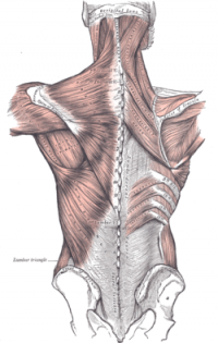 [edit | edit source]

The TLF is a complex of several layers that separates the paraspinal muscles from the muscles of the posterior abdominal wall, quadratus lumborum (QL) and psoas major.
Numerous descriptions of this structure have presented either a two-layered model or a three-layered model.
These insert on the transverse and spinous processes of the lumbar vertebrae. They also attach to the iliac crest and fuse with the posterior surface of the sacrum.
The illustration opposite shows the superficial muscles of the back. The thoracolumbar fascia is the gray area at bottom centre.
The TLF is a girdling structure consisting of several aponeurotic and fascial layers that separates the paraspinal muscles from the muscles of the posterior abdominal wal[1]l.
The superficial lamina of the posterior layer of the TLF (PLF) is dominated by the aponeuroses of the latissimus dorsi and the serratus posterior inferior. The deeper lamina of the PLF forms an encapsulating retinacular sheath around the paraspinal muscles.
The middle layer of the TLF (MLF) appears to derive from an intermuscular septum that developmentally separates the epaxial from the hypaxialmusculature.
Function[edit | edit source]
The unyielding character of the deep fascia enables it to serve as a means of containing and separating groups of muscles into relatively well-defined spaces called ‘compartments’. The deep fascia integrates these compartments and transmits load between them.
The TLF is a critical part of a myofascial girdle that surrounds the lower portion of the torso, playing an important role in the following functions[1]:
- posture
- load transfer
- respiration (Bogduk &Macintosh, 1984; Mier et al. 1985; Tesh et al. 1987; De Tro-yer et al. 1990; Vleeming et al. 1995; Hodges, 1999; Barkeret al. 2004; Gatton et al. 2010).
[edit | edit source]
Pathology/Injury[edit | edit source]
Techniques[edit | edit source]
Palpation[edit | edit source]
Examination[edit | edit source]
Treatment[edit | edit source]
Resources[edit | edit source]
Recent Related Research (from Pubmed)[edit | edit source]
Extension:RSS -- Error: Not a valid URL: Feed goes here!!|charset=UTF-8|short|max=10
References[edit | edit source]
References will automatically be added here, see adding references tutorial.

