PPIVMs of the Cervical Spine - Assessment
Introduction[edit | edit source]
Passive assessment of segmental movements includes Passive Physiological Intervertebral Movements (PPIVMs) and
Assessment of passive intervertebral movements form an important part of the physical assessment of people with musculoskeletal spinal conditions and serves to localize and characterize impairments of mobility of individual motion segments. Assessment of passive intervertebral movements can be divided into Passive Physiological Intervertebral movements (PPIVMs) and Passive Accessory Intervertebral Movements (PAIVMs). Although these terms originated with the Maitland approach, they can be used to describe assessment techniques used in a variety of approaches.
There is some contention about whether limitations of segmental mobility actually exist and, if they do, whether their assessment is relevant. At least in the cervical spine, specifically targeted treatment produces superior results to general treatment.[1] In order to specifically target treatment, it is necessary to localize impairments of individual motion segments by assessing their mobility.
PPIVMs are defined as passively producing movements in directions that can be performed actively. Like active movements, PPIVMs usually involve rotation around at least one axis
PAIVMs are defined as producing movements in directions that can not be produced actively in isolation but nonetheless need to be available for active movements to occur. In the periphery, these movements are generally glides produced by a linear force and result in translational rather than rotational movement. In the spine, PAIVMs are produced by applying a translational force, but unlike in the periphery, the movements that are produced are not purely translational.
This section will primarily consider two axes of PPIVMS of the cervical spine which will be referred to as rotation and lateral flexion.
Nomenclature[edit | edit source]
Although PPIVM techniques are frequently described as being around the three cardinal axes, the segmental movements in the cervical spine are best described as being around two rather than three axes.[2]
One axis accounts for flexion/extension and the other accounts for a movement that lies somewhere between, and includes both, rotation and lateral flexion). The directions would therefore perhaps be more accurately described as being around an axis perpendicular to the plane of the facets in extension or flexion respectively.
Nonetheless, the directions of PPIVMs will be referred to here as being in the cardinal directions to be consistent with convention.
Rotation and lateral flexion PPIVM assessment (and treatment) are often described as opening the contralateral side and closing the ipsilateral side. It is unclear what is meant to be opening or closing during these movements – the facets or the intervertebral foramina.
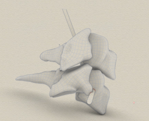
During active rotation and lateral flexion the facets largely remain in contact and glide either in anterior/superiorly or posterior/inferior directions. If opening refers to facet separation, this can be produced, but generally requires combinations of movements similar to what is done with positioning for manipulations .[3]
During rotation and lateral flexion, the intervertebral foramina increase in size on the contralateral sider and reduce on the ipsilateral side, but these differences in size are not the primary effect of the movement and are not as great as what occurs during flexion and extension.[4] The authors therefore consider that describing the direction of the movements is more accurate than using terms such as opening and closing.
PPIVMs are generally intended to be pain-free and used to assess mobility rather than symptom provocation. Symptom provocation with PPIVMs can occur and may provide useful information for localization and characterization of impairments but symptom provocation with PPIVMs is considered to be less reliable than with PAIVMs.
Technique[edit | edit source]
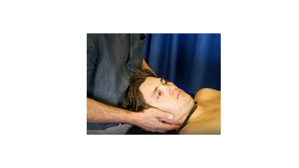
There are several variations in how PPIVMs are taught and performed. Most commonly while passively moving the neck, the therapist feels for the movement of the facets directly with their fingers. The movement of the whole region can be performed while feeling the movements which provides an indication of how individual segments move within the context of the movement of the whole neck. Alternatively the movement can be localized by adjusting the extent of flexion while feeling different levels. Regardless of whether the movement is regional or more localized, it can be difficult for therapists to learn to perceive the relevant differences in facet movement using this approach. The video below demonstrates this approach.
PPIVMs of the cervical spine resource videos, Catherine Moore & Lizzie Nile - Helm Open Repository (CC BY-NC 2.0 UK Deed). Helm – School of Health Sciences, Faculty of Medicine, University of Nottingham.
The second approach is similar to what would be done with accessory movements of most peripheral joints - hold one vertebra and localize the passive movement as much as possible to the motion segment of interest. In this instance the therapist controls the axis of movement without allowing translation thereby localizing the movement to the intended motion segment. This approach is intended to provide a clearer indication of the available segmental movement by, rather than attempting to feel the gliding of the facets, the therapist feels the movement of the motion segment directly.
The Cervical Flexion-Rotation Test which is used to assess rotation at the C1/2 level is a well known example of this type of technique . It has good reliability meaning the test is repeatable both within and between examiners, good validity meaning that it accurately isolates the movement to the intended C1/2 level [5], and is clinically relevant because it is significantly different in people with headaches than in an asymptomatic population.[6]
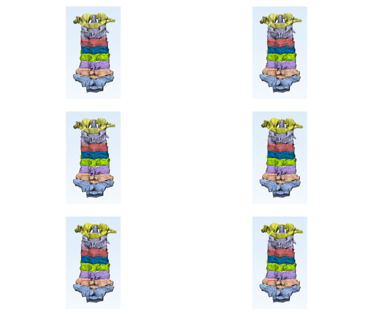
The same principle can be applied for other intervertebral levels where the therapist controls the upper vertebra such that it and all vertebrae above this level move with the head one piece. By adding a small amount of distraction, not allowing rotation outside of the intended axis, nor allowing translation to the side, the movement can be localized to the one motion segment . The intended movement is shown in the two Gifs below.
An animation of the movement that would be expected to occur with a C4-C5 rotation PPIVM to the left. Note that these images were not taken while performing an actual technique. The contours of the vertebrae were extracted from MRI images from a study participant.[7] The animation was produced from images of C1 to C4 and C5 to T1 in neutral. The two positions of C1 to C4 are: 1) in a neutral position and 2) moved by to the movement that occurred at C4-5 at the limit of active rotation.
Rotation PPIVM at C4-5. If the movement is well controlled, the amount of movement is small and can be perceived as a distinct stop.
It is very easy to not have sufficient control of the axis of movement and allow greater range of movement. When done correctly, there is typically around 5 degreed of movement to where the therapist feels a distinct stop similar to what occurs in the cervical flexion rotation test. An additional advantage of this approach for educators is that they, as well as other students, can see whether the technique is being done effectively and both educator and other students can see the relative amount of movement at each level and direction.
A more complete description of the techniques are shown in the video below.
Clinical implications[edit | edit source]
PPIVMs can be performed in a variety of ways, each with advantages, disadvantages and often providing somewhat different information. The translation of findings on PPIVMs to treatment is considered elsewhere. Briefly, since the direction segmental impairment does not necessarily correspond with imitation of of active range of movement (AROM), so too limitations found with PPIVM assessments do not necessarily occur in the same direction as limitation of AROM. It is therefore proposed that treatmetn decisions are based on the limitations found on PPIVM assessment rather than the pattern of impairments of active movement.
Resources[edit | edit source]
Manual Therapy for Cervical Spine PPIVM
References[edit | edit source]
- ↑ Slaven EJ, Goode AP, Coronado RA, Poole C, Hegedus EJ. The relative effectiveness of segment specific level and non-specific level spinal joint mobilization on pain and range of motion: results of a systematic review and meta-analysis. Journal of Manual & Manipulative Therapy (Maney Publishing). 2013;21(1):7-17.
- ↑ Bogduk, N., & Mercer, S. (2000). Biomechanics of the cervical spine. I: Normal kinematics. Clinical Biomechanics, 15(9), 633-648. Retrieved from http://www.ncbi.nlm.nih.gov/entrez/query.fcgi?cmd=Retrieve&db=PubMed&dopt=Citation&list_uids=10946096
- ↑ Salem, W., & Klein, P. (2013). In vivo 3D kinematics of the cervical spine segments during pre-manipulative positioning at the C4/C5 level. Manual Therapy
- ↑ Chang, V., Basheer, A., Baumer, T., Oravec, D., McDonald, C. P., Bey, M. J., . . . Yeni, Y. N. (2017). Dynamic measurements of cervical neural foramina during neck movements in asymptomatic young volunteers. Surgical and Radiologic Anatomy, 39, 1069-1078
- ↑ Hall TM, Robinson KW, AkasakaK. Intertester Reliability and Diagnostic Validity of the Cervical Flexion-Rotation Test. J Manipulative Physiol Ther. 2008;31:293-300
- ↑ Ogince M, Hall T, Robinson K. The diagnostic validity of the cervical flexion-rotation test in C1/2 related cervicogenic headache. Man Ther 2007;12:256-262
- ↑ Tuttle, N., Rocha, C. S. d. S., Sheehan, B., Kennedy, B. A., & Evans, K. (2019). Measurement of three-dimensional cervical segmental kinematics: Reliability of whole vertebrae and facet-based approaches. Musculoskeletal Science and Practice, 44, 102039.


