
Leprosy
Top Contributors - Manisha Shrestha, Stacy Schiurring, Jess Bell, Kim Jackson, Nikhil Benhur Abburi and Nupur Smit Shah
Aetiology
Leprosy (also known as Hansen's disease) is caused by Mycobacterium leprae (M. leprae) and M. lepromatosis bacterium. These mycobacterium are highly infectious but have minimal pathogenicity. They multiply extremely slowly, with an incubation period ranging from 2 to 12 years, but averaging 5 years.[1][2][3]
Leprosy is transmitted primarily through person-to-person spread, via nasal droplets. However, the exact mode of transmission is not conclusively proven. Humans serve as the principal reservoir of infection worldwide. In the Americas, nine-banded armadillos provide a secondary reservoir—two-thirds of cases in the southern United States are armadillo-derived strains. Most people infected with M. leprae do not develop clinical disease, and currently no tools exist to diagnose subclinical infection.[1]
Leprosy can be treated with a combination of antibiotics known as multidrug therapy. With early diagnosis and proper treatment, leprosy can be completely cured and its devastating complications prevented.
Risk factors for developing leprosy include close, prolonged contact with untreated persons—household contacts face the highest transmission risk. Host genetic factors significantly influence leprosy susceptibility and disease presentation. Genome-wide studies have identified specific gene variants that regulate immune responses, affecting both infection risk and clinical manifestations. Additional risk factors include immunocompromising conditions, residence in endemic areas, and socioeconomic factors, such as poverty and overcrowding.[1]
Epidemiology
Current global epidemiology shows that leprosy remains a significant public health problem despite substantial progress. Approximately 250,000 new cases are detected annually worldwide. 94% of these cases occur in 17 countries that report more than 1,000 new cases per year.[1] The number of new cases reported globally to World Health Organization (WHO) in 2023 was 182,815 from 184 countries.[4] Brazil and India continue to have high transmission rates. Surveys in Maharashtra, India, revealed rates of 3-9 cases per 10,000 population, with 30% of cases occurring in children. While 15 million people have been treated with multidrug therapy and an estimated 2 million have been prevented from developing disabilities, leprosy continues to be the leading infectious cause of disability globally.[1]
Clinical Presentation
Leprosy is a chronic granulomatous inflammation of the skin and peripheral nerves. The clinical manifestation is entirely determined by the host's cell-mediated immune response to infection. The disease should be understood primarily as a peripheral neuropathy with secondary skin manifestations, existing along an immunological spectrum, from strong immune response (tuberculoid forms) to weak or absent response (lepromatous forms).[1][2]
Patients with strong cell-mediated immunity develop the tuberculoid form of leprosy, which is characterised by effective bacterial control, few lesions with low bacterial load, and granuloma formation. People with this form of leprosy do risk nerve damage from the inflammatory response itself. Conversely, patients with weak or absent cell-mediated immunity develop the lepromatous form of leprosy with poor bacterial control, multiple lesions with high bacterial loads, systemic bacterial dissemination, and an anergic immune response to the pathogen.[1][2][3]
A leprosy reaction is an acute inflammatory episode that can cause rapid, permanent nerve damage. It affects approximately 30% of multibacillary patients during or after multidrug therapy. Type 1 reactions involve the acute inflammation of existing lesions and nerves. They occur in borderline forms during immune system changes and require immediate corticosteroid intervention. Type 2 reactions (also known as erythema nodosum leprosum) occur in about 50% of patients with lepromatous leprosy. They involve systemic inflammation of the skin, nerves, eyes, and testes. These episodes are difficult to manage and may remit and relapse over several years. There are limited treatment options for type 2 reactions and an insufficient evidence base for optimal management strategies.[1][2]
Classification Systems
Two major classification systems guide clinical management. The WHO operational classification, used primarily for treatment decisions, divides patients into paucibacillary (≤5 skin lesions) and multibacillary (>5 skin lesions) categories.[5] The more detailed Ridley-Jopling histopathological classification recognises five forms of leprosy based on immune response and bacterial load: (1) tuberculoid, (2) borderline tuberculoid, (3) mid-borderline, (4) borderline lepromatous, and (5) lepromatous. Research indicates that up to 60% of borderline tuberculoid patients may be overtreated when classified as multibacillary using the WHO criteria, highlighting the importance of accurate classification in referral centres.[3][6]
The WHO disability grading system for leprosy provides a standardised method for assessing and documenting impairments. It includes separate evaluations for the hands, feet, and eyes that are graded individually based on the severity of functional loss and visible deformity. The WHO system uses a three-point scale.
Grade 0 represents no impairment—there is no anaesthesia, visible deformity, or damage present. There are no eye problems due to leprosy with no evidence of visual impairment.
Grade 1 indicates impairment is present but without visible deformity. It is characterised by anaesthesia in the hands and feet but no visible deformity or damage. Eye problems due to leprosy are present but they do not severely affect vision; visual acuity remains 6/60 or better and the ability to count fingers at 6 metres is maintained.
Grade 2 is the most severe category. It indicates visible deformity or severe impairment is present—there is visible deformity or damage present in the hands and feet. There is severe visual impairment with vision worse than 6/60 or an inability to count fingers at 6 metres.[7]
Signs and Symptoms
Nerve damage is the most serious complication of leprosy. It can occur before, during, and after treatment. Epidemiological data reveals that 47% of patients already have established nerve impairment at diagnosis, 8% show recent nerve damage at presentation, and 12% develop nerve damage after treatment initiation. Only 33% of people have no nerve involvement throughout their disease course. The degree of nerve damage at diagnosis reflects the often years-long delay between symptom onset and diagnosis, during which time neuropathy can develop almost unnoticed. The inflammatory process in nerves is driven by mycobacterial antigens that activate a destructive immune response mediated by CD4+ cells and macrophages. It also invovles multiple pro-inflammatory cytokines including tumor necrosis factor alpha.[1][2]
Common neurological symptoms include partial loss of mobility, unilateral facial paralysis, sensory deficits, motor dysfunction, and autonomic irregularities. A substantial proportion of patients with leprosy experience neuropathic pain, with sensory symptoms such as numbness and tingling being frequently reported.[8]
Physical deformities/impairments in leprosy result primarily from peripheral nerve damage. They can affect multiple anatomical regions, creating significant functional impairments and contributing to ongoing stigma. Hand deformities are some of the most common and debilitating complications, including claw hand deformity, where ulnar and median nerve involvement lead to flexion contractures of the fingers, "ape thumb" deformity, where median nerve damage leads to a loss of thumb opposition, and wrist drop, where radial nerve involvement prevents wrist extension. Sensory loss over the palm significantly increases the risk of unnoticed injuries and burns, which further compound functional limitations.[9]
Foot deformities/impairments include foot drop, where common peroneal nerve damage causes a characteristic high-stepping gait, claw toes, where there are flexion deformities of the digits, and plantar anaesthesia, which leads to loss of protective sensation on the sole of the foot. This sensory loss frequently results in plantar ulceration, with chronic wounds developing due to repeated pressure and trauma that goes unnoticed by patients. These foot complications significantly impact mobility and independence and often require specialised footwear and ongoing wound care management.[9]
Facial and eye deformities pose serious risks to vision and facial function. Lagophthalmos, where there is an inability to close the eyelids due to facial nerve involvement, is one of the most concerning complications. This condition leads to corneal anaesthesia and dryness, which creates substantial risk for corneal ulceration and potential blindness if left untreated. Additional facial manifestations include the loss of eyebrows and eyelashes, and partial or complete facial paralysis that is typically unilateral. Nasal deformities involve the destruction of nasal cartilage, resulting in a collapsed nose appearance and perforated nasal septum. This can affect breathing and contribute to the stigmatising appearance associated with advanced leprosy.[9]
-
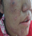 Skin lesions on the face
 -
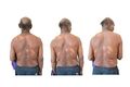 Skin lesions on the back of a patient with leprosy
 -
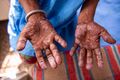 Trophic changes of the hands
 -
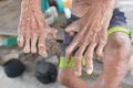 Claw finger deformities, palmer view
 -
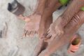 Claw finger deformities, dorsal view
 -
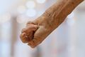 Severe hand contracture related to leprosy
 -
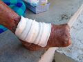 Leprosy-related amputation of the toes
 -
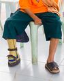 Leprosy-related amputations on multiple limbs


Diagnosis Process
The World Health Organization recommends diagnosing leprosy when patients present with at least one of the following: hypopigmented or reddish skin lesions with sensory loss; thickened peripheral nerves causing weakness or sensory deficits; or when acid-fast bacilli is detected in a split skin smear or biopsy. When all three criteria are met, diagnostic accuracy reaches 95%. Clinical examination should focus on assessing temperature sensation, nerve thickening, and neurological changes. Particular attention should be paid skin lesions with hypo/anaesthetic sensations on the face, buttocks, trunk, and earlobes.[2]
Laboratory confirmation relies primarily on bacteriological methods, including slit skin smear examination and histopathological analysis. While slit skin smears have nearly 100% specificity, their sensitivity is low at only 18-30%, limiting their utility in paucibacillary cases. Polymerase chain reaction testing offers improved sensitivity and specificity. This type of testing is particularly valuable for patients with low bacterial loads, though it is expensive and labour-intensive. Serological testing provides a less invasive diagnostic option, with sensitivities ranging from 34-77% depending on the specific antigen tested, though it is most effective in multibacillary cases.[2]
Advanced diagnostic techniques include electrophysiological nerve studies, ultrasonography, and magnetic resonance neurography, which help evaluate nerve involvement and monitor disease progression. Nerve conduction studies and warm detection threshold testing can identify early neuropathy, while high-resolution ultrasonography detects nerve thickening with greater sensitivity than clinical palpation (47% versus 20%). Magnetic resonance neurography provides detailed assessment of proximal nerve involvement and can identify central nervous system complications in some patients. These advanced techniques are particularly valuable for early diagnosis and monitoring treatment response, though their availability remains limited in resource-constrained endemic regions where leprosy is most prevalent.[2]
Medical Treatment Considerations
The medication treatment of leprosy has evolved significantly since the 1950s when dapsone monotherapy was first introduced. However, the emergence of dapsone-resistant M. leprae after nearly 30 years of single-drug treatment led the WHO to recommend multidrug therapy (MDT) in 1981.
Multidrug therapy combines dapsone, rifampicin, and clofazimine. Current WHO guidelines, standardised globally in 2018, recommend treating patients with paucibacillary leprosy with all three drugs for 6 months. They recommend treating patients with multibacillary leprosy for 12 months, with monthly supervised administration in endemic regions. This approach has dramatically reduced leprosy prevalence, decreased drug resistance rates, and lowered recurrence and reaction rates, though rifampicin-resistant M. leprae remains a concern, with an 11% incidence rate.[2] While early multidrug therapy can prevent new or further nerve damage, it cannot reverse nerve damage that have already occurred.
For drug-resistant cases, the WHO recommends alternative second-line therapies including ofloxacin, minocycline, and clarithromycin, with treatment periods extending up to 24 months depending on the resistance pattern. Rifampicin-resistant cases require initial intensive therapy with ofloxacin, minocycline, and clofazimine for 6 months, followed by 18 months of maintenance therapy. When both rifampicin and ofloxacin resistance is present, treatment relies on clarithromycin, minocycline, and clofazimine combinations. Treatment monitoring increasingly incorporates serological testing, as bacterial load correlates with immunoglobulin M (IgM) antibody titres against M. leprae-specific PGL-1 antigens.[2]
Prevention strategies focus on prophylactic immunisation and chemoprophylaxis for high-risk contacts. The Bacillus Calmette-Guérin (BCG) vaccination provides 20-90% protection against leprosy infection, with effectiveness varying depending on regional factors and infectious agent characteristics. Studies from Paraguay demonstrate that comprehensive BCG vaccination programmes, combined with contact tracing and early diagnosis, can significantly reduce leprosy incidence rates. For chemoprophylaxis, single-dose rifampicin (SDR) treatment of household contacts has proven effective in reducing infection risk, with the WHO recommending this approach since 2018. Whilst concerns exist about rifampicin resistance development, studies suggest the benefits of rifampicin chemoprophylaxis substantially outweigh the risks, particularly when combined with BCG vaccination and comprehensive contact management strategies.[2]
Pain management for leprosy neuropathy requires a multidimensional approach, combining pharmacological and surgical interventions. Tricyclic antidepressants, particularly amitriptyline, remain the mainstay of neuropathic pain treatment. However, treatment failures often occur due to misdiagnosis and improper dosing. Gabapentin and pregabalin show limited benefit, leading to investigations into combination therapies for adequate pain relief. Selective serotonin-norepinephrine reuptake inhibitors (SSRIs) demonstrate potential and have fewer side effects than tricyclic antidepressants. Corticosteroids are frequently used to manage leprosy reactions (which affect approximately 40% of patients), though their efficacy depends heavily on dosage and treatment duration. There are also concerns about the side effects of corticosteroids, including increased infection risk, weight gain, elevated blood sugar, and mood disturbances.[8]
Leprosy neuropathy can also lead to the formation of unnoticed injuries and wounds that can become severely infected and require amputation of the affected digit or limb. Reconstructive surgery for patients with leprosy address deformities in the hands, feet, eyes, and nose that result from nerve damage, aiming to restore both function and form while reducing stigma and disability. Hand procedures include claw correction, opponenplasty for thumb opposition, wrist drop correction, arthrodesis for stabilisation, and tissue reconstruction through contracture release and flap coverage. Foot surgeries include foot drop correction, claw toe correction, soft tissue reconstruction of the sole, and stabilisation procedures, while eye surgeries focus on correcting lagophthalmos (the inability to close the eyelids) and nasal procedures to reconstruct collapsed noses. Tendon transfer procedures are also common to improve functional range of motion. Pre- and post-operative physical rehabilitation is considered a crucial component of the surgical process.[10]
Rehabilitation Treatment Considerations
Patients with leprosy can experience significant physical disabilities primarily due to the peripheral nerve damage caused by the disease's immunological responses and inflammatory reactions. People with lepromatous leprosy are at particularly high risk, with 5- to 12-fold greater odds of developing disabilities (because their immune system fails to control bacterial growth in Schwann cells, leading to nerve injury).[11]
Assessment and monitoring: regular evaluation of nerve function impairment (NFI) through nerve palpation, sensory testing, using Semmes-Weinstein monofilaments, and motor testing using the Medical Research Council (MRC) grading scale is required. These assessments should be conducted before starting treatment, monthly during multidrug therapy, every two weeks during steroid courses, and at treatment completion.
Prevention of further deterioration: comprehensive interventions are required to prevent deterioration, including exercise programmes based on muscle strength grading, splinting for neuritis and deformity correction, and self-care education.[12]
The primary goal of physical rehabilitation is to restore an individual to maximal physical wellbeing, while secondary goals involve customising lifestyle modifications for patients, families, and communities.
Specific Therapeutic Interventions
Exercise therapy: patients should receive tailored exercise programmes based on muscle strength—passive exercises for paralysed muscles (Grade 0), active and active-assisted exercises for weak muscles (Grades 1-3), and active-resisted exercises for strong muscles (Grades 4-5). Muscle weakness of lesser duration tends to recover with appropriate exercise and a course of steroids.
Splinting programmes: various splints can be prescribed, including ulnar neuritis slabs for acute nerve inflammation, functional foot slabs for foot drop, and specialised splints for pre- and post-operative care. These devices provide rest, warmth, and support to affected nerves and muscles while preventing contractures.
Occupational therapy: includes the provision of assistive devices, like padded utensils, grip aids, and adaptive equipment to compensate for impairments and increase independence in activities of daily living. The goal of occupational therapy is to enhance or enable meaningful participation in activities of daily living and work- and leisure-related activities.
Self-care education: patients should be provided intensive training on protective behaviours, including daily inspection of anaesthetic areas, proper wound care, use of protective eyewear and footwear, and lifestyle modifications. Every patient with a leprosy-related impairment needs to be educated about self-care.
Surgical rehabilitation: comprehensive pre- and post-operative rehabilitation protocols for tendon transfer procedures aim to restore function and include muscle strengthening, range of motion exercises, and functional retraining, typically over a structured 4 week programme.[12]
Addressing Leprosy-Related Stigma
Leprosy-related stigma remains one of the most enduring and devastating forms of health-related discrimination, despite the disease being entirely curable with modern treatment. The stigma is as old as the disease itself, with writings that date back to 2000 BC reporting on perceptions about those affected to be unclean, untrustworthy, and morally corrupt. Today, this ancient prejudice continues to manifest in profound ways that extend far beyond medical concerns. In many countries where leprosy remains endemic, affected individuals are still forced to live as outcasts. This can lead to a multitude of mental health issues associated with isolation and discrimination, thereby perpetuating a cycle of ill health. The consequences are devastating: patients delay seeking treatment due to fear of social rejection, families face dissolution, employment opportunities vanish, and entire communities maintain discriminatory practices that prevent early diagnosis and effective treatment. This stigma not only causes immense personal suffering, it actively undermines global leprosy elimination efforts—it may prevent people from seeking treatment, which, in turn, can exacerbate issues surrounding the allocation of funding and infrastructure to provide treatment.[13]
A 2024 systematic review identified six methods to address leprosy-related stigma.
Information, education, and communication (IEC) interventions include counselling programmes using peer and lay counsellors, social skills training, awareness packages, resilience training, and participatory video production. Counselling from peers (those also currently or previously affected by leprosy) reduced stigma more than with lay counsellors by allowing those affected to relate to their counsellor. IEC packages delivered door-to-door showed significant results, with the proportion of the population holding stigmatising views about those with leprosy falling from 86% to 61% post-intervention.
Community-led projects focus on changing influential community members' attitudes through programmes like the "leprosy friendly village (LFV)" model, which targets religious leaders, village leaders, and other community influencers. Participants in the LFV reported perceiving less stigma and receiving better acceptance from their community members post-implementation.
Socioeconomic rehabilitation involves mainstreaming affected individuals into existing microfinance businesses while creating alternative financing options. This "twin-track" approach showed that pre- and post-intervention self-stigma score decreased by 8.5 points.
Mixed interventions combine multiple approaches, such as integrating people into communities while providing educational materials to address misconceptions. Contact interventions showed substantial impact, with the adjusted mean total score of the Explanatory Model Interview Catalogue Community Stigma Scale (EMIC-CSS) reducing by 4.95 points among respondents who had attended a contact event.
Integration within health systems involves treating leprosy through the entire health system rather than just by designated staff. Studies found stigma was minimal in the integrated areas when compared to the vertical areas.
Cosmetic or surgical care includes cosmetic camouflage for visible lesions and reconstructive surgery. Cosmetic interventions showed dramatic improvements, with quality of life scores improving from 16.67 to 2.23, while surgical correction increased patient satisfaction with societal acceptance from 1% to 51%.[13]
Resources
Clinical Resources
- Physical Rehabilitation In Leprosy ( International textbook of Leprosy)
- SWCR Guidelines for Leprotic Ulcers (The Society for Wound Care and Research)
Optional Video
The following video provides an overview of the pathology and aetiology of leprosy, and explores the on-going stigma associated with this condition.
References
- ↑ 1.0 1.1 1.2 1.3 1.4 1.5 1.6 1.7 1.8 Rodrigues LC, Lockwood DN. Leprosy now: epidemiology, progress, challenges, and research gaps. The Lancet infectious diseases. 2011 Jun 1;11(6):464-70.
- ↑ 2.00 2.01 2.02 2.03 2.04 2.05 2.06 2.07 2.08 2.09 2.10 Huang CY, Su SB, Chen KT. An update of the diagnosis, treatment, and prevention of leprosy: A narrative review. Medicine. 2024 Aug 23;103(34):e39006.
- ↑ 3.0 3.1 3.2 Silva MJ, Silva CS, Brasil TP, Alves AK, Dos Santos EC, Frota CC, Lima KV, Lima LN. An update on leprosy immunopathogenesis: systematic review. Frontiers in Immunology. 2024 Sep 6;15:1416177.
- ↑ World Health Organization. Leprosy/Hansen Disease: Management of reactions and prevention of disabilities. Available from: https://www.who.int/data/gho/data/themes/topics/leprosy-hansens-disease (accessed 16 August 2025).
- ↑ World Health Organization. Leprosy. Available from: https://www.who.int/news-room/fact-sheets/detail/leprosy#:~:text=After%20detailed%20consultations%20with%20countries,at%20WHA78%2026%20May%202025 Lasted accessed: 16/August/2025.
- ↑ Alrehaili J. Leprosy classification, clinical features, epidemiology, and host immunological responses: failure of eradication in 2023. Cureus. 2023 Sep 6;15(9).
- ↑ The World Health Organization. Guidelines for the Diagnosis, Treatment and Prevention of Leprosy. Available from: https://iris.who.int/bitstream/handle/10665/274127/9789290226383-eng.pdf (accessed 16/August/2025).
- ↑ 8.0 8.1 Calderone A, Aloisi MC, Casella C, Fiannacca S, Cosenza B, Quartarone A, Calabrò RS. The neurological impact of leprosy: manifestations and treatment approaches. Neurology International. 2024 Nov 16;16(6):1492-508.
- ↑ 9.0 9.1 9.2 Mowla MR, Angkur DM, Hasan Z, Sultana MN, Afrin S, Akhter MS. Leprosy patients with deformities at post-elimination stage: The Bangladesh experience. Skin Health and Disease. 2021 Mar;1(1):ski2-5.
- ↑ Shrivastava K, Ray S, Rathod H, Wahegaonkar AL, Jadhav SL, Mahajan A, Verma P, Palal D, Sengupta S, Srivastava K, Johnson S. Transforming Lives: A Qualitative Study Seeking Insights Into Reconstructive Surgery for Leprosy Patients in Western Maharashtra. Cureus. 2024 Sep 1;16(9).
- ↑ De Paula HL, De Souza CD, Silva SR, Martins-Filho PR, Barreto JG, Gurgel RQ, Cuevas LE, Santos VS. Risk factors for physical disability in patients with leprosy: a systematic review and meta-analysis. JAMA dermatology. 2019 Oct 1;155(10):1120-8.
- ↑ 12.0 12.1 Pradhan S, Rout AN. Physical rehabilitation in leprosy. InClinical Cases in Leprosy 2022 Sep 21. Cham: Springer International Publishing.
- ↑ 13.0 13.1 Willis M, Fastenau A, Penna S, Klabbers G. Interventions to reduce leprosy related stigma: A systematic review. PLOS Global Public Health. 2024 Aug 22;4(8):e0003440.
- ↑ YouTube. Leprosy: A Horrifying History | Into the Shadows. Available from: https://www.youtube.com/watch?v=odXh00mz6dw [last accessed 16/August/2025]