Lateral Pterygoid
Original Editor - Alyssa Brooks-Wells
Top Contributors - Alyssa Brooks-Wells and Kim Jackson
Description[edit | edit source]
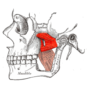
The lateral pterygoid muscle is a craniomandibular muscle that helps control movement of the temporomandibular joint (TMJ). It has two heads, superior and inferior, and the inferior is three times larger than the superior. The lateral pterygoid muscle's fibers are horizontally directed. Dysfunction of this muscle may possibly contribute to temporomandibular disorders.[1][2]
Origin[edit | edit source]
Each head of the lateral pterygoid originates from separate locations and converges to its insertion point. The origins of each head include:
- Superior Head (Sphenoid Head): the infratemporal surface of the greater wing of the sphenoid and the superior quarter of the lateral wing of the pterygoid process[2]
- Inferior head: lateral pterygoid plate of the sphenoid[3]
Insertion[edit | edit source]
The lateral pterygoid muscle inserts onto the pterygoid fossa of the mandible, the capsule of the temporomandibular joint, and the articular disc on their respective sides. [3]
The insertion onto the disc has been debated among researchers and professionals, but there is agreement that some fibers of the superior head of the lateral pterygoid attach to the anteromedial border and medial aspect of the articular disc. [2]
Nerve[edit | edit source]
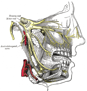
The lateral pterygoid muscle is innervated by the muscular branches from the anterior division of the mandibular nerve. The mandibular nerve is the third branch of the trigeminal nerve also known as the fifth cranial nerve (CN V3). [3]
In variations, the lateral pterygoid may also be innervated by the deep temporal nerves, the masseteric nerve, and the buccal nerve.[2]
Artery[edit | edit source]
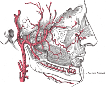
Muscular branches of the maxillary artery supply the lateral pterygoid muscles. [3] The second portion of the maxillary artery travels between the two heads of the pterygoid muscle and enters the pterygomaxillary fissure. [2]
Function[edit | edit source]
The function of the lateral pterygoid muscle has been debated, and recent studies suggest the two heads of the muscle function more similarly than previously believed. This change in belief of its function may be due to errors in electromyography interpretation, as the position of the sensors was not able to be exactly determined.[2]
Overall the lateral pterygoid is a muscle of mastication. It is the only muscle of mastication that depresses the mandible. The muscle has the following actions:
- Bilaterally: anteriorly protrudes the mandible
- Inferior head: depresses, protrudes, and contralaterally deviates the mandible
- Superior head: may be active with clenching to prevent the posterior displacement of the mandibular condyle to avoid exertion on the post-condylar structures
- Lateral pterygoid with the anterior head of digastric and mylohyoid muscles: mandibular depression and mouth opening
- Unilateral lateral pterygoid with the medial pterygoid ipsilaterally: lateral excursion to the contralateral side (observed during chewing and clenching) [2]
Clinical relevance[edit | edit source]
Both the superior and inferior heads of the lateral pterygoid generate horizontal forces required for producing forward and lateral motions of the mandible required for chewing and grinding. Dysfunction of this muscle can therefore affect mastication.
Spasms of the lateral pterygoid can cause lockjaw. Poor coordination of the superior and inferior heads can disrupt the position of the articular disc of the TMJ which may contribute to temporomandibular disorders (TMD). Typically, patients with TMD present with pain of the lateral pterygoid with palpation and mandibular movements. [2]
Resources[edit | edit source]
- ↑ Murray GM, Bhutada M, Peck CC, Phanachet I, Sae-Lee D, Whittle T. The human lateral pterygoid muscle. Archives of Oral Biology. 2007;52:377-380.
- ↑ 2.0 2.1 2.2 2.3 2.4 2.5 2.6 2.7 Rathee M, Jain P. Anatomy, Head and Neck, Lateral Pterygoid Muscle. In: StatPearls. Treasure Island: StatPearls Publishing, 2022.
- ↑ 3.0 3.1 3.2 3.3 Netter FH. Head and Neck. In: Hansen JT editor. Atlas of Human Anatomy. Philadelphia: Saunders Elsevier, 2014. p170.
- ↑ Human Anatomy Lessons. Lateral Pterygoid muscle | Origin | Insertion | Nerve Supply | Actions | Relations. Available from: https://www.youtube.com/watch?v=Mid-cKmTUM0 [last accessed 26/11/2023]


