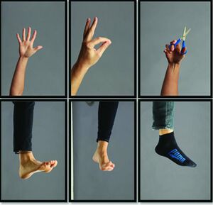
Graded Motor Imagery
Original Editor - Melissa Coetsee
Top Contributors - Melissa Coetsee and Kim Jackson
Description[edit | edit source]
Graded Motor Imagery (GMI) is a treatment program that aims to sequentially activate the premotor and primary motor cortices through three steps[1]:
- Laterality recognition: left/right discrimination
- Explicit Motor Imagery: imagined movements of the affected extremity
- Mirror therapy: view reflected movement of the unaffected extremity
GMI was developed by Moseley et al. (NOI group) as a treatment approach to address pain and function in patients with chronic CRPS, but it has since been applied to other complex pain and movement disorders.
Mechanism of Action[edit | edit source]
GMI is basically brain exercises aimed at addressing cortical changes associated with pain and movement dysfunction[2]. Each component targets a different brain area/function. Researchers believe the effectiveness is related to forced attention given to the affected extremity, reduced fear of movement, increased large fibre inhibition and the reorganisation of sensorimotor incongruence[1].
The sequence allows for the pre-motor cortex to be activated without setting off other cortical networks involved with movement.[1]Imagined movements utilises mirror neurons which start firing when we think of movement or observe a movement.
GMI is a 'top-down' treatment paradigm that emphasises the importance of addressing cortical changes[3].
Indication[edit | edit source]
GMI can be used for any condition where the sensorimotor cortex is affected. The sensorimotor cortex of an affected body part has been shown to be less active in both CRPS and phantom limb pain[3]. In chronic pain, changes in cortical mapping of the affected body part has also been observed, as well as poor functioning of mirror neurons[3]. It has also been proposed that elements of GMI can be used as a tool to prevent sensorimotor changes during periods of immobilisation (eg following a radius fracture)[4].
Although GMI was originally developed to treat chronic CRPS, studies have proven its effectiveness in treating the following conditions:
- CRPS-1[5][6]
- Phantom limb pain[7][8]
- Post-stroke limb pain[9][10]
- Distal radius fracture [4]
- Post lumbar surgery[11] - limited evidence, but promising results from a case report
- Frozen shoulder[12][13] - limited evidence, but promising results from a case report and RCT, with potential added benefits when used in conjunction with PNE and tactile discrimination training.
- Musician's Focal Dystonia[14] - limited evidence for modified GMI
- Knee osteoarthritis[15]
- Knee arthroplasty[16]
- Cancer neuropathic pain - some evidence that GMI may improve cancer pain in children[17]
Contra-indications & Precautions[edit | edit source]
GMI should not be approached with caution in the following cases[1]:
- The patient has an inability to establish ownership of the mirrored extremity
- If pain increases or movement disorders worsen during/after treatment
- Avoid explicit motor imagery if patient has experienced significant trauma or has symptoms of PTSD[18]
- If a patient experiences pain/sweating of the affected side during MVF, the frequency/complexity of the movement needs to be reduced
Components[edit | edit source]
Each step is applied for about 2 weeks (or until the patient meets the criteria to progress) before progressing to the next step[1]. The sequence of components is important to systematically target the cortical system to allow for reorganisation. It is important that each step should be adapted in order to ensure the patient experiences minimal pain during the task[3].
1. Laterality Training[edit | edit source]
Left/right discrimination is the process of recognising one side of the body as district from the other and is trained by reviewing images of left and right limbs[19]. The inability to recognise the affected limb accurately is related to disrupted sensory and motor pathways. Laterality training aims to establish an accurate cortical representation of the body part - this helps to ensure optimal effects with subsequent cortical training[3].
- Method: The patient is shown a sequence of images of the affected limb, and asked to identify each image as a right or left limb. Recognise Apps[20] or flash cards (NOI group) or magazines can be used.
- For the spine (neck/back) the direction of rotation/bending should be identified
- Frequency: Should practise x4 per day
- Progression: Increase the number of cards/images or increase the difficulty of the images[3]
- Normal responses:
2. Explicit Motor Imagery[edit | edit source]
Explicit motor imagery is the process of thinking about moving without actually moving[2]. By imagining movements, similar brain areas to those involved in actual movement, are activated. Thus, imaginary movements can help train the brain towards movement. This is very useful for patients who struggle to initiate movement, or who have poor quality of movement[18].
- Indications: Patient reports being unsure about where the limb is, experiences pain by just thinking about moving the limb and/or pain increases with movement initiation
- Method: The patient should imagine themselves moving by matching specific positions/movement queues. Begin imagined movements on the opposite side, or same side away from the painful site and gradually move towards the painful area. Begin with small amplitude imagined movements, gradually progressing to larger movements.
- Progress from a quiet place to busier environments
- Add more elements to the imagined experience (eg. warmth, smells, sounds)
- Frequency: Should be practised 7 times per day[18]
3. Mirror Visual Feedback[edit | edit source]
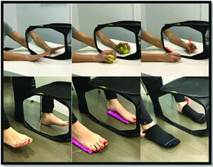
MVF uses movements of the unaffected body part to 'trick' the brain into thinking the affected part is moving[2]. By putting the affected limb behind a mirror, and looking at the reflection of movements caused by the the unaffected side, the affected side is actually being 'exercised' in the brain. Although mirror therapy can be used in isolation, it's effectiveness can be enhanced by first establishing good left/right discrimination and imagined movements.
- Method: Completely conceal the affected limb, and the patient should be well supported and comfortable. Both limbs should start in the same position.
- Start with exercises that only involve looking at the reflection (no movement)
- Slowly start congruent movements of both limbs whilst looking at the reflection. The affected side moves only to the point where pain starts, while the unaffected side (and as a result the reflected movement) continues to full ROM.
- Start with simple, pain free movements and progress to more complex movements[21]
- Frequency: At least 5min, max 10min; 4-9 times daily
The following video demonstrates the process of MVF:
| Do's | Dont's |
|---|---|
| Warn patient that is may feel strange and they can stop any time | Sit too far back, resulting in both arms being visible |
| Remove jewellery/ watch | Continue if symptoms worsen |
| Allow time for patient to look at the reflection until they are convinced of the illusion and feel comfortable with it | Perform unilateral/ asynchronous movements |
Resources[edit | edit source]
References[edit | edit source]
- ↑ 1.0 1.1 1.2 1.3 1.4 Harden RN, McCabe CS, Goebel A, Massey M, Suvar T, Grieve S, Bruehl S. Complex regional pain syndrome: practical diagnostic and treatment guidelines. Pain Medicine. 2022 May 1;23(Supplement_1):S1-53.
- ↑ 2.0 2.1 2.2 2.3 2.4 NOI group. Graded Motor Imagery. Available from: https://www.noigroup.com/graded-motor-imagery/. (accessed 2 Oct 2023).
- ↑ 3.0 3.1 3.2 3.3 3.4 3.5 Priganc VW, Stralka SW. Graded motor imagery. Journal of hand therapy. 2011 Apr 1;24(2):164-9.
- ↑ 4.0 4.1 Dilek B, Ayhan C, Yagci G, Yakut Y. Effectiveness of the graded motor imagery to improve hand function in patients with distal radius fracture: A randomized controlled trial. Journal of Hand Therapy. 2018 Jan 1;31(1):2-9.
- ↑ Méndez-Rebolledo G, Gatica-Rojas V, Torres-Cueco R, Albornoz-Verdugo M, Guzmán-Muñoz E. Update on the effects of graded motor imagery and mirror therapy on complex regional pain syndrome type 1: A systematic review. Journal of back and musculoskeletal rehabilitation. 2017 Jan 1;30(3):441-9.
- ↑ Strauss S, Barby S, Härtner J, Pfannmöller JP, Neumann N, Moseley GL, Lotze M. Graded motor imagery modifies movement pain, cortical excitability and sensorimotor function in complex regional pain syndrome. Brain Communications. 2021 Oct 1;3(4):fcab216.
- ↑ Limakatso K, Madden VJ, Manie S, Parker R. The effectiveness of graded motor imagery for reducing phantom limb pain in amputees: a randomised controlled trial. Physiotherapy. 2020 Dec 1;109:65-74.
- ↑ Limakatso K, Cashin AG, Williams S, Devonshire J, Parker R, McAuley JH. The Efficacy of Graded Motor Imagery and Its Components on Phantom Limb Pain and Disability: A Systematic Review and Meta-Analysis. Canadian Journal of Pain. 2023 Dec 31;7(1):2188899.
- ↑ Polli A, Moseley GL, Gioia E, Beames T, Baba A, Agostini M, Tonin P, Turolla A. Graded motor imagery for patients with stroke: a non-randomized controlled trial of a new approach. European journal of physical and rehabilitation medicine. 2016 Jul 21;53(1):14-23.
- ↑ Ji EK, Wang HH, Jung SJ, Lee KB, Kim JS, Jo L, Hong BY, Lim SH. Graded motor imagery training as a home exercise program for upper limb motor function in patients with chronic stroke: A randomized controlled trial. Medicine. 2021 Jan 1;100(3).
- ↑ Louw A, Schmidt SG, Louw C, Puentedura EJ. Moving without moving: immediate management following lumbar spine surgery using a graded motor imagery approach: a case report. Physiotherapy Theory and Practice. 2015 Oct 3;31(7):509-17.
- ↑ Sawyer EE, McDevitt AW, Louw A, Puentedura EJ, Mintken PE. Use of pain neuroscience education, tactile discrimination, and graded motor imagery in an individual with frozen shoulder. journal of orthopaedic & sports physical therapy. 2018 Mar;48(3):174-84.
- ↑ Gurudut P, Godse AN. Effectiveness of graded motor imagery in subjects with frozen shoulder: a pilot randomized controlled trial. The Korean Journal of Pain. 2022 Apr 1;35(2):152-9.
- ↑ Ramella M, Borgnis F, Giacobbi G, Castagna A, Baglio F, Cortesi M, Converti RM. Modified graded motor imagery for musicians’ focal dystonia: a case series. Medical Problems of Performing Artists. 2021 Mar 1;36(1):10-7.
- ↑ Galonski T, Mansfield C, Moeller J, Miller R, Rethman K, Briggs MS. Does graded motor imagery benefit individuals with knee pain: A systematic review and meta-analysis. Journal of Bodywork and Movement Therapies. 2023 May 3.
- ↑ Candiri B, Talu B, Guner E, Ozen M. The effect of graded motor imagery training on pain, functional performance, motor imagery skills, and kinesiophobia after total knee arthroplasty: randomized controlled trial. The Korean Journal of Pain. 2023 Jul 1;36(3):369-81.
- ↑ Casanova-García C, Lara SL, Ruiz MP, Domínguez DR, Sosa ES. Non-pharmacological treatment for neuropathic pain in children with cancer. Medical Hypotheses. 2015 Dec 1;85(6):791-7.
- ↑ 18.0 18.1 18.2 European Pain Federation. Explicit Motor Imagery. Available from: https://crps.europeanpainfederation.eu/where-do-i-start-with-managing-crps/symptoms-to-manage/motor-symptoms/treatment/explicit-motor-imagery/ (accessed 15 Sept 2023).
- ↑ 19.0 19.1 European Pain Federation. Left/Right Discrimination. Available from:https://crps.europeanpainfederation.eu/where-do-i-start-with-managing-crps/symptoms-to-manage/motor-symptoms/treatment/leftright-discrimination/(accessed 15 Sept 2023).
- ↑ Wajon A. Recognise™ Hands app for graded motor imagery training in chronic pain. Journal of physiotherapy. 2014 Jun 1;60(2):117.
- ↑ European Pain Federation. Mirror Visual Feedback. Available from: https://crps.europeanpainfederation.eu/#/3/0/4/0/1(accessed 15 Sept 2023).


