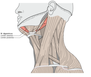
Digastric Muscle
Original Editor -Priyanka Chugh
Lead Editors - Priyanka Chugh, Kim Jackson, Wendy Snyders, Vidya Acharya, 127.0.0.1 and Chrysolite Jyothi Kommu
Description[edit | edit source]
The digastric muscle (also digastricus) has two bellies[1], namely the anterior and posterior belly. It is a small, important muscle in the neck[2]. It has many variations but these variations do not necessarily produce clinical symptoms[2]. It belongs to the suprahyoid muscles group which includes the mylohyoid, geniohyoid and stylohyoid muscles[1].
Origin[edit | edit source]
Anterior belly - Mandible's digastric fossa, close to the midline [2][1]
Posterior belly - Notch of the mastoid process of the temporal bone[2][1]
Insertion[edit | edit source]
The two join, forming the intermediate tendon which inserts onto the body and greater cornu of the hyoid bone[2][1]. The intermediate tendon can sometimes penetrate the stylohyoid muscle[2].
Nerve Supply[edit | edit source]
Anterior belly - mandibular division (V3) of the trigeminal (CN V) via the mylohyoid nerve[2]. In some cases, the anterior belly can be innervated by both the mylohyoid nerve and facial nerve[2].
Posterior belly - facial nerve (CN VII)[2]
Blood Supply[edit | edit source]
Anterior belly - submental branch of facial artery[2]
Posterior belly - posterior auricular and occipital arteries[2]
Function[edit | edit source]
- Elevates the hyoid bone to stabilize it during swallowing (protection of the airway)[2][1]
- Opens the jaw by depressing the mandible[2][1]
- Plays a role during speech and chewing[2]
Structure[edit | edit source]
The digastricus (digastric muscle) consists of two muscular bellies united by an intermediate rounded tendon. The two bellies of the digastric muscle have different embryological origins, and are supplied by different cranial nerves. Each person has a right and left digastric muscle. The digastric muscle stretches between the mastoid process of the cranium to the mandible at the chin, and part-way between, it becomes a tendon which passes through a tendinous pulley attached to the hyoid bone. It
Posterior belly
The posterior belly, longer than the anterior belly, arises from the mastoid notch which is on the inferior surface of the skull, medial to the mastoid process of the temporal bone. The mastoid notch is a deep groove between the mastoid process and the styloid process. The mastoid notch is also referred to as the digastric groove or the digastric fossa. The posterior belly is supplied by the digastric branch of the facial nerve.
Anterior belly
The anterior belly arises from a depression on the inner side of the lower border of the mandible called the digastric fossa of mandible, close to the symphysis, and passes downward and backward. The anterior body is supplied by the trigeminal nerve via the mylohyoid nerve, a branch of the inferior alveolar nerve, itself a branch of the mandibular division of the trigeminal nerve. It originates from the first pharyngeal arch.
Intermediate tendon
The two bellies end in an intermediate tendon which perforates the stylohyoideus muscle, and is held in connection with the side of the body and the greater cornu of the hyoid bone by a fibrous loop, which is sometimes lined by a mucous sheath.
Triangles
The digastric muscle divides the anterior triangle of the neck into three smaller triangles:
- The submandibular triangle (also called digastric triangle) is formed by the mandible superiorly, laterally but the digastric posterior belly and medially by the digastric anterior belly[2].
- The carotid triangle is formed by the posterior belly of the digastric muscle superiorly, the sternocleidomastoid muscle laterally and medially but the superior belly of the omohyoid muscle[2].
- The submental triangle is formed by the hyoid bone inferiorly and by both anterior bellies of the digastric muscles
Trigger Point Referral Pattern[edit | edit source]
The anterior and posterior bellies of digastric may contain a myofascial trigger point (MTrP). The MTrP in the posterior belly may refer pain over the mastoid process and occasionally to the throat under the chin[3] . The anterior belly refers to the lower central incisors and to the alveolar ridge below.[3]
Palpation[edit | edit source]
Direct external palpation of posterior digastric is difficult due to the depth of the muscle. The anterior digastric is examined by identifying the lateral margins of the hyoid, and then palpating the inferior surface of the mandible by placing the thumbs on either side of the midline. To confirm the location of the anterior digastric, the patient is requested to swallow; a prominence of the anterior belly can be palpated under the thumb tips as the hyoid is drawn superiorly. <be>
Resources[edit | edit source]
References[edit | edit source]
- ↑ 1.0 1.1 1.2 1.3 1.4 1.5 1.6 Kenhub 2022, Kenhub: Digastric, Viewed 16/12/22,https://www.kenhub.com/en/library/anatomy/digastric-muscle
- ↑ 2.00 2.01 2.02 2.03 2.04 2.05 2.06 2.07 2.08 2.09 2.10 2.11 2.12 2.13 2.14 2.15 Kim SD, Loukas M. Anatomy and variations of digastric muscle. Anatomy & Cell Biology. 2019 Mar;52(1):1-1.
- ↑ 3.0 3.1 http://www.sciencedirect.com/topics/page/Digastric_muscle
- ↑ Kenhub - Learn Human Anatomy Digastric muscle - Origin, Insertion, Innervation & Function - Anatomy | Kenhub.Available fromhttps://www.youtube.com/watch?v=CxqCWyindCI. Accessed on 10/03/23


