When refering to evidence in academic writing, you should always try to reference the primary (original) source. That is usually the journal article where the information was first stated. In most cases Physiopedia articles are a secondary source and so should not be used as references. Physiopedia articles are best used to find the original sources of information (see the references list at the bottom of the article).
If you believe that this Physiopedia article is the primary source for the information you are refering to, you can use the button below to access a related citation statement.
Clubfoot also known as CTEV (Congenital Talipes Equino Varus) is a congenital physical deformity in which a newborn’s baby foot is internally rotated at the ankle, soles of the feet face each other while the toes are twisted downward and inwards. In 50% of cases, both feet are affected. Male babies are two times more likely to get this deformity. According to National Institute of Health every 1.2 in 1000 babies are born with clubfoot.[2][3] It has been estimated that every year at least 150,000 are born with clubfoot.[4] It may occur alone or in combination with multiple neuromuscular pathologies. The exact cause of clubfoot is still yet to be found but studies have shown many possible risk factors which may contribute to clubfoot.[5]See... “Ponseti method” is considered the Gold standard for the treatment of clubfoot. For information on Clubfoot management, See...
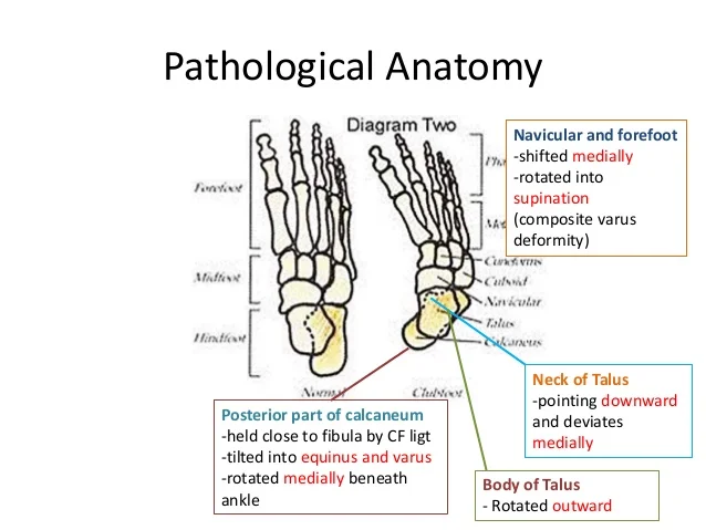
PATHOANATOMY
Bones
The bones of the foot are smaller than usual.
The neck of the talus is angled, causing the head to point down and inward.
The calcaneus (heel bone) is also small and has a concave shape on its inner side.
Joints
Deformities result from misalignment in different joints:
Equinus Deformity: The ankle joint points downwards, limiting upward movement.
Inversion Deformity: The subtalar joint causes the heel to tilt inward, turning the sole of the foot medially.
Forefoot Adduction Deformity: The front of the foot turns inward due to changes at the mid tarsal joints.
Forefoot Cavus Deformity: The foot has an excessive arch due to mid-tarsal joint changes.
Muscles and Tendons
The calf muscles are underdeveloped, leading to tightness in these areas:
Posteriorly: Tendo Achilles
Medially:
Tibialis Posterior
Flexor Digitorum Longus
Flexor Hallucis Longus
Capsules and Ligaments
Ligaments on the back and inner sides of the foot are shortened:
Posterior Structures:
Posterior Capsule of the Ankle Joint
Posterior Capsule of the Subtalar Joint
Talo-Fibular and Calcaneo-Fibular Ligaments
Medial Ligaments:
Talo-Navicular Ligament
Spring Ligament
Deltoid Ligament
Plantar Structures:
Plantar Fascia
Plantar Ligaments
Other: Interosseous ligament between the talus and calcaneum
Skin Changes
The skin on the inner side of the foot shortens and develops deep creases.
There are dimples on the outer side of the ankle and midfoot.
Secondary Changes
If a child walks on deformed feet, these issues can worsen:
Weight bearing can increase the deformity.
Calluses and bursae may form over bony areas on the outer side of the foot.
Management
Principles of Treatment: The main goal is to correct the deformity and maintain that correction until the foot grows to a normal size, preventing recurrence.
Methods of Correction
1. Non-Operative Methods:
Manipulation Alone:
For newborns, parents are taught to gently manipulate the foot after every feeding.
The foot is flexed (dorsiflexed) and turned outward (everted) with enough pressure to blanch the fingers.
This manipulation lasts about 5 seconds and is repeated several times for about 5 minutes.
Minor deformities may be corrected this way, while major ones often require further treatment.
Manipulation and Plaster of Paris (PoP):
In this method, the child is sedated, and the surgeon manipulates the foot into the correct position.
The corrected foot is then held in place with plaster casts.
Treatment Philosophies:
Kite Method
- Developed by Dr. Robert Kite, this method focuses on using a series of manipulations and plaster casts to correct clubfoot.
- The emphasis is on gentle, repeated manipulations of the foot to achieve alignment over time.
- Typically involves less structured casting, relying more on the clinician's experience and judgment.
Ponseti Method
- Developed by Dr. Ignacio Ponseti, this method is more systematic and involves specific steps.
- It includes gentle manipulation followed by a series of well-defined plaster casts to maintain the corrected position.
- The Ponseti method often incorporates a final Achilles tendon lengthening procedure to address equinus (downward pointing foot).
- Maintenance with specific braces (like the Denis-Brown splint) is crucial to prevent recurrence.
Key Differences:
Approach: Kite's method is more reliant on manipulation, while Ponseti emphasizes a structured casting process.
Outcome Focus: Ponseti’s method has a strong emphasis on long-term maintenance and prevention of recurrence.
Operative Methods for Foot Deformities
In cases of severe deformities that don’t improve with non-operative methods, or if they recur, surgery is needed. The type of surgery depends on the child's age:
Younger Children (under 3 years): Soft tissue release operations may be enough.
Older Children: More complex bony surgeries are usually required.
Types of Surgeries
1. Postero-Medial Soft Tissue Release (PMSTR):
This surgery involves cutting and releasing tight soft tissue structures (like tendons, ligaments, and capsules) on the back and inner side of the foot.
Structures Released During Surgery
1. On the Posterior Side:
Lengthening of the Tendo Achilles: Achieved using a technique called Z-plasty.
Release of Posterior Capsules: Involves cutting the capsules of the ankle and subtalar joints.
Release of Ligaments: Includes the posterior talo-fibular and calcaneo-fibular ligaments.
2. On the Medial Side:
Lengthening of Tendons: This includes the tibialis posterior, flexor digitorum longus, and flexor hallucis longus. Their thickened sheaths are also removed.
Release of Ligaments: Involves the talo-navicular ligament, the superficial part of the deltoid ligament, and the spring ligament.
Additional Releases: In severe cases, the interosseous talo-calcaneal ligament and the capsules of the naviculo-cuneiform and cuneiform-first metatarsal joints are also released.
3. On the Plantar Side:
Release of Plantar Fascia: This helps relieve tension on the foot.
Release of Short Flexors: Involves cutting the flexor digitorum brevis and abductor hallucis from their origins on the calcaneus.
Limited Soft Tissue Release
In some cases where the foot is only partially corrected after conservative treatment, a limited release may be enough:
For Equinus Deformity: A posterior release is performed.
For Adduction Deformity: A medial release is done.
Additional Operative Methods for Foot Deformities
1. Limited Soft Tissue Release for Cavus Deformity:
For cavus (high arch) deformity, a plantar release is performed to alleviate tension.
2. Tendon Transfers:
In cases where the tibialis anterior and tibialis posterior (which pull the foot inward) are too strong compared to the weaker peroneal muscles (which pull the foot outward), tendon transfers may be needed.
The tibialis anterior can be transferred to the outer side of the foot to act as an everter.
The minimum age for this procedure is 5 years.
3. Dwyer's Osteotomy:
This is an open-wedge osteotomy of the calcaneus (heel bone) used to correct heel varus (inward angling of the heel).
The operation can be performed starting at 3 years of age, as the calcaneus is still mostly cartilaginous at this stage.
Some surgeons may prefer a closed-wedge osteotomy on the outer side instead.
4. Dilwyn Evan's Procedure:
This involves a thorough soft tissue release (PMSTR) along with calcaneo-cuboid fusion.
It is typically used for neglected or recurring clubfoot in children aged 4 to 8 years.
The fusion of the calcaneo-cuboid joint stabilizes the lateral side of the foot.
Additional Operative Methods for Foot Deformities
5. Wedge Tarsectomy:
This procedure involves removing a wedge of bone from the mid-tarsal area.
The wedge is cut with its base on the outer (dorso-lateral) side of the foot.
Once the wedge is removed, the foot can be adjusted to a normal (plantigrade) position.
This surgery is typically performed on neglected clubfeet in children aged 8 to 11 years.
6. Triple Arthrodesis:
This surgery fuses three key joints in the foot: the subtalar, calcaneo-cuboid, and talo-navicular joints.
Wedges are used to correct the deformity before fusion.
It is performed on children aged 12 years and older, as the bones are still cartilaginous in younger children, making fusion difficult.
Among these joints, achieving fusion of the talo-navicular joint is the most challenging.
7. Ilizarov's Technique:
This technique uses an external fixator to gradually stretch and correct different components of the foot deformity.
Once correction is achieved, it is maintained with plaster casts.
Ilizarov's technique is indicated for neglected clubfeet and cases where previous treatments have failed.
Methods of Maintenance of Correction
Once the foot deformity is corrected, it’s important to maintain that correction through various methods:
1. CTEV Splints:
These are plastic splints shaped to keep the foot in the corrected position when secured with straps.
2. Denis-Brown Splint (DB Splint):
This splint is used to hold the foot in the corrected position, particularly before the child starts walking.
After walking begins, the DB splint is used at night while CTEV shoes are worn during the day.
3. CTEV Shoes:
These are specially modified shoes that are used once the child begins walking. The modifications help maintain proper foot alignment.
CTEV Shoes Modifications
CTEV shoes have specific features to help maintain foot correction:
Straight Inner Border: Helps prevent forefoot adduction (inward turning of the front of the foot).
Outer Shoe Raise: Prevents foot inversion (tilting inward of the heel).
No Heel: Helps prevent equinus (downward pointing of the foot).
These shoes are typically used until the child is 5 years old.
Plan of Treatment
Most cases treated early respond well to non-operative methods. However, surgical methods may be needed in certain situations:
1. Resistant Clubfeet:
If a child does not respond to non-operative treatment, it may indicate severe deformities.
These feet might appear "chubby" or may be associated with conditions like arthrogryposis multiplex congenita (AMC), which requires a more intensive approach.
2. Recurrent Clubfeet:
A child may experience a recurrence of deformities, often if the correction is not properly maintained.
The first deformity to return is usually equinus (downward pointing of the foot).
3. Neglected Clubfeet:
This occurs in children who have been treated inadequately or presented late for treatment.
Once a clubfoot is corrected, it must be maintained in the corrected position using the methods mentioned earlier (like CTEV splints, Denis-Brown splint, and CTEV shoes).
For a comprehensive treatment plan for CTEV, refer to the flow chart (Flow chart-25.1) that outlines the steps involved.
Barriers to Clubfoot Treatment in Underdeveloped Countries
Beliefs and Lack of Awareness
According to some studies, smoking by mothers during pregnancy may be one of the most possible causes of clubfoot.[6] It is also believed that if one parent is having this deformity, the newborn baby has the risk of getting it. The risk is even higher if both parents are affected.[7] Many researches have been taken place and still ongoing in developed countries but in underdeveloped countries, many false perceptions and beliefs are found. In many underdeveloped countries of the world, clubfoot and such physical deformities are still linked with solar and lunar eclipses.[8] while some people claim it is punishment of God to the parents. Many false perceptions and beliefs are still present about clubfoot and generally all physical deformities. Parents don’t have any awareness of this deformity; they don’t know when is the most appropriate time to start treatment.[9]
Lack of Knowledge and Skilled Clinicians
In underdeveloped countries, many clinicians are unqualified, inexperienced and untrained in Ponseti method. Some clinicians who do not know anything about this deformity advised parents to keep away from any treatment as it will get better as the child grows up. Some call it a deficiency of vitamins. Some parents seek religious healers. Some seek surgery. And many think that just exercises and massage can correct it. Many children are left untreated as it is believed that it can’t be treated. In short, there is still too much work to be done about this deformity. We need to know what this deformity is. How and when it can be treated? And if not treated what complications can occur in the future?
The most widely accepted method being used for clubfoot treatment is the “Ponseti Method”, developed by Ignacio Ponseti (1942-2009). It includes Manipulations by an expert physiotherapist, several serial castings and a minor surgical operation in which the Achilles tendon is released. If applied correctly and in time, individuals with clubfoot can recover up to 90% of normal foot alignment with minor differences when compared to normal foot.[10] In developed countries, a lot of research, annual conferences and training sessions are arranged to educate people about this deformity and to train medical professionals in successful treatment of clubfoot. But in underdeveloped countries, there is no such a system. Many medical professionals and clinicians do not know about clubfoot, many don’t have the idea of treating it with a method of most accurate outcome. If there are any hospitals or facilities available to treat clubfoot, they don’t have any qualified or expert medical professionals. As seen in general practice, such children are brought for medical treatment when the deformity has worsened.
Affordability and Availability of Treatment
Generally, at Government hospitals, there is a dearth of medical professionals who can perform Ponseti method. In rare situations where it is available in private clinics, the cost of treatment is high. Many parents don’t have enough money to continue the treatment for many sessions as in most cases correction cannot be gained in a single or two sessions. As seen in many cases, even if there is a free Ponseti treatment available, it does not achieve full correction through serial castings; thus surgical interventions are needed.
Refusal of Surgical Intervention
As suggested by some studies, 80% of cases require a minor surgical operation to release the rigid equinous.[11] But often seen in underdeveloped countries due to lack of awareness, many parents refuse to perform surgery, which causes a hurdle in complete recovery of the equinous deformity. Their refusals are most times due to their inherent beliefs and sometimes as a result of financial constraints.
Preventive braces are not used correctly
It has been noted that after serial castings when Abduction foot brace shoes and other corrective braces are applied, many parents don’t know how to fit them properly; Which is also a hurdle in gaining proper correction of the deformity. Some parents due to their ignorance do not adhere to the continued use of AFAB for up to four years of age[12]
No proper follow up
Many parents never visit the clinics in proper time after correction as proper follow-up is a basic part of clubfoot treatment. Many children are brought to the clinic so late for follow-up, as the deformity has already recurred.
Conclusion
“Ponseti method” as a Gold standard for the treatment of clubfoot can be implemented accurately in underdeveloped countries only if the barriers are removed.
↑Wang H, Barisic I, Loane M, Addor MC, Bailey LM, Gatt M, Klungsoyr K, Mokoroa O, Nelen V, Neville AJ, O'Mahony M. Congenital clubfoot in Europe: A population‐based study. American Journal of Medical Genetics Part A. 2019 Apr;179(4):595-601.
↑Parker SE, Mai CT, Strickland MJ, Olney RS, Rickard R, Marengo L, Wang Y, Hashmi SS, Meyer RE. Multistate study of the epidemiology of clubfoot. Birth Defects Research Part A: Clinical and Molecular Teratology. 2009 Nov;85(11):897-904.
↑Africa Clubfoot Training Project. Chapter 2 Africa Clubfoot Training Basic & Advanced Clubfoot Treatment Provider Courses - Participant Manual. University of Oxford: Africa Clubfoot Training Project, 2017.