
Anatomy of the Equine Hind Limb
Introduction
The equine hind limb is also referred to as the pelvic hind limb. When working with horses, it is important to be able to accurately assess, diagnose and manage an equine patient. To do this, a good understanding of equine anatomy is essential.
Anatomy
Pelvic hind limb bears 40-45% of the weight and provides the majority of propulsion for locomotion.
Bones

- Os coxae
- The Tuber coxae and tuber sacrale both palpable
- Tuber ischii is located underneath the hamstrings
- Femur
- Greater trochanter has a cranial and caudal part for gluteal attachments (deep & middle)
- Third trochanter is prominent laterally for superior gluteal attachment
- Medial trochlear ridge is enlarged
- Patella
- Parapatellar fibrocartillage forms hook medially
- Tibia
- Large and only weight bearing component of crus (stifle/ knee)
- Large tibial tuberosity – patellar ligament
- Medial tibia is subcutaneous
- Cochlea is inclined craniolaterally. This causes the lower limb to move laterally on flexion

- Fibula is greatly reduced
- Distally incorporated into tibia
- Proximally tightly articulated with tibia
- It has a short shaft
- Tarsals
- Proximal row (calcaneus & talus)
- Calcaneus is enlarged for muscle attachments
- Distal sustentaculum tali forms the plantar groove
- Talus has oblique trochlea
- Middle row (central talus)
- Distal row
- 1st & 2nd bones fused (medially)
- 3rd wedged shaped
- 4th large and deep
- Proximal row (calcaneus & talus)
Joints
Sacro iliac
- Type:
- Synchondrosis (synovial joint – sacropelvic surface of illium)
- ROM:
- Minimal. Designed for stability
- Accessory movements - rotation
- Supporting structures:
- Dorsal and ventral Sacroiliac ligaments
- Sacrotuberous

Hip
- Type:
- Ball and socket
- Femoral head and acetabulum of the illium, ischium and pubis
- A band of fibrocartilage on the rim of the acetabulum deepens acetabulum
- ROM:
- Flexion and extention
- Minimal – adduction and abduction
- Supporting structures:
- Acetabular lip (fibrocartilage) continues as transverse ligament
- Ligament of the femoral head
- Accessory ligament (horse)
- Synovial structures and tendon sheaths:
- Large joint capsule
- Rectus femoris has a bursa at illium
- Internal obturator
- Bursa between gluteals and greater trochanter
Stifle
- Type:
- Hinge joint with two cartilages/menisci
- Femur & tibia – femorotibial (condylar)
- Femur & patella – femoropatella (gliding joint)
- ROM:
- Femur and tibia – Flexion and extension
- End flexion there is internal rotation
- End extension there is external rotation
- Supporting structures:
- Meniscal ligaments
- Menisicii attached cranially by Transverse ligament
- Cranial tibial ligament (medial and lateral meniscus) attaches menisci to tibial spine
- Lateral meniscus has extra ligament – meniscofemoral
- Seven other ligaments
- Lateral and medial collateral
- Cranial and caudal cruciate ligament
- Lateral, middle and medial patellar ligaments
- Synovial structures and tendon sheaths:
- Three synovial sacs
- Femoropatellar
- Medial and lateral femorotibial
- Link between femoropatellar and femorotibial
- Joint capsule – extends to femoral extensor fossa
- Bursa between tibial crest and semitendinosus attachment
- Three synovial sacs
Hock/Tarsal
Click on this link to see the sideview of the hock joint
- Type:
- Tarsocrucal joint (TCJ) has the greatest movement
- Proximal and distal intertarsal joint
- Tarsometatarsal joint
- Intertarsal
- ROM:
- TCJ – flexion and extension and Lateral & rotatory accessory movements
- Others – small amount of translatory and rotatory
- Supporting structures:
- Medial and lateral collaterals – each have long and short pair
- Annular ligament
- Dorsal and plantar ligaments
- Intertarsal ligaments
- Proximal & distal transverse ligaments
- Synovial Structures and tendon sheaths
- Each of 4 joints has own synovial sac
- Bursa with most tendons crossing joints
- Bursa separates SDF from gastrocnemius tendon at tuber calcanei
- Most tendons have sheaths in this region
Muscles
| Muscle | Origin | Insertion | Action | Nerve |
Pectineus  |
Pubis | Femur | Adducts the limb and flexes the hip | Obturator |
Psoas Major  |
Ventral surface of lumbar vertebrae | Lesser trochanter of femur | Flexes the hip and externally rotates the thigh | Ventral branches of thoracic and lumbar nerves and femoral nerves |
Iliacus  |
Ventral surface of wing of the ilium | |||
Gracilis  |
Pubic symphysis | Medial tibia | Adducts the limb | Obturator |
Adductor  |
Pubic symphysis | Caudal surface of femur | Adducts the limb | Obturator |
Internal Obturator  |
Pelvic floor | Trochanteric fossa of femur | Lateral rotation of the femur | Ischiatic |
| Gemelli | Ischium | Trochanteric fossa of femur | Lateral rotation of the femur | Ischiatic |
External Obturator  |
Ventral surface of pubis and ischium | Trochanteric fossa of femur | Adducts the thigh | Ischiatic |
| Quadratus femoris | Ischium | Caudal surface of femur | Extends the hip and adducts the thigh | Obturator |
Deep gluteal  |
Body of ilium | On or near greater trochanter | Extension and abduction | Gluteal |
Superficial Gluteal  |
Dorsal to hip joint | Third Trochanter | Abducts the hip | Gluteal |
Tensor fascia latae  |
Tuber coxae | Lateral femoral fascia | Hip flexion and stifle extension via tension of lateral femoral fascia | Gluteal |
Biceps femoris  |
Ischiatic tuberosity (Tuber Ischii) | Greater Trochanter | Extends and abducts the hip | Gluteal |
Semitendinosus  |
Ischiatic tuberosity | Tibia and calcaneal tub. | Extends the hip
Flexes the stifle Extends the tarsus |
Ischiatic |
Semimembranosus  |
Ischiatic tuberosity | Femur and tibia | Extends the hip
Flexes or extends the stifle |
Ischiatic |
Middle Gluteal _.jpeg) |
Wing of ilium | Greater trochanter of femur | Extension and abduction | Gluteal |
Rectus femoris  |
Ilium (cranioventral iliac spine) | Patella and patella tuberosity | Flexion of the hip
Extension of the stifle |
Femoral |
| Vastus lateralis | Proximal femur | Extends the stifle | ||
| Vastus medialis | ||||
| Vastus intermedius |
Table of muscles of the Hip and Stifle
| Muscle | Origin | Insertion | Action | Nerve |
Gastrocnemius  |
Distocaudal surface of the femur | Calcaneal tuberosity | Extends the tarsus
Flexes the stifle |
Tibial |
Long digital extensor (combined in this image with long digital extensor) |
Extensor fossa of femur | Extensor processes of digital phalanges | Flexes the hip
Extends the stifle |
Peroneal |
| Soleus | Head of fibula | Tendon and lateral head of gastrocnemius | Extends the tarsus | Tibial |
| Lateral digital extensor (see long digital extensor image above) | Fibula | Lateral aspect of digit | Extends the digits
Flexes the tarsus |
Peroneal |
Deep digital flexor  |
Tibia and fibula | Distal Phalanx | Extends the tarsus
Flexes the digits |
Tibial |
Superficial digital flexor  |
Caudal distal femur, deep to gastrocnemius | Calcaneal tuberosity
Middle phalanx |
Flexes the stifle
Extends the tarsus Flexes the digits |
Tibial |
Table of muscles of the Stifle, hock and pes
Nerves to the Pelvic Limb

- Femoral nerve (L4, L5 sometimes L3, L6)
- Muscle Innervation – Stifle extensors (quadriceps), Iliopsoas
- Cutaneous Innervation – Medial aspect of limb
- Obturator nerve (L5, L6)
- Muscle Innervation– Adductors (obturator, adductors, gracilis)
- Cutaneous Innervation– nil
- Gluteal nerve (L6, L7, S1)
- Muscle Innervation– gluteals, TFL, bicaps femoris, semitendinosus
- Cutaneous Innervation– nil
- Sciatic nerve (L6, L7, S1, S2)
- Muscle Innervation– biceps femoris, semitendinosus, semimembranosis
- Cutaneous Innervation– see tibial and fibula branches
- Tibial nerve (S1, S2)
- Muscle Innervation– extensors of hock, flexors of digits
- Cutaneous Innervation– caudal aspects of the limb below stifle
- Fibular nerve
- Muscle Innervation– flexors of the hock, extensors of the digits
- Cutaneous Innervation– cranial and lateral aspects of limb
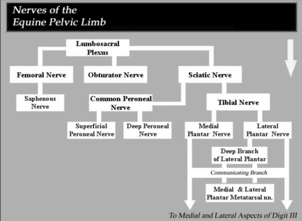
Nerves of the Equine Limb

Pelvic Limb Stay Apparatus
Function
The pelvic limb stay apparatus is used by the horse to support the weight of the caudal end of its body while using a minimal amount of muscular activity. When employed by one pelvic limb, the stay apparatus allows the other pelvic limb to be placed in a "resting" position with just the tip of the hoof touching the ground. Although the stay apparatus reduces the amount of energy required to remain standing, the amount of muscular effort is not reduced. This explains horses can be observed switching their weight from one hind limb to another.
How does the stay apparatus function?
The pelvic limb stay apparatus has three essential elements.
- The stifle joint locking mechanism, which allows the weight of the caudal body to rest, essentially, on the locked joint
- The reciprocal mechanism, which ensures that the stifle and hock joints will move in unison, and the leg will move in a smooth, coordinated manner
- All other ligaments / tendons in the distal limb
References
- ↑ The Horse's Skeleton: Hind Limbs. Available from https://www.youtube.com/watch?v=Y7xbcWGWLc4
- ↑ Equine Pelvic Limb Stay and Reciprocal Apparatus. Available from https://www.youtube.com/watch?v=Iw3lkbC1pPY