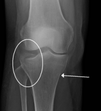
Tibial Plateau Fractures
Original Editors - Jentel Van De Gucht
Top Contributors - Jentel Van De Gucht, Abbey Wright, Sarah Harnie, Admin, Kim Jackson, Jean Yves Lieveld, Robin Petroons, Rachael Lowe, Ulrike Lambrix, Oyemi Sillo, WikiSysop, Evan Thomas, Karen Wilson, Claire Knott, Wanda van Niekerk, 127.0.0.1, Lauren Lopez and Peter Vaes
Definition/Description [edit | edit source]
Tibial plateau fractures are complex injuries of the knee. The tibial plateau is one of the most critical load-bearing areas in the human body. Early detection and appropriate treatment of these fractures are essential in minimizing patient's disability in range of motion and stability and reducing the risk of documented complications. [1]
The fractures are classified according the Schatzker classification system. It divides tibial plateau fractures into six types:
• Schatzker I: lateral tibial plateau fracture without depression
• Schatzker II: lateral tibial plateau fracture with depression
• Schatzker III: compression fracture of the lateral (IIIa) or central (IIIb) tibial plateau
• Schatzker IV: medial tibial plateau fracture
• Schatzker V: bicondylar tibial plateau facture
• Schatzker VI: tibial plateau fracture with diaphyseal discontinuity [2][1]
A type I fracture is a wedge-shaped pure cleavage fracture of the lateral tibial plateau, with a displacement or depression less than 4mm. They are caused by the lateral femoral condyle being driven into the articular surface of the tibial plateau.
Type II is a fracture with a combined cleavage and compression of the lateral tibial plateau, a type I fracture with a depressed component. There is a depression greater than 4mm.
A Schatzker type III fracture is a pure compression fracture of the lateral tibial plateau in which the articular surface of the tibial plateau is depressed and driven into the lateral tibial metaphysis by axial forces. Type III fractures are divided into two subgroups: those with lateral depression (type IIIA) and those with central depression (type IIIB).
Type IV is a medial tibial plateau fracture with a split or depressed component. These fractures occur as a result of varus forces combined with axial loading in a hyperflexed knee. Type IV fractures have the worst prognosis.
Type V fracture consists of a wedge fracture of the medial and lateral tibial plateau, often with an inverted “Y” appearance. Articular depression is typically seen in the lateral plateau and might be associated with a fracture of the intercondylar eminence.
Type VI is a bicondylar fracture with a dislocation of the metaphysis from the diaphysis.This pattern results from high-energy trauma and diverse combinations of forces.[1] [2]
The first three types are mostly the result of low energy injury, the three others of high energy injury. The magnitude of the force determined the degree of fragmentation and the degree of displacement.
Tibial plateau fractures are often associated with anterior cruciate ligament, collateral ligaments (MCl and/or LCL), menisci and articular cartilage injuries[2]
Clinically Relevant Anatomy[edit | edit source]
Tibial plateau of the knee.
Epidemiology /Etiology [edit | edit source]
The fractures are caused by a strong force on the lower leg with the leg in varus or valgus or simultaneously vertical stress and flexion of the knee.
The reason of tibial plateau fractures are mostly car- or motor accidents and sometimes sportaccidents, mostly sports with a high velocity such as skiing, horse riding and certain water sports.
The symptoms of tibial plateau fractures are swelling, unable to weight bear on the injured side, stiffness of the knee and a recent history of trauma.[1]
Characteristics/Clinical Presentation[edit | edit source]
• Trauma to the knee area followed by swelling and pain in the joint.
• The patient may also complain of stiffness of the knee and may not be able to bear weight.
• Bruising may be seen over the skin.
Differential Diagnosis[edit | edit source]
add text here
Diagnostic Procedures [edit | edit source]
Most of the tibial plateau fractures are easy to identify on standard anteroposterior and lateral projections of the knee. Sometimes minimally displaced vertical split fractures are not visible on anteroposterior and lateral radiographs. Between Schatzker I and II there isn’t a clear visible difference on radiographs.
An anteroposterior radiograph with the knee angled 15° caudally (tibial plateau view) can provide a more accurate assessment of the depth of plateau surface depression. Traction radiographs give a clearer image of the fracture configuration after anatomic alignment is restored. CT scans give more detailed information in 3D of the fracture. This helps to choose the best treatment.
When there is a presumption of damage of the soft tissue, an MRI scan must be done.[1]
Outcome Measures[edit | edit source]
add links to outcome measures here (also see Outcome Measures Database)
Examination[edit | edit source]
add text here related to physical examination and assessment
Medical Management
[edit | edit source]
For these fractures, preoperative planning is fundamental. The clinical history, trauma mechanism, age and associated comorbidities influence the treatment decisions. In the physical examination, the soft-tissue envelope, neurovascular functioning and associated lesions should be assessed so that the intervention will be appropriate.
Physical Therapy Management
[edit | edit source]
After 10 to 12 weeks the bone is expected to be healed. The goals of the physical therapy are to restore the range of motion as early as possible, to improve and restore the strength of the muscles and to restore the stability of the knee. After surgical treatment the indications to start passive mobilization are 0° extension and 90° flexion of the knee. The patient may not bear weight on his leg for 3 months.
From the day of the injury to one week after, early range of motion is very important. Active and active-assistive flexion and extension of the knee are allowed while protecting the knee from varus and valgus, once the pain has subsides. This is done in a sitting position. Initially 40° to 60° flexion are allowed. After one week it is possible to increase to 90° flexion. Sometimes a continuous passive motion machine is used. Gentle ankle isotonics without resistance and gluteal exercises are prescribed to strength muscles.
After 2 weeks the patient must be able to do active and active-assistive range of motion exercises, obtaining 0° to at least 90° of knee flexion. The patient may start with isometric exercises to the quadriceps at the end of 2 weeks and continue gluteal exercises.
Also from the fourth to the sixth week active and active-assistive range of motion exercises, obtaining 0° to at least 90° of knee flexion must be done. At the end of week 6 gentle passive range of motion is allowed. Active and passive range of motion of the ankle and hip can begin. From now on we can start with isometric hamstring exercises and continue with isometric exercises of the quadriceps and isotonics to strength the muscles of the ankle.
From the eighth to the twelfth week, when the fracture appears stable and there is no collateral ligament injury or instability, we can start bearing partial weight on crutches. The patient must be able to do full extension and at least 90° of flexion. From now on resistive exercises to the quadriceps, hamstrings and ankle musculature are prescribed. The number of repetitions can increase gradually. At the end of the twelfth week, weight bearing activities are started.
From the twelfth to the sixteenth week the patient is fully weight bearing and should be weaned off assistive devices. Muscle strength exercises must be continued and resistive exercise are increased progressively. The range of motion is still at least full extension and 90° of flexion.[1][3][4]
Key Research[edit | edit source]
add links and reviews of high quality evidence here (case studies should be added on new pages using the case study template)
Resources
[edit | edit source]
http://www.sportsinjuryclinic.net/cybertherapist/front/knee/tibial-plateua-fracture.htm
Clinical Bottom Line[edit | edit source]
add text here
Recent Related Research (from Pubmed)
[edit | edit source]
Failed to load RSS feed from http://www.ncbi.nlm.nih.gov/entrez/eutils/erss.cgi?rss_guid=12QQbiNmM99bUQG1V2M-KE3CXMAqSkipzD_PEt96qHBOpVckH5|charset=UTF-8|short|max=10: Error parsing XML for RSS
References[edit | edit source]
- ↑ 1.0 1.1 1.2 1.3 1.4 1.5 Vidyadhara S, Tibial Plateau Fractures, eMedicine Specialties, 2009
- ↑ 2.0 2.1 2.2 B. Keegan Markhardt, Jonathan M. Gross, Johnny Monu, Schatzker Classification of Tibial Plateau Fractures: Use of CT and MR Imaging Improves Assessment, the journal of continuing medical education in radiology, 2009, 29, 585-597
- ↑ Hoppenfeld S, L. Murthy V., treatment & rehabilitation of fractures, lippincott williams and wilkins, 2000
- ↑ Chien-Jen Hsu, Wei-Ning Chang, Chi-Yin Wong, The results of surgical management of displaced tibial plateau fractures in the elderly, Arch Orthop Trauma Surg, 2001, 121 :67–70


