Tibial Nerve: Difference between revisions
(added image) |
No edit summary |
||
| Line 5: | Line 5: | ||
</div> | </div> | ||
== Description == | == Description == | ||
The Tibial Nerve is one of the two main muscular branches of the Sciatic Nerve | The Tibial Nerve is one of the two main muscular branches of the Sciatic Nerve . TheTibial nerve is the larger terminal branch with root values of L4, L5, S1, S2, and S3. | ||
The Tibial Nerve provides innervation to the muscles of the lower leg and foot. Specifically: Triceps Surae ( the two headed Gastocnemius and Soleus); Plantaris, Popliteus; Tibialis Posterior; Flexor Digitorum Longus; and Flexor Hallucis Longus muscles.<ref name=":0">KenHub. [[Tibial Nerve]]. Available from: https://www.kenhub.com/en/library/anatomy/tibial-nerve (last accessed 17.3.2019)</ref><ref name=":1">Healthline. [https://www.healthline.com/human-body-maps/tibial-nerve#1 Tibial Nerve.] Available from: https://www.healthline.com/human-body-maps/tibial-nerve#1 (last accessed 17.3.2019)</ref> It also has articular and cutaneous branches.<ref name=":2">Wikipedia. [https://en.wikipedia.org/wiki/Tibial_nerve The Tibial Nerve.] Available from: https://en.wikipedia.org/wiki/Tibial_nerve (last accessed 18.3.2019)</ref> | |||
{{#ev:youtube|https://www.youtube.com/watch?v=yPJ9sxUubRI|width}}<ref>nabi lebraheim. Nerves of the lower leg 3D. Available from: https://www.youtube.com/watch?v=yPJ9sxUubRI (last accessed 17.3.2019) </ref> | {{#ev:youtube|https://www.youtube.com/watch?v=yPJ9sxUubRI|width}}<ref>nabi lebraheim. Nerves of the lower leg 3D. Available from: https://www.youtube.com/watch?v=yPJ9sxUubRI (last accessed 17.3.2019) </ref> | ||
=== Branches === | === Terminal Branches === | ||
At the foot level (just after the heel) the Tibial Nerve divides into the Medial Plantar Nerve (MPN) and the Lateral Plantar Nerve (LPN).<ref name=":0" /> The MPN supplies muscular branches to the big toe and the two toes next to it, and the LPN the other two toes. The Sural Nerve is a cutaneous branch of the Tibial nerve that supplies the skin of the legs and feet.<ref name=":1" /> | At the foot level (just after the heel) the Tibial Nerve divides into the Medial Plantar Nerve (MPN) and the Lateral Plantar Nerve (LPN).<ref name=":0" /> The MPN supplies muscular branches to the big toe and the two toes next to it, and the LPN the other two toes. The Sural Nerve is a cutaneous branch of the Tibial nerve that supplies the skin of the legs and feet.<ref name=":1" /> | ||
[[File:Tibial Nerve.png|right|frameless]] | [[File:Tibial Nerve.png|right|frameless]] | ||
=== Motor === | === Motor === | ||
* At Popliteal Fossa region branches of the Tibial Nerve supply medial and lateral heads of gastrocnemius, soleus, plantaris and popliteus muscles. The popliteus branch goes on to supply tibialis posteriormuscle, superior tibiofibular joint, tibia bone, interosseous membrane of leg, and the inferior tibiofibular joint.<ref name=":2" /> | |||
* Back of the Leg branches of the Tibial Nerve supply tibialis posterior, flexor digitorum longus, flexor hallucis longus, and deep part of soleus.<ref name=":2" /> | |||
* from | |||
=== Sensory === | === Sensory === | ||
Branches of the Tibial nerve are | |||
* Medial Sural Nerve supplies skin on lower half back of leg and skin of foot laterally till little toe. | |||
* Medial Calcaneal Nerve supplies skin on posterior and inferior surface calcaneous. | |||
* Articular branches are to the knee joint ( 3 in total) and ankle joint.<ref name=":2" /> | |||
== Clinical relevance == | === Clinical relevance === | ||
Injury to the Tibial Nerve can cause motor loss and altered sensation and pain to any of the areas it supplies, depending on site of involvement. | |||
== | # Popliteal Fossa region. Injury may occur due to eg. Space occupying lesion; Laceration injury; Posterior dislocation of the knee<ref name=":3">Earth's Lab. [https://www.earthslab.com/anatomy/tibial-nerve/ Tibial Nerve.] Available from: https://www.earthslab.com/anatomy/tibial-nerve/ (last accessed 18.3.2019)</ref>; Entrapement in Soleus arch. [[Soleus|Soleu]]<nowiki/>s arch entrapment neuropathy can occur with sports that make special demands on the calf muscles. Swelling and hypertrophy of the soleus muscle may cause its tendinous arch to compress the popliteal artery and vein as well as the tibial nerve. This can cause chronic mechanical damage to the nerve and the artery and vein may become occluded.<ref>Thetter O. (1989) [https://link.springer.com/chapter/10.1007/978-3-642-72942-3_48 Entrapment Syndrome by the Tendinous Arch of the Soleus Muscle] (“Soleus Syndrome”). In: Heberer G., van Dongen R.J.A.M. (eds) Vascular Surgery. Springer, Berlin, Heidelberg. Available from: https://link.springer.com/chapter/10.1007/978-3-642-72942-3_48 (last accessed 18.3.2019)</ref> | ||
# Medial Malleous Level. Compression of the tibial nerve in the Osseo fibrous tunnel below the flexor retinaculum of the ankle causes a condition termed [[Tarsal Tunnel Syndrome|Tarsal Tunnel Syndrome.]] On examination it presents as pain and paresthesia in the sole of the foot, For a full assessment and treatment of this condition se [[Tarsal Tunnel Syndrome]].<ref name=":3" /> | |||
# Sole of foot. Abnormal pressure at the ball of the foot can irritate the first plantar digital nerve causing [[Morton's Neuroma]]/[[Metatarsalgia]]. | |||
== Treatment == | == Assessment and Treatment == | ||
'''see''' | |||
* [[Tarsal Tunnel Syndrome]] | |||
* [[Morton's Neuroma]]/[[Metatarsalgia]]. | |||
== Resources == | == Resources == | ||
Revision as of 07:52, 18 March 2019
Original Editor - Lucinda hampton
Top Contributors - Lucinda hampton, Leana Louw, Nehal Shah and Rucha Gadgil
Description[edit | edit source]
The Tibial Nerve is one of the two main muscular branches of the Sciatic Nerve . TheTibial nerve is the larger terminal branch with root values of L4, L5, S1, S2, and S3.
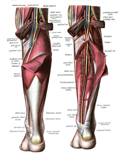
The Tibial Nerve provides innervation to the muscles of the lower leg and foot. Specifically: Triceps Surae ( the two headed Gastocnemius and Soleus); Plantaris, Popliteus; Tibialis Posterior; Flexor Digitorum Longus; and Flexor Hallucis Longus muscles.[1][2] It also has articular and cutaneous branches.[3]
Terminal Branches[edit | edit source]
At the foot level (just after the heel) the Tibial Nerve divides into the Medial Plantar Nerve (MPN) and the Lateral Plantar Nerve (LPN).[1] The MPN supplies muscular branches to the big toe and the two toes next to it, and the LPN the other two toes. The Sural Nerve is a cutaneous branch of the Tibial nerve that supplies the skin of the legs and feet.[2]
Motor[edit | edit source]
- At Popliteal Fossa region branches of the Tibial Nerve supply medial and lateral heads of gastrocnemius, soleus, plantaris and popliteus muscles. The popliteus branch goes on to supply tibialis posteriormuscle, superior tibiofibular joint, tibia bone, interosseous membrane of leg, and the inferior tibiofibular joint.[3]
- Back of the Leg branches of the Tibial Nerve supply tibialis posterior, flexor digitorum longus, flexor hallucis longus, and deep part of soleus.[3]
- from
Sensory[edit | edit source]
Branches of the Tibial nerve are
- Medial Sural Nerve supplies skin on lower half back of leg and skin of foot laterally till little toe.
- Medial Calcaneal Nerve supplies skin on posterior and inferior surface calcaneous.
- Articular branches are to the knee joint ( 3 in total) and ankle joint.[3]
Clinical relevance[edit | edit source]
Injury to the Tibial Nerve can cause motor loss and altered sensation and pain to any of the areas it supplies, depending on site of involvement.
- Popliteal Fossa region. Injury may occur due to eg. Space occupying lesion; Laceration injury; Posterior dislocation of the knee[5]; Entrapement in Soleus arch. Soleus arch entrapment neuropathy can occur with sports that make special demands on the calf muscles. Swelling and hypertrophy of the soleus muscle may cause its tendinous arch to compress the popliteal artery and vein as well as the tibial nerve. This can cause chronic mechanical damage to the nerve and the artery and vein may become occluded.[6]
- Medial Malleous Level. Compression of the tibial nerve in the Osseo fibrous tunnel below the flexor retinaculum of the ankle causes a condition termed Tarsal Tunnel Syndrome. On examination it presents as pain and paresthesia in the sole of the foot, For a full assessment and treatment of this condition se Tarsal Tunnel Syndrome.[5]
- Sole of foot. Abnormal pressure at the ball of the foot can irritate the first plantar digital nerve causing Morton's Neuroma/Metatarsalgia.
Assessment and Treatment[edit | edit source]
see
Resources[edit | edit source]
References[edit | edit source]
- ↑ 1.0 1.1 KenHub. Tibial Nerve. Available from: https://www.kenhub.com/en/library/anatomy/tibial-nerve (last accessed 17.3.2019)
- ↑ 2.0 2.1 Healthline. Tibial Nerve. Available from: https://www.healthline.com/human-body-maps/tibial-nerve#1 (last accessed 17.3.2019)
- ↑ 3.0 3.1 3.2 3.3 Wikipedia. The Tibial Nerve. Available from: https://en.wikipedia.org/wiki/Tibial_nerve (last accessed 18.3.2019)
- ↑ nabi lebraheim. Nerves of the lower leg 3D. Available from: https://www.youtube.com/watch?v=yPJ9sxUubRI (last accessed 17.3.2019)
- ↑ 5.0 5.1 Earth's Lab. Tibial Nerve. Available from: https://www.earthslab.com/anatomy/tibial-nerve/ (last accessed 18.3.2019)
- ↑ Thetter O. (1989) Entrapment Syndrome by the Tendinous Arch of the Soleus Muscle (“Soleus Syndrome”). In: Heberer G., van Dongen R.J.A.M. (eds) Vascular Surgery. Springer, Berlin, Heidelberg. Available from: https://link.springer.com/chapter/10.1007/978-3-642-72942-3_48 (last accessed 18.3.2019)


