Talar tilt: Difference between revisions
m (Text replace - 'Read more.' to ' ') |
No edit summary |
||
| (21 intermediate revisions by 7 users not shown) | |||
| Line 1: | Line 1: | ||
<div class="editorbox"> | |||
'''Original Editor '''- [[User:Laurel|Laurel]] | '''Original Editor '''- [[User:Laurel|Laurel]] | ||
''' | '''Top Contributors''' - {{Special:Contributors/{{FULLPAGENAME}}}} | ||
</div> | </div> | ||
== Purpose | |||
== Purpose == | |||
To examine the ankle for injury of the Anterior Talofibular ligament and the Calcaneofibular ligament. | To examine the ankle for injury of the Anterior Talofibular ligament and the Calcaneofibular ligament. | ||
== Technique | == Technique == | ||
Patient is seated with foot and ankle unsupported. | Patient is seated with foot and ankle unsupported. The foot is positioned in 10-20 degrees of plantarflexion. The distal lower leg is stabilized with one hand just proximal to the malleloi and the hindfoot is inverted with the other hand. The lateral aspect of the talus is palpated to determine if tilting occurs. The laxity is compared to the contralateral side. <ref>Cook CE and Hegedus EJ. Orthopedic Physical Examination Tests: An Evidence-Based Approach. Prentice Hall. New Jersey. 2007.</ref> <ref name="Wheeless">Wheeless Online Textbook of Orthopaedics. Talar Tilt: Physical Exam. http://www.wheelessonline.com/ortho/talar_tilt_physical_exam</ref> | ||
[[Image:Ankle.png|350px]] | |||
{{#ev:youtube|SDGU7cqxN6s}} | |||
== Evidence == | |||
In a prospective study of 244 patients with ankle lesions, a comparison between the talar tilt and the anterior drawer sign was made, leading to the following conclusions; <ref>Johannsen, A. 1978. Acta Orthopaedica. Radiological diagnosis of lateral ligament lesion of the ankle: a comparison between talar tilt and anterior drawer sign.</ref> | |||
*Ligament lesions that are not identified by the talar tilt examination may be diagnosed by the anterior drawer sign | |||
*The anterior drawer sign cannot replace the talar tilt examination, or vice versa. The two methods are complementary. | |||
*It is not possible to differentiate between an isolated lesion of the anterior talofibular ligament and a combined lesion of the anterior talofibular and the calcaneofibular ligaments by the two methods.<br> | |||
The reliability of the radiographic talar tilt test was evaluated using MRI in 112 athletes with injuries to the lateral ligaments of the ankle. Twenty-five athletes with a talar tilt of 15" were treated operatively. lntraoperative findings and the talar tilt test were compared with MR imaging results. The results suggested that MRI is a reliable method for diagnosing injuries of the lateral ankle ligaments. The talar tilt test cannot evaluate the specific pathology of lateral ankle ligaments, but it was reliable in indicating complete double-ligament ruptures (anterior talofibular and calcaneo-fibular ligaments), when talar tilt was 15" or more than on the uninjured side. Sensitivity 67, Specificity 75, LR+ 2.7, LR- 0.44.<ref>Hertel et al. Extracted from Orthopedic Physical Examination Tests: An Evidence-Based Approach: "Medial Talar Tilt Stress Test."</ref> | |||
The recent cross-sectional diagnostic study found out talar tilt test to be a valuable method of identifying mechanical ankle instabilities.<ref>Wenning M, Gehring D, Lange T, Fuerst-Meroth D, Streicher P, Schmal H, Gollhofer A. [https://www.ncbi.nlm.nih.gov/pmc/articles/PMC7890850/ Clinical evaluation of manual stress testing, stress ultrasound and 3D stress MRI in chronic mechanical ankle instability.] BMC Musculoskeletal Disorders. 2021 Dec;22(1):1-3.</ref> | |||
== References == | == References == | ||
<references /> | |||
<references / | |||
[[Category:Ankle]] [[Category:Assessment]] | [[Category:Ankle]] | ||
[[Category:Assessment]] | |||
[[Category:Ankle - Assessment and Examination]] | |||
[[Category:EIM_Residency_Project]] | |||
[[Category:Musculoskeletal/Orthopaedics]] | |||
[[Category:Special_Tests]] | |||
[[Category:Sports Medicine]] | |||
[[Category:Athlete Assessment]] | |||
[[Category:Ankle - Special Tests]] | |||
Latest revision as of 15:06, 1 April 2021
Original Editor - Laurel
Top Contributors - Admin, Laura Ritchie, Kim Jackson, WikiSysop, Tony Lowe, Laurel, Evan Thomas, Vidya Acharya and Wanda van Niekerk
Purpose[edit | edit source]
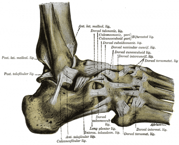
To examine the ankle for injury of the Anterior Talofibular ligament and the Calcaneofibular ligament.
Technique[edit | edit source]
Patient is seated with foot and ankle unsupported. The foot is positioned in 10-20 degrees of plantarflexion. The distal lower leg is stabilized with one hand just proximal to the malleloi and the hindfoot is inverted with the other hand. The lateral aspect of the talus is palpated to determine if tilting occurs. The laxity is compared to the contralateral side. [1] [2]
Evidence[edit | edit source]
In a prospective study of 244 patients with ankle lesions, a comparison between the talar tilt and the anterior drawer sign was made, leading to the following conclusions; [3]
- Ligament lesions that are not identified by the talar tilt examination may be diagnosed by the anterior drawer sign
- The anterior drawer sign cannot replace the talar tilt examination, or vice versa. The two methods are complementary.
- It is not possible to differentiate between an isolated lesion of the anterior talofibular ligament and a combined lesion of the anterior talofibular and the calcaneofibular ligaments by the two methods.
The reliability of the radiographic talar tilt test was evaluated using MRI in 112 athletes with injuries to the lateral ligaments of the ankle. Twenty-five athletes with a talar tilt of 15" were treated operatively. lntraoperative findings and the talar tilt test were compared with MR imaging results. The results suggested that MRI is a reliable method for diagnosing injuries of the lateral ankle ligaments. The talar tilt test cannot evaluate the specific pathology of lateral ankle ligaments, but it was reliable in indicating complete double-ligament ruptures (anterior talofibular and calcaneo-fibular ligaments), when talar tilt was 15" or more than on the uninjured side. Sensitivity 67, Specificity 75, LR+ 2.7, LR- 0.44.[4]
The recent cross-sectional diagnostic study found out talar tilt test to be a valuable method of identifying mechanical ankle instabilities.[5]
References[edit | edit source]
- ↑ Cook CE and Hegedus EJ. Orthopedic Physical Examination Tests: An Evidence-Based Approach. Prentice Hall. New Jersey. 2007.
- ↑ Wheeless Online Textbook of Orthopaedics. Talar Tilt: Physical Exam. http://www.wheelessonline.com/ortho/talar_tilt_physical_exam
- ↑ Johannsen, A. 1978. Acta Orthopaedica. Radiological diagnosis of lateral ligament lesion of the ankle: a comparison between talar tilt and anterior drawer sign.
- ↑ Hertel et al. Extracted from Orthopedic Physical Examination Tests: An Evidence-Based Approach: "Medial Talar Tilt Stress Test."
- ↑ Wenning M, Gehring D, Lange T, Fuerst-Meroth D, Streicher P, Schmal H, Gollhofer A. Clinical evaluation of manual stress testing, stress ultrasound and 3D stress MRI in chronic mechanical ankle instability. BMC Musculoskeletal Disorders. 2021 Dec;22(1):1-3.


