Stress tests for Ankle ligaments: Difference between revisions
No edit summary |
No edit summary |
||
| Line 19: | Line 19: | ||
CFL is an extra-articular component of the complex. Its function is to resist torsion and inversion stresses in a dorsi-flexed foot. The PTFL resists posterior movement of the talus. It is the strongest and least injured part of the complex. Injuries to PTFL usually occur in severe ankle sprains which also involve the ATFL and CFL.<ref name="Lynch">Lynch S. Assessment of the injured ankle in the athlete. Journal of Athletic training. 2002 Dec;37(4):406-412</ref> | CFL is an extra-articular component of the complex. Its function is to resist torsion and inversion stresses in a dorsi-flexed foot. The PTFL resists posterior movement of the talus. It is the strongest and least injured part of the complex. Injuries to PTFL usually occur in severe ankle sprains which also involve the ATFL and CFL.<ref name="Lynch">Lynch S. Assessment of the injured ankle in the athlete. Journal of Athletic training. 2002 Dec;37(4):406-412</ref> | ||
[[Image:Lateral-ankle-ligaments.jpg|thumb|center|500x300px|Lateral Ankle Ligaments]] | |||
<u>''Medial Ankle Ligaments''</u> | |||
The deltoid ligament is the only ligamentous complex stabilizing the medial side of the ankle. This complex is composed of two layers: superficial and deep. The superficial layer includes the tibinavicular, tibiocalcaneal and the posterior talotibial ligament. The deep layer is composed of deep posterior talotibial and deep anterior talotibial ligament. The deep layer has greater contribution to the stbaility of the ankle.<ref name="Aslan" /> The deltoid ligament as a whole has a dual function of providing stability to the talotibial joint as well as transferring forces between tibia and tarsus. It fixates the tibia above the talus and restricts the talus from shifting into a valgus postion, translating antero-laterally or rotating externally.<ref name="Stufkens">Stufkens SAS, van den Bekerom MPJ, Knupp M, Hintermann B, van Dijk CN. The diagnosis and treatment of deltoid ligament lesions in supination-external rotation ankle fractures:a review. Strat Traum Limb Recon. 2012 July 6;7:73-85</ref> | |||
<br> | |||
<br> | |||
<br> | <br> | ||
Revision as of 15:56, 26 February 2017
Original Editor - Your name will be added here if you created the original content for this page.
Top Contributors - Vidhu Sindwani, Evan Thomas, Wanda van Niekerk, Rucha Gadgil, Kim Jackson and WikiSysop
Introduction[edit | edit source]
Ankle sprains are one of the most common musculoskeletal and sports-related injury, constituting nearly 25% of all musculoskeletal trauma cases and almost 40% of all sports-related trauma cases.[1] 40%-50% of these case have reported to have long term residual symptoms with almost 20% of acute ankle sprains developing chronic ankle instability. [2] Individuals with chronic instability often report recurrent sprains and 'giving-way' sensation at the ankle joint, a condition clinicall referred to as Functional Ankle Instability (FAI). [3] Several clinical tests can be used to assess FAI and the respective ligament involved in the acute sprain or chronic instability.
Relevant Anatomy[edit | edit source]
Ligaments of the ankle
Lateral ankle ligaments
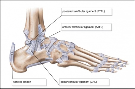
The lateral side of the ankle has three supporting ligaments: the anterior talofibular (ATFL), the posterior talofibular (PTFL) and the calcaneofibular (CFL). The three ligaments are together called the Lateral Collateral Ligament Complex. The ATFL resists torsion and inversion stresses in a plantar flexed foot. It is the weakest and hence the most commonly injured part of the Lateral Collateral Ligament Complex. [1]
CFL is an extra-articular component of the complex. Its function is to resist torsion and inversion stresses in a dorsi-flexed foot. The PTFL resists posterior movement of the talus. It is the strongest and least injured part of the complex. Injuries to PTFL usually occur in severe ankle sprains which also involve the ATFL and CFL.[4]
Medial Ankle Ligaments
The deltoid ligament is the only ligamentous complex stabilizing the medial side of the ankle. This complex is composed of two layers: superficial and deep. The superficial layer includes the tibinavicular, tibiocalcaneal and the posterior talotibial ligament. The deep layer is composed of deep posterior talotibial and deep anterior talotibial ligament. The deep layer has greater contribution to the stbaility of the ankle.[1] The deltoid ligament as a whole has a dual function of providing stability to the talotibial joint as well as transferring forces between tibia and tarsus. It fixates the tibia above the talus and restricts the talus from shifting into a valgus postion, translating antero-laterally or rotating externally.[5]
Sub Heading 3[edit | edit source]
Recent Related Research (from Pubmed)[edit | edit source]
Extension:RSS -- Error: Not a valid URL: Feed goes here!!|charset=UTF-8|short|max=10
References[edit | edit source]
References will automatically be added here, see adding references tutorial.
- ↑ 1.0 1.1 1.2 Aslan A, Sofu H, Kirdemir V. Ankle Ligament Injury: Current concept. OA Orthopaedics. 2014 March 11; 2(1):5
- ↑ Chan KE. Acute and Chronic Lateral Ankle Instability in the Athlete. Bulletin of the NYU hospital for joint diseases. 2011;69(1):17-26
- ↑ Ross SE, Guskiewicz KM, Gross MT. Assessment tools for identifying functional limitations associated with functional ankle instability. Journal of athletic training. 2008 Feb;43(1):44-50.
- ↑ Lynch S. Assessment of the injured ankle in the athlete. Journal of Athletic training. 2002 Dec;37(4):406-412
- ↑ Stufkens SAS, van den Bekerom MPJ, Knupp M, Hintermann B, van Dijk CN. The diagnosis and treatment of deltoid ligament lesions in supination-external rotation ankle fractures:a review. Strat Traum Limb Recon. 2012 July 6;7:73-85


