Stork Test: Difference between revisions

No edit summary
No edit summary
 
(27 intermediate revisions by 6 users not shown)
Line 4: Line 4:
'''Top Contributors''' - {{Special:Contributors/{{FULLPAGENAME}}}}    
</div>  
</div>  
== Search Strategy ==
== Purpose ==
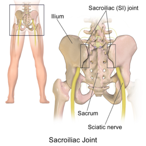
[[File:Sacroiliac Joint human.png|thumb]]
The [[sacroiliac Joint|sacroiliac joint]] (SIJ) is the joint connection between the spine and the pelvis. It can easily be palpated in the low back region in the posterior pelvic area. The sacroiliac joint accounts for 10-27% of the causes of low back pain or buttock pain<ref>Nejati P, Safarcherati A, Karimi F. Effectiveness of exercise therapy and manipulation on sacroiliac joint dysfunction: a randomized controlled trial. Pain physician. 2019;22(1):53-61.</ref>, with a common complaint of localized pain at joint itself.  SIJ dysfunction refers to misalignment or abnormal movement of the SIJ, which can cause pain in or around the SIJ.<ref><span style="line-height: 1.5em;">Daniel L Riddle, Janet K Freburger and North American Orthopaedic Rehabilitation Research Network, “Evaluation of the presence of sacroiliac joint region dysfunction using a combination of test: a multicenter intertester reliability study.” , 2002. Journal of the amercian Physical Therapy association, 2002; 87:879-887. (2B)</span></ref>


University’s Library (Vrije Universiteit Brussel) and databases: Pubmed, Pedro, Sciencedirect and google.<br>Keywords: stork test, Gillet test, sacroiliac joint technique, rucklauf test<br><br>  
The Stork Test, also known as the Gillet Test, the Step Test, and the One-Legged Stance Test, assesses the movement of the SIJ between the innominate and sacrum through the clinician's palpation of the [[Sacroiliac Joint Syndrome|posterior superior iliac spine]] (PSIS). This may be a useful test for clinical evaluation of a subject's ability to stabilize intrapelvic motion.<ref name=":1">Barbara A Hungerford et al., “Evaluation of the ability of physical therapists to palpate intrapelvic motion with the stork test on the support side.”, Journal of American Physical therapy Association, 2007; 87:879-887. (2B)</ref>


== Definition/Description  ==
== Technique ==
<div>
To perform the Stork Test, begin by standing behind the patient and palpate the posterior superior iliac spines (PSIS) on the right and left side of the hip. The person is asked to stand on one leg while flexing the opposite knee and bring the knee closer to the chest.<ref>Ribeiro RP, Guerrero FG, Camargo EN, Beraldo LM, Candotti CT. Validity and Reliability of Palpatory Clinical Tests of Sacroiliac Joint Mobility: A Systematic Review and Meta-analysis. Journal of manipulative and physiological therapeutics. 2021 May;44(4):307-18.</ref> The test is then repeated on the other side and compared bilaterally<ref name="null">Dutton M. Orthopaedic examination, evaluation, and intervention. 2nd ed. New York: McGraw Hill, 2008.</ref><ref name="null2">Konin J, Wiksten D, Isear J, Brader H. Special test for orthopedic examination. New Jersey: Slack, 2002.</ref>. The examiner should compare each side for quality and amplitude of movement<ref name="Lee">Lee D. The pelvic girdle: an approach to the examination and treatment of the lumbo-pelvic-hip region. 3rd ed. Edinburgh: Churchill Livingstone, 2004.</ref>. 


Sacroiliac joint (SIJ) mobility tests are a large number of clinical test to asses movement or asymmetry of the SIJ.<sup>10</sup>&nbsp;(LOE 2B) Sacroiliac joint region dysfunction is a term frequently used to describe the cause of pain in or around the region of the joint that is presumed to be due to misalignment or abnormal movement of the SI-joints.<sup>4</sup> (LOE 2B)[[Image:Book008 sec5 fig257 l1337814891017.jpg|center|250x250px]]  
In a normal functioning pelvis, the PSIS should move caudally, or inferiorly, when the leg is flexed towards the chest. If the PSIS remains level or moves superiorly, this indicates a positive test and warrants further examination of the SIJ.  A positive test is an indication of sacroiliac joint hypomobility.<ref>Magee DJ. Orthopedic physical assessment. 5th ed. St. Louis: Saunders, 2008. (LOE 1A)</ref> {{#ev:youtube|pvsDU6IJoSc|400}}<ref>PhysioTutors. The Gillet Test for SI-Joint Dysfunction. Available from: http://www.youtube.com/watch?v=dvhvKXnXAac[last accessed 19/10/2023]</ref>


<br>  
== Evidence ==
<div>


<br>The assessment of the Stork test involves palpation of the posterior superior iliac spine (PSIS).<br>  
The range of motion in the SIJ is small, less than 4° of rotation and up to 1.6 mm of translation. There is a minimal palpable difference between the symptomatic and asymptomatic sides for patient with presumed SIJ pain due to the minimal movement the joint allows.<ref name=":1" />Rather than use one test during an examination, it is recommended that a cluster of tests be used to confirm a diagnosis. The recommended [[Sacroiliac Joint Special Test Cluster|SIJ Test Item Cluster]] are:<ref>KJ Stuber. Specificity, sensitivity, and predictive values of clinical tests of the sacroiliac joint: a systematic review of the literature. J Can Chiropr Assoc. 2007; 51(1): 30-41 ( LOE 1A)</ref>
# Distraction Test
# Compression Test
# Thigh Thrust Test
# Gaenslen’s Test
# Sacral Thrust Test
In order to confirm a diagnosis of SIJ pain, 3 of 5 of the tests must be positive. At least 1 of the 3 positive results must be the Thigh Thrust Test or the Compression Test.<ref>Karolina M. Szadek, et al.; Diagnostic validity of criteria for sacroiliac joint pain: a systematic review.; The Journal of Pain: Official Journal of the American Pain Society 10, no. 4 (April 2009): 354-368. (LOE 1A)</ref><br>A positive Stork Test, combined with other positive sacroiliac mobility tests, indicates a valid impairment of mobility of the sacroiliac joint (SIJ). Springing tests, by means of which a passive mobility ("joint play") is being tested, are most valuable in dysfunction diagnostics<ref>Grgić V.; The sacroiliac joint dysfunction: clinical manifestations, diagnostics and manual therapy; Lijec Vjesn. 2005 Jan-Feb;127(1-2):30-5. (LOE 3A)</ref>. However, the clinical use of these clusters has yet to been validated. Multiple studies confirm that these tests have no significance to determine SIJ dysfunction nor pain.<ref name=":0">Hungerford B, Evaluation of the ability of physical therapists to palpate intrapelvic motion with the Stork test on the support side; Physical Therapy; 2007; Jul;87(7):879-87. (LOE 2A)</ref><ref name=":2">van der Wurff P, Hagmeijer RH, Meyne W., “Clinical tests of the sacroiliac joint. A systematic methodological review. Part 1: Reliability.”, Man Ther. 2000 Feb;5(1):30-6. (1A)</ref><ref>Riddle DL, Freburger JK., “Evaluation of the presence of sacroiliac joint region dysfunction using a combination of tests: a multicenter intertester reliability study.”, PHYS THER., 2002 Aug; 82(8):772-782. (2B)</ref>.
</div>


<span style="font-size: 13.2799997329712px; line-height: 1.5em;">The therapist places one thumb directly on the PSIS and the other thumb is placed medial to the PSIS, on the sacral base. Ask the patient to raise one knee up, that the hip and knee are flexed to 90°. Assess the posterior rotation of the innominate on the side of the lifted knee. Then a</span><span style="font-size: 13.2799997329712px; line-height: 1.5em;">sk the patient to raise the other knee up while assessing the anterior rotation of the innominate on single leg support side. The direction of the bone motion is palpated as the contralateral foot is lifted off the ground.<sup>6</sup> (LOE 5)</span>
== Reliability ==
<div>
<div>
Various studies show that men are less mobile than women and elderly people starting from the age of 65 are less mobile than younger people.<sup>23</sup> <br>Women who gave birth are more mobile with an increase in contra nutation of the pelvic joint than women who have not.<sup>17</sup>. (LOE 2A)<br><br>
The Stork Test should not be the sole test used to diagnose SIJ dysfunction. <ref name=":2" /><ref>Wilco Meijne, Katinka van Neerbos, Geert Aufdemkampe, Peter van der Wurff d, Intraexaminer and interexaminer reliability of the Gillet test, Journal of Manipulative &amp; Physiological Therapeutics Volume 22, Issue 1, Pages 4–9, January 1999. (LOE 1B)</ref>The Stork Test demonstrates high reliability when a group of mobility and provocation tests are performed along with it.<ref>Arab AM, Abdollahi I, Joghataei MT, Golafshani Z, Kazemnejad A.,”Inter- and intra-examiner reliability of single and composites of selected motion palpation and pain provocation tests for sacroiliac joint., Man Ther. 2009 Apr;14(2):213-21. (1B)</ref> When used alone, the Stork Test's reliability is low (0.22) and is not recommended to be used individually.<ref>Dreyfuss P, Dreyer S, Griffin J, Hoffman J, Walsh N. Positive sacroiliac screening test in asymptomatic adults. Spine. 1994;19(10):1138-1143.</ref>
 
== Clinically Relevant Anatomy  ==
 
The sacroiliac joints are essential for effective load transfer between the spine and the lower extremities. The sacrum, pelvis and spine-, are functionally interrelated through muscles, fascia and ligamentous interconnections. <br>This joint is the largest axial joint in our body and has an average surface of 17, 5 cm<sup>2</sup>, <sup>14</sup>. (LOE 2B, 3A)
 
The SIJ is uniquely composed of a synovial joint, which permits very little, to no movement and a diarthrodial joint or free moveable joint. The capsular portion of the sacrum is made up of hyaline cartilage (diarthrosis). The capsular portion of the ilium consists of fibrocartilage, which provides considerable internal stability. <sup>13</sup> (LOE 3A) Other characteristics include strong weight bearing, paired C-shaped joints. <sup>14</sup> (LOE 3A)
 
<br>Essentially, the SIJ is encased in a capsule that has a smooth anterior wall but there is an absence of a rudimentary posterior capsule. Hence the SI ligaments are more developed dorsally, functioning as a connecting band between the sacrum and ilia. These ligaments are represented in figure 1. The sacroiliac ligament complex prevents a great mobility of the sacrum between the two ilia. The ligaments of the SI ligament complex are weaker in women than in men, allowing the ability to give birth.<sup>16</sup> (LOE 2A)
 
<br>Low back pain at the level of the SIJ is not always linked to force closure, but can also be linked to form closure with the passive structures. There is some evidence18 (LOE 3A) that the long dorsal sacroiliac ligament should not be overlooked in causes of low back pain. <br>The sacrotuberous ligaments are directly connected to the long head of the biceps femoris, the piriformis and the gluteus maximus, thus they can have an influence on the SIJ mobility.16 (LOE 2A) There are 32 other muscles, which attach on the sacrum and can be found in: [http://www.physio-pedia.com/Sacroiliac_joint#Muscles SACRO-ILIAC JOINT MUSCLES]<br><br>
 
== Differential Diagnosis<br> ==
 
== Purpose  ==
 
The relationship between the SIJ and low back pain has been a subject of debate. Some researchers regard SIJ pain as a major contributor to the problem of low back pain while others regard it as unimportant or irrelevant. It is nowadays generally accepted that about 13&nbsp;% of patients with chronic low back pain have the origin of pain confirmed as the SIJ.<sup>2</sup>&nbsp;(LOE 2B)<br>
 
There are two clinical perspectives to consider. The first is the SIJ as a load-transferring mechanical junction between the pelvis and the spine that may cause either the SIJ or other structures to produce painful stimuli. The second perspective considers the SIJ as a source of pain.
 
The first perspective proposes that the joint is malfunctioning. Therefore the word dysfunction is commenly used to encapsulate the complexity of abberations believed to occur. The evidence favoring that mechanical SIJ dysfunctions are related to the experience of back and referred pain is less than convincing. The range of motion in the SIJ is small, less than 4° of rotation and up to 1,6 mm of translation. Hence in patients presumed to have an SIJ source of pain it is difficult and doubtful to found differences in range of motion between the symptomatic and asymptomatic sides.<sup>2</sup>(LOE 2B)
 
Due to the poor reliability and validity of many SIJ mobility assesment tests. And with an increased understanding of the role of the pelvis in load transfer has shifted the focus of clinical assessment procedures for SIJ function from SIJ mobility testing to functional assessment procedures that test the ability of the pelvis to maintain stability during load transfer between the spine and the lower limbs.<sup>2</sup>  
 
An investigation of motion between the innominate bone and the sacrum on the side of single-leg support during a standing hip flexion movement revealed that the innominate bone on the side of single-leg support rotated posteriorly relative to the sacrum in subjects who were healthy. This pattern was altered reliably in the presence of PGP. Here the innominate bone rotated anteriorly relative to the sacrum, which was an indication of a failure of the self-bracing mechanism to maintain the SIJ in its closed pack position. Therefore the Stork test may be a useful test for clinical evaluation of a subject's ability to stabilize intrapelvic motion.<sup>2</sup>&nbsp;(LOE 2B)
 
== Technique  ==
 
The assessment of the Stork test involves palpation of the posterior superior iliac spine (PSIS). The therapist places one thumb directly on the PSIS and the other thumb is placed medial to the PSIS, on the sacral base. Ask the patient to raise one knee up, that the hip and knee are flexed to 90°. Assess the posterior rotation of the innominate on the side of the lifted knee. Then ask the patient to raise the other knee up while assessing the anterior rotation of the innominate on single leg support side. The direction of the bone motion is palpated as the contralateral foot is lifted off the ground<sup>.6</sup> (LOE 5)
 
<br>In a normally functioning pelvis, the pelvis of the side being palpated should rotate posteriorly, causing the PSIS to drop or move inferiorly. The test is positive when the PSIS on the ipsilateral side (same side of the body) of the knee flexion moves minimally in the inferior direction or doesn’t move. A positive test is an indication of sacroiliac joint hypomobility<sup>.19</sup> (LOE 1A)
 
<br>The test is negative when the SI-joint is not blocked on the side of the lifted leg, the ilium of the leg will rotate in dorso-caudal direction. The PSIS will move a fraction below the PSIS of the supporting leg in dorso-caudal direction.
 
<br>Hungerford et al. concluded that the ability of the physiotherapist to reliably palpate and recognize an altered pattern of intrapelvic motion during Stork Test on the support side was substantiated. The ability to distinguish between no relative movement and anterior rotation of the innominate bone during a load-bearing task was good. Although further research is needed to determine the validity of this test for detecting pelvic girdle dysfunction<sup>2</sup>(LOE 2B).<br><br><br>
 
== Outcome Measures  ==
 
Several studies concluded that not a single test but a cluster of tests should be used to confirm diagnosis. Other sacroiliac pain provocation tests to evaluate the mobility of the sacroiliac joint are the Distraction test, Compression test, Thigh thrust test, Gaenslen’s test and Sacral thrust test. <sup>21</sup> ( LOE 1A)
 
<br>A positive Stork test (Gillet test), combined with other positive sacroiliac mobility tests, indicates an valid impairment of mobility of the sacroiliac joint (SIJ). Springing tests, by means of which a passive mobility ("joint play") is being tested, are most valuable in dysfunction diagnostics<sup>22</sup>.&nbsp;;The clinical use of these clusters however has yet to been validated .Multiple studies confirm that these tests have no significance to determine SIK dysfunction nor pain.<sup>17,11,9</sup>&nbsp;.1 study determined that the stork test, together with the examniation of irritation points and irritation points during functinal testing could asses SIJ dysfunction.Therefore, the physical therapy management for this dysfunction was Mobilizing techniques without impulse as well as manipulative techniques with high-velocity impulses were applied to resolve the dysfunction <sup>5</sup>. (LOE 2B).
 
The thigh thrust test, compression test, and three or more positive stressing tests showed discriminative power for diagnosing SI joint pain. 3 of 5 must be positive (Thigh Thrust, Compression, Gaenslen, FABER, Distraction). 1 of 3 positive results must be Thigh Thrust or Compression.<sup>23</sup>.
 
Manual High-Velocity-Low-Amplitude (HVLA) manipulation is an efficient method of treatment for patients with sacroiliac dysfunction. In a study<sup>7</sup> (LOE 1B), improvements in pain (VAS-scale) and function were detected 2 days and 1 month after a single session. No consensus is found about the underlying mechanisms of treating SIJ dysfunctions. A few plausible explanations for improvements are increased ROM, normalization of muscle tone and disrupting articular adhesions.
 
<br>The following is a description of the HVLA manipulation technique used in the LOE 1B study. The patient is supine and the therapist stands contralateral to the side which is to be manipulated (e.g. right) (Fig. 2). The patient is passively moved into side bending toward the side to be manipulated. The patient should inter-lock their fingers behind their head. The therapist passively rotates the patient, and then delivers a quick thrust to the Anterior Superior Iliac Spine (ASIS) in a posterior and inferior direction.<sup>3</sup>(LOE 1B)<br>  
 
== Examination<br>  ==
 
== Reliability<br>  ==
 
1) The outcomes of individual mobility tests, including the Gillet’s test, are not reliable in diagnosing SIJ dysfunction<sup>11</sup>(LOE 1A)<br>2) The outcomes of individual mobility tests, including the Gillet’s test, are not valid in diagnosing SIJ dysfunction<sup>12</sup>(LOE 1A)<br>3) A cluster of mobility tests and provocation tests have an average to high reliability in the assessment of the SIJ <sup>1</sup>(LOE2B)<br>4) A single manual HVLA manipulation session is proven to reduce pain and mobility impairment for people suffering from SIJ dysfunction<sup>1</sup>(LOE 2B)<br>5) Physical therapists can recognize an altered pattern of intrapelvic motion during the Stork Test. Physical therapists can distinguish between no relative movement and anterior rotation of the innominate bone during a Stork test<sup>17</sup>(LOE 2A).<br>6) According to a study, The Gillet test, as performed in this study, does not appear to be reliable.<sup>20</sup> (LOE 1B)<br>The Diagnostic Accuracy of the Gillet test (stork test) is described below:<br>Sensitivity is the proportion of people with a positive test result who have the target disorder, essentially true positives.<sup>21</sup> (LOE 1A)  
 
[[Image:T6-jcca51 1p030.png|center|250x250px]]<br>Specificity is the proportion of people with a negative test result who do not have the target disorder, essentially true negatives.21 (LOE 1A)
 
[[Image:4.jpg|center|250x250px]]<br>
 
== Visual example of test  ==
 
[https://www.youtube.com/watch?v=RJNXg_CQz4M Kinetic Test of SI Joint: Stork Test]<br>[https://www.youtube.com/watch?v=tdACVi825gY How to assess motion of the Sacroiliac Joint - Stork / Gillett test]<br><br>  
 
[[Image:Test2.jpg|center|250x250px]]Figure 2. Subject palpation for the Stork Test. (Left) Positive right-side support-phase test - no movement. (Right) Negative left-side support-phase test with anterior motion of the left posterior superior iliac spine relative to central S2. <br>
 
== Resources <br>  ==


&nbsp;Pubmed, Pedro, Web of Knowledge<br>
A meta analysis on intra-rater reliability reported the Stork test to have moderate to good agreement (κ = 0.46)<ref>Ribeiro RP, Guerrero FG, Camargo EN, Beraldo LM, Candotti CT. [https://doi.org/10.1016/j.jmpt.2021.01.001. Validity and Reliability of Palpatory Clinical Tests of Sacroiliac Joint Mobility: A Systematic Review and Meta-analysis]. Journal of Manipulative and Physiological Therapeutics, 2021; 44(4):307-318. <nowiki>https://doi.org/10.1016/j.jmpt.2021.01.001</nowiki>.</ref> Hungerford et al. concluded that the ability of the physiotherapist to reliably palpate and recognize an altered pattern of intrapelvic motion during Stork Test on the support side was substantiated. The ability to distinguish between no relative movement and anterior rotation of the innominate bone during a load-bearing task was good. The validity of the test is 55.%.<ref>Stuber KJ. Specificity, sensitivity, and predictive values of clinical tests of the sacroiliac joint: a systematic review of the literature. The Journal of the Canadian Chiropractic Association. 2007 Mar;51(1):30.</ref> <br>
 
== Recent Related Research (from pubmed) ==
 
see tutorial on Adding PubMed Feed
<div class="researchbox">
<span>Feed goes here!!|</span>  
</div>  
== References  ==
== References  ==
 
</div>
1. Arab AM, Abdollahi I, Joghataei MT, Golafshani Z, Kazemnejad A.,”Inter- and intra-examiner reliability of single and composites of selected motion palpation and pain provocation tests for sacroiliac joint.”, Man Ther. 2009 Apr;14(2):213-21. (1B)<br>2. Barbara A Hungerford et al., “Evaluation of the ability of physical therapists to palpate intrapelvic motion with the stork test on the support side.”, Journal of American Physical therapy Association, 2007; 87:879-887. (2B)
<references />
 
3. Cleland, J., Fritz, J., et al., “Comparison of the effectiveness of three manual physical therapy techniques in a subgroup of patients with low back pain who satisfy a clinical prediction rule: a randomized clinical trial.” Spine (2009)34 (25), 2720. (1B)
 
<span style="line-height: 1.5em;">4. Daniel L Riddle, Janet K Freburger and North American Orthopaedic Rehabilitation Research Network, “Evaluation of the presence of sacroiliac joint region dysfunction using a combination of test: a multicenter intertester reliability study.” , 2002. Journal of the amercian Physical Therapy association, 2002; 87:879-887. (2B)</span>  
 
5. Galm R, Fröhling M, Rittmeister M, Schmitt E., “Sacroiliac joint dysfunction in patients with imaging-proven lumbar disc herniation.”, Eur Spine J. 1998;7(6):450-3. (1B)
 
<span style="line-height: 1.5em;">6. Jeffrey Gross, Wiley-Blackwell, Musculoskeletal Examination 3rd edition, 2009, p. 114.</span>
 
7. Kamali F, Shokri E., “The effect of two manipulative therapy techniques and their outcome in patients with sacroiliac joint syndrome.”, Journal of Bodywork &amp; Movement Therapies, 2012 16, 29e35. (1B)
 
<span style="line-height: 1.5em;">8. Kamali F, Shokri E., “The effect of two manipulative therapy techniques and their outcome in patients with sacroiliac joint syndrome.”, 2007; 87:879-887.PHYS THER.  (3B)</span>
 
9. Riddle DL, Freburger JK., “Evaluation of the presence of sacroiliac joint region dysfunction using a combination of tests: a multicenter intertester reliability study.”, PHYS THER., 2002 Aug; 82(8):772-782. (2B)
 
10. Sturesson, Bengt Md, Uden Alf MD, PhD, Vleeming, Andry PhD., “A radiostereometric analysisof movements of the sacroiliac joints during the standing hip flexion test.”, Spine, 2000; 25(2):214. (2B)
 
11. van der Wurff P, Hagmeijer RH, Meyne W., “Clinical tests of the sacroiliac joint. A systematic methodological review. Part 1: Reliability.”, Man Ther. 2000 Feb;5(1):30-6. (1A)
 
12. van der Wurff P, Hagmeijer RH, Meyne W., “Clinical tests of the sacroiliac joint. A systematic methodological review. Part 1: Reliability.”, Manual Therapy, 2000; 5(2):89±96. (1A)
 
13. A. Vleeming;M. D. Schuenke;A. T. Masi;J. E. Carreiro;L. Danneels and F. H. Willard. The sacroiliac joint: an overview of its anatomy, function and potential clinical implications; Journal of Anatomy Volume 221, Issue 6, pages 537–567, December 2012. (LOE 3A)<br>14. Cohen S., Steven P., Sacroiliac joint pain: a comprehensive review of anatomy, diagnosis and treatment, IARS, November 2005, volume 101, issue 5, pp 1440-1453 (LOE 3A)<br>15. Bernard TN, Cassidy JD. The sacroiliac syndrome. Pathophysiology, diagnosis and management. In: Frymoyer JW, ed. The adult spine: principles and practice. New York: Raven, 1991;2107–30. (LOE 2A)<br>16. Calvillo O., Skaribas I., Turnispeed J., Anatomy and pathophysiology of the SIJ, current science, 2000 (LOE 2A)<br>17. Hungerford B, Evaluation of the ability of physical therapists to palpate intrapelvic motion with the Stork test on the support side; Physical Therapy; 2007; Jul;87(7):879-87. (LOE 2A)
 
18. Vleeming A, Pool-Goudzwaard AL, Hammudoghlu D, Stoeckart R, Snijders CJ, Mens JM. The function of the long dorsal sacroiliac ligament: its implication for understanding low back pain. Spine 1996;21:556-62 (LOE 3A)
 
19. Magee DJ. Orthopedic physical assessment. 5th ed. St. Louis: Saunders, 2008. (LOE 1A)
 
20. Wilco Meijne, Katinka van Neerbos, Geert Aufdemkampe, Peter van der Wurff d, Intraexaminer and interexaminer reliability of the Gillet test, Journal of Manipulative &amp; Physiological Therapeutics Volume 22, Issue 1, Pages 4–9, January 1999. (LOE 1B)
 
21. KJ Stuber. Specificity, sensitivity, and predictive values of clinical tests of the sacroiliac joint: a systematic review of the literature. J Can Chiropr Assoc. 2007; 51(1): 30-41 ( LOE 1A)
 
22. Grgić V.; The sacroiliac joint dysfunction: clinical manifestations, diagnostics and manual therapy; Lijec Vjesn. 2005 Jan-Feb;127(1-2):30-5. (LOE 3A)
 
23. Karolina M. Szadek, et al.; Diagnostic validity of criteria for sacroiliac joint pain: a systematic review.; The Journal of Pain: Official Journal of the American Pain Society 10, no. 4 (April 2009): 354-368. (LOE 1A)
 
24. © 2015 Davis et al. Mobility predicts change in older adults’ health-related quality of life: evidence from a Vancouver falls prevention prospective cohort study. Health and Quality of Life Outcomes 2015
 
<br><br>
<br></div>
[[Category:Special_Tests]]
[[Category:Special_Tests]]
[[Category:Sacroiliac Examination]]
[[Category:Sacroiliac Conditions]]
[[Category:Sports Medicine]]
[[Category:Athlete Assessment]]
[[Category:Pelvis]]
[[Category:Pelvis - Special Tests]]
[[Category:Vrije Universiteit Brussel Project]]

Latest revision as of 14:18, 10 March 2024

Purpose[edit | edit source]

Sacroiliac Joint human.png

The sacroiliac joint (SIJ) is the joint connection between the spine and the pelvis. It can easily be palpated in the low back region in the posterior pelvic area. The sacroiliac joint accounts for 10-27% of the causes of low back pain or buttock pain[1], with a common complaint of localized pain at joint itself. SIJ dysfunction refers to misalignment or abnormal movement of the SIJ, which can cause pain in or around the SIJ.[2]

The Stork Test, also known as the Gillet Test, the Step Test, and the One-Legged Stance Test, assesses the movement of the SIJ between the innominate and sacrum through the clinician's palpation of the posterior superior iliac spine (PSIS). This may be a useful test for clinical evaluation of a subject's ability to stabilize intrapelvic motion.[3]

Technique[edit | edit source]

To perform the Stork Test, begin by standing behind the patient and palpate the posterior superior iliac spines (PSIS) on the right and left side of the hip. The person is asked to stand on one leg while flexing the opposite knee and bring the knee closer to the chest.[4] The test is then repeated on the other side and compared bilaterally[5][6]. The examiner should compare each side for quality and amplitude of movement[7].

In a normal functioning pelvis, the PSIS should move caudally, or inferiorly, when the leg is flexed towards the chest. If the PSIS remains level or moves superiorly, this indicates a positive test and warrants further examination of the SIJ. A positive test is an indication of sacroiliac joint hypomobility.[8]
[9]

Evidence[edit | edit source]

The range of motion in the SIJ is small, less than 4° of rotation and up to 1.6 mm of translation. There is a minimal palpable difference between the symptomatic and asymptomatic sides for patient with presumed SIJ pain due to the minimal movement the joint allows.[3]Rather than use one test during an examination, it is recommended that a cluster of tests be used to confirm a diagnosis. The recommended SIJ Test Item Cluster are:[10]

  1. Distraction Test
  2. Compression Test
  3. Thigh Thrust Test
  4. Gaenslen’s Test
  5. Sacral Thrust Test

In order to confirm a diagnosis of SIJ pain, 3 of 5 of the tests must be positive. At least 1 of the 3 positive results must be the Thigh Thrust Test or the Compression Test.[11]
A positive Stork Test, combined with other positive sacroiliac mobility tests, indicates a valid impairment of mobility of the sacroiliac joint (SIJ). Springing tests, by means of which a passive mobility ("joint play") is being tested, are most valuable in dysfunction diagnostics[12]. However, the clinical use of these clusters has yet to been validated. Multiple studies confirm that these tests have no significance to determine SIJ dysfunction nor pain.[13][14][15].

Reliability[edit | edit source]

The Stork Test should not be the sole test used to diagnose SIJ dysfunction. [14][16]The Stork Test demonstrates high reliability when a group of mobility and provocation tests are performed along with it.[17] When used alone, the Stork Test's reliability is low (0.22) and is not recommended to be used individually.[18]

A meta analysis on intra-rater reliability reported the Stork test to have moderate to good agreement (κ = 0.46)[19] Hungerford et al. concluded that the ability of the physiotherapist to reliably palpate and recognize an altered pattern of intrapelvic motion during Stork Test on the support side was substantiated. The ability to distinguish between no relative movement and anterior rotation of the innominate bone during a load-bearing task was good. The validity of the test is 55.%.[20]

References[edit | edit source]

  1. Nejati P, Safarcherati A, Karimi F. Effectiveness of exercise therapy and manipulation on sacroiliac joint dysfunction: a randomized controlled trial. Pain physician. 2019;22(1):53-61.
  2. Daniel L Riddle, Janet K Freburger and North American Orthopaedic Rehabilitation Research Network, “Evaluation of the presence of sacroiliac joint region dysfunction using a combination of test: a multicenter intertester reliability study.” , 2002. Journal of the amercian Physical Therapy association, 2002; 87:879-887. (2B)
  3. 3.0 3.1 Barbara A Hungerford et al., “Evaluation of the ability of physical therapists to palpate intrapelvic motion with the stork test on the support side.”, Journal of American Physical therapy Association, 2007; 87:879-887. (2B)
  4. Ribeiro RP, Guerrero FG, Camargo EN, Beraldo LM, Candotti CT. Validity and Reliability of Palpatory Clinical Tests of Sacroiliac Joint Mobility: A Systematic Review and Meta-analysis. Journal of manipulative and physiological therapeutics. 2021 May;44(4):307-18.
  5. Dutton M. Orthopaedic examination, evaluation, and intervention. 2nd ed. New York: McGraw Hill, 2008.
  6. Konin J, Wiksten D, Isear J, Brader H. Special test for orthopedic examination. New Jersey: Slack, 2002.
  7. Lee D. The pelvic girdle: an approach to the examination and treatment of the lumbo-pelvic-hip region. 3rd ed. Edinburgh: Churchill Livingstone, 2004.
  8. Magee DJ. Orthopedic physical assessment. 5th ed. St. Louis: Saunders, 2008. (LOE 1A)
  9. PhysioTutors. The Gillet Test for SI-Joint Dysfunction. Available from: http://www.youtube.com/watch?v=dvhvKXnXAac[last accessed 19/10/2023]
  10. KJ Stuber. Specificity, sensitivity, and predictive values of clinical tests of the sacroiliac joint: a systematic review of the literature. J Can Chiropr Assoc. 2007; 51(1): 30-41 ( LOE 1A)
  11. Karolina M. Szadek, et al.; Diagnostic validity of criteria for sacroiliac joint pain: a systematic review.; The Journal of Pain: Official Journal of the American Pain Society 10, no. 4 (April 2009): 354-368. (LOE 1A)
  12. Grgić V.; The sacroiliac joint dysfunction: clinical manifestations, diagnostics and manual therapy; Lijec Vjesn. 2005 Jan-Feb;127(1-2):30-5. (LOE 3A)
  13. Hungerford B, Evaluation of the ability of physical therapists to palpate intrapelvic motion with the Stork test on the support side; Physical Therapy; 2007; Jul;87(7):879-87. (LOE 2A)
  14. 14.0 14.1 van der Wurff P, Hagmeijer RH, Meyne W., “Clinical tests of the sacroiliac joint. A systematic methodological review. Part 1: Reliability.”, Man Ther. 2000 Feb;5(1):30-6. (1A)
  15. Riddle DL, Freburger JK., “Evaluation of the presence of sacroiliac joint region dysfunction using a combination of tests: a multicenter intertester reliability study.”, PHYS THER., 2002 Aug; 82(8):772-782. (2B)
  16. Wilco Meijne, Katinka van Neerbos, Geert Aufdemkampe, Peter van der Wurff d, Intraexaminer and interexaminer reliability of the Gillet test, Journal of Manipulative & Physiological Therapeutics Volume 22, Issue 1, Pages 4–9, January 1999. (LOE 1B)
  17. Arab AM, Abdollahi I, Joghataei MT, Golafshani Z, Kazemnejad A.,”Inter- and intra-examiner reliability of single and composites of selected motion palpation and pain provocation tests for sacroiliac joint.”, Man Ther. 2009 Apr;14(2):213-21. (1B)
  18. Dreyfuss P, Dreyer S, Griffin J, Hoffman J, Walsh N. Positive sacroiliac screening test in asymptomatic adults. Spine. 1994;19(10):1138-1143.
  19. Ribeiro RP, Guerrero FG, Camargo EN, Beraldo LM, Candotti CT. Validity and Reliability of Palpatory Clinical Tests of Sacroiliac Joint Mobility: A Systematic Review and Meta-analysis. Journal of Manipulative and Physiological Therapeutics, 2021; 44(4):307-318. https://doi.org/10.1016/j.jmpt.2021.01.001.
  20. Stuber KJ. Specificity, sensitivity, and predictive values of clinical tests of the sacroiliac joint: a systematic review of the literature. The Journal of the Canadian Chiropractic Association. 2007 Mar;51(1):30.