Spring Ligament: Difference between revisions
No edit summary |
(Changed the headings and subheadings to title case) |
||
| (5 intermediate revisions by the same user not shown) | |||
| Line 1: | Line 1: | ||
= | <div class="editorbox"> '''Original Editor '''- [[User:Priyanka Chugh|Priyanka Chugh]] '''Top Contributors''' - {{Special:Contributors/{{FULLPAGENAME}}}}</div> | ||
== Description == | |||
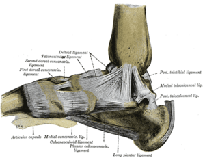
[[File:Foot ligaments.png|thumb|Calcaneonavicular Ligament ]] | |||
The plantar calcaneonavicular ligament also referred to as spring ligament is a thick wide band of cartilaginous connective tissue that supports the medial longitudinal arch of the foot, failure in the spring ligament leads to [[Pes Planus|flat foot]] deformity. The spring ligament despite its name does not possess spring like properties as it is highly collagenous.<ref name=":0">Neumann DA. Kinesiology of the musculoskeletal system-e-book: foundations for rehabilitation. Elsevier Health Sciences; 2013 Aug 7.</ref><ref name=":1">Steginsky B, Vora A. What to do with the spring ligament. Foot and Ankle Clinics. 2017 Sep 1;22(3):515-27.</ref> | |||
== | == Anatomy == | ||
The spring ligament fills the gap between the [[calcaneus]] and the navicular bone, it attaches from the sustentaculum tali of the calcaneus to the medial-plantar surface of the navicular.<ref name=":0" /> | |||
The | The spring ligament is supported medially by the superficial deltoid ligament, posteriorly by the [[Tibialis Posterior|posterior tibial]] tendon and laterally by the bifurcate ligament.<ref>Rule J, Yao L, Seeger LL. Spring ligament of the ankle: normal MR anatomy. AJR. American journal of roentgenology. 1993 Dec;161(6):1241-4.</ref> | ||
= | The superior surface of the spring ligament is covered with synovial membrane, which articulates with the head of talus.<ref name=":0" /> | ||
The | == Function == | ||
The spring ligament functions as static restraint of the medial longitudinal arch, it supports the head of the [[talus]] from planter and medial subaxation against the body weight during standing. <ref name=":0" /><ref name=":1" /> | |||
== Clinical Relevance == | |||
The | === Flat Foot Deformity Secondary to Posterior Tibial Tendon Insufficiency === | ||
The posterior tibial tendon is the main dynamic stabilizer of the medial longitutnal arch, functional loss of the posterior tibial tendon increases the stresses on the medial soft tissue structures of the foot including the spring ligament, which could result in their attenuation leading to the development of acquired [[Pes Planus|flat foot]] deformity. Deland and coworkers studied the MRI finding in patients with posterior tibial tendon insufficiency found that, all patients with posterior tibial tendon insufficiency showed some degree of spring ligament attenuation in MRI and 74% were diagnosed with spring ligament tear. | |||
Although posterior tibial tendon dysfunction is the main cause of flat foot deformity, It should be noted that flat foot deformity can’t result from the failure of the posterior tibial tendon alone, the medial soft tissue structures must also fail to develop the deformity.<ref name=":1" /> | |||
=== Isolated Spring Ligament Rupture === | |||
Acute isolated spring ligament injuries (without posterior tibial tendon involvement) are uncommon, they result in acquired flat foot deformity. Patients with spring ligament rupture are often misdiagnosed as medial ankle sprain before developing flat foot deformity components as hindfoot valgus, loss of midfoot arch and forefoot abduction. | |||
Spring ligament insufficiency has a characteristic clinical picture that can differentiate it from posterior tibial tendon insufficiency. In spring ligament insufficiency the patient can stand on his tip toes on the affected side with partial restoration on the medial arch, but with forefoot abduction and heel valgus. | |||
Also. patients with spring ligament insufficiency reported tenderness in anterior to the medial malleolus or between the sustentaculum tali and the navicular bone while patients with posterior tibial tendon insufficiency reported tenderness posterior and inferior to the medial malleolus. | |||
Differenial diagnosis include superficial deltoid ligament tear but it can’t be differentiated from spring ligament insufficiency clinically.<ref>Bastias GF, Dalmau-Pastor M, Astudillo C, Pellegrini MJ. Spring ligament instability. Foot and ankle clinics. 2018 Dec 1;23(4):659-78.</ref><ref name=":1" /> | |||
== References == | == References == | ||
[[Category:Anatomy]] | [[Category:Anatomy]] | ||
<references /> | |||
Latest revision as of 15:22, 19 July 2020
Description[edit | edit source]
The plantar calcaneonavicular ligament also referred to as spring ligament is a thick wide band of cartilaginous connective tissue that supports the medial longitudinal arch of the foot, failure in the spring ligament leads to flat foot deformity. The spring ligament despite its name does not possess spring like properties as it is highly collagenous.[1][2]
Anatomy[edit | edit source]
The spring ligament fills the gap between the calcaneus and the navicular bone, it attaches from the sustentaculum tali of the calcaneus to the medial-plantar surface of the navicular.[1]
The spring ligament is supported medially by the superficial deltoid ligament, posteriorly by the posterior tibial tendon and laterally by the bifurcate ligament.[3]
The superior surface of the spring ligament is covered with synovial membrane, which articulates with the head of talus.[1]
Function[edit | edit source]
The spring ligament functions as static restraint of the medial longitudinal arch, it supports the head of the talus from planter and medial subaxation against the body weight during standing. [1][2]
Clinical Relevance[edit | edit source]
Flat Foot Deformity Secondary to Posterior Tibial Tendon Insufficiency [edit | edit source]
The posterior tibial tendon is the main dynamic stabilizer of the medial longitutnal arch, functional loss of the posterior tibial tendon increases the stresses on the medial soft tissue structures of the foot including the spring ligament, which could result in their attenuation leading to the development of acquired flat foot deformity. Deland and coworkers studied the MRI finding in patients with posterior tibial tendon insufficiency found that, all patients with posterior tibial tendon insufficiency showed some degree of spring ligament attenuation in MRI and 74% were diagnosed with spring ligament tear.
Although posterior tibial tendon dysfunction is the main cause of flat foot deformity, It should be noted that flat foot deformity can’t result from the failure of the posterior tibial tendon alone, the medial soft tissue structures must also fail to develop the deformity.[2]
Isolated Spring Ligament Rupture[edit | edit source]
Acute isolated spring ligament injuries (without posterior tibial tendon involvement) are uncommon, they result in acquired flat foot deformity. Patients with spring ligament rupture are often misdiagnosed as medial ankle sprain before developing flat foot deformity components as hindfoot valgus, loss of midfoot arch and forefoot abduction.
Spring ligament insufficiency has a characteristic clinical picture that can differentiate it from posterior tibial tendon insufficiency. In spring ligament insufficiency the patient can stand on his tip toes on the affected side with partial restoration on the medial arch, but with forefoot abduction and heel valgus.
Also. patients with spring ligament insufficiency reported tenderness in anterior to the medial malleolus or between the sustentaculum tali and the navicular bone while patients with posterior tibial tendon insufficiency reported tenderness posterior and inferior to the medial malleolus.
Differenial diagnosis include superficial deltoid ligament tear but it can’t be differentiated from spring ligament insufficiency clinically.[4][2]
References[edit | edit source]
- ↑ 1.0 1.1 1.2 1.3 Neumann DA. Kinesiology of the musculoskeletal system-e-book: foundations for rehabilitation. Elsevier Health Sciences; 2013 Aug 7.
- ↑ 2.0 2.1 2.2 2.3 Steginsky B, Vora A. What to do with the spring ligament. Foot and Ankle Clinics. 2017 Sep 1;22(3):515-27.
- ↑ Rule J, Yao L, Seeger LL. Spring ligament of the ankle: normal MR anatomy. AJR. American journal of roentgenology. 1993 Dec;161(6):1241-4.
- ↑ Bastias GF, Dalmau-Pastor M, Astudillo C, Pellegrini MJ. Spring ligament instability. Foot and ankle clinics. 2018 Dec 1;23(4):659-78.


