Splenius Capitis: Difference between revisions
Rachel Vogel (talk | contribs) m (I changed the formatting of the anatomical image) |
No edit summary |
||
| (7 intermediate revisions by 2 users not shown) | |||
| Line 3: | Line 3: | ||
'''Top Contributors''' - {{Special:Contributors/{{FULLPAGENAME}}}} | '''Top Contributors''' - {{Special:Contributors/{{FULLPAGENAME}}}} | ||
</div> | </div> | ||
== Description == | == Description == | ||
[[File: | [[File:Semispinalis capitis02.png|thumb|Spenius capitis]] | ||
Splenius capitis is a thick, flat muscle at the posterior aspect of the neck arising from the midline and extending superolaterally to the cervical vertebrae and, along with the [[Splenius Cervicis|splenius cervicis]], comprise the superficial layer of intrinsic back muscles.<ref name=":0">Moore KL. Dalley AF. Agur AMR. Clinically Orientated Anatomy. 7<sup>th</sup> edition. Philadelphia: Lippincott Williams & Wilkins, 2014.</ref> | Splenius capitis is a thick, flat muscle at the posterior aspect of the neck arising from the midline and extending superolaterally to the [[Cervical Vertebrae|cervical vertebrae]] and, along with the [[Splenius Cervicis|splenius cervicis]], comprise the superficial layer of intrinsic [[Back Muscles|back muscles]].<ref name=":0">Moore KL. Dalley AF. Agur AMR. Clinically Orientated Anatomy. 7<sup>th</sup> edition. Philadelphia: Lippincott Williams & Wilkins, 2014.</ref> | ||
In relation to its surrounding musculature, it sits: | In relation to its surrounding musculature, it sits: | ||
| Line 16: | Line 14: | ||
* Forms the floor of the posterior neck triangle, positioned between the [[Sternal Precautions|sternocleidomastoid]] and the [[trapezius]] | * Forms the floor of the posterior neck triangle, positioned between the [[Sternal Precautions|sternocleidomastoid]] and the [[trapezius]] | ||
== Origin | == Origin == | ||
[[File:Occiput attachments.png|thumb|Occiput attachments]] | |||
Lower half of [[ligamentum nuchae]] (C4-C6) and spinous process of C7-T3<ref name=":0" /> | Lower half of [[ligamentum nuchae]] (C4-C6) and spinous process of C7-T3<ref name=":0" /> | ||
== Insertion | == Insertion == | ||
Superior nuchal line, mastoid process of temporal bone, and rough surface adjoining occipital bone<ref name=":0" /> | Superior nuchal line, mastoid process of [[Skull|temporal bone]], and rough surface adjoining [[Occipital Bone|occipital bone]]<ref name=":0" /> | ||
== Nerve Supply | == Nerve and Supply == | ||
Lateral branches of the posterior rami of the middle and lower cervical spinal nerves.<ref name=":0" /> | Nerve: Lateral branches of the posterior rami of the middle and lower cervical spinal nerves.<ref name=":0" />Blood: Muscular branches of the occipital artery from the [[External Carotid Artery|external carotid artery]].<ref name=":0" /> | ||
== | == Action == | ||
[[File:Musculus_splenius_capitis_marked.png|alt=|thumb]]Splenius capitis assists in supporting the head in the erect position.<ref name=":0" /> | |||
Acting bilaterally: extension of the head and cervical spine <br>Acting unilaterally: lateral flexion of the head and neck and rotation the head to the same side (when working synergistically with [[sternocleidomastoid]]). <ref name=":0" /> | |||
The muscle tightens with mandibular protrusive movement and in the wide opening movement of the lower jaw.<ref name=":1">PPM Splenius Capitis Muscle Syndrome Available: https://www.practicalpainmanagement.com/pain/maxillofacial/splenius-capitis-muscle-syndrome (accessed 3.2.2022)</ref> | |||
Synergists: [[Splenius Cervicis|splenius cervicis]], [[Semispinalis Capitis|semispinalis capitis]], [[Semispinalis Cervicis|semispinalis cervicis]], superior portion of [[trapezius]]. | Synergists: [[Splenius Cervicis|splenius cervicis]], [[Semispinalis Capitis|semispinalis capitis]], [[Semispinalis Cervicis|semispinalis cervicis]], superior portion of [[trapezius]]. | ||
== | == Physiotherapy == | ||
Dysfunction of splenius capitis may be found in those with mechanical chronic neck pain<ref>Bonilla-Barba L. Lima Florencio L. Rodriguez-Jimenez J. Falla D. Fernandez-de-las-Penas C. Ortega-Santiago R. Women with mechanical neck pain exhibit increased activation of their superficial neck extensors when performing the cranio-cervical flexion test. Musculoskeletal Science and Practice. 2020;49:102222.</ref> or | Dysfunction of splenius capitis may be found in those with mechanical [[Chronic Neck Pain|chronic neck pain]]<ref>Bonilla-Barba L. Lima Florencio L. Rodriguez-Jimenez J. Falla D. Fernandez-de-las-Penas C. Ortega-Santiago R. [https://www.sciencedirect.com/science/article/abs/pii/S2468781220300655 Women with mechanical neck pain exhibit increased activation of their superficial neck extensors when performing the cranio-cervical flexion test.] Musculoskeletal Science and Practice. 2020;49:102222.</ref> or [[Whiplash Associated Disorders|whiplash]] disorders. In people with neck pain there may also be overactivity of superficial muscles, splenius capitis and inhibition of [[Semispinalis Cervicis]].<ref>Schomacher J. Erlenwein J. Dieterich A. Petzke F. Petzkeb F. Falla D. [https://www.sciencedirect.com/science/article/abs/pii/S1356689X1500079X Can neck exercises enhance the activation of the semispinalis cervicis relative to the splenius capitis at specific spinal levels?] Manual therapy. 2015;20:694-702.</ref> Effective management of this condition should include exercises that focus on activating [[Semispinalis Cervicis]], as well as stretching and [[Myofascial Release|myofascial]] release of the splenius capitis. | ||
[[File:Neck Pain Diagram.png|thumb|Hyeadache]] | |||
The most common cause of muscle [[Tension-type headache|tension headache (]]MTH) results from inflammatory changes at the site of muscular attachment on the occipital ridge. In the adult, this occurs most often at the attachment of the Splenius Capitis Muscles causing Splenius Capitis Muscle Syndrome. This is a very painful and commonly occurring syndrome. It typically mimics the respective pain reference patterns of temporal tendinitis and [[Migraine Headache|migraine]] headache. The painful headache starts at the lateral margin of the superior nuchal line and medial to the mastoid process. As inflammation develops, entrapment and irritation of Greater Occipital Nerve results. The onset of pain is often caused by eg motor vehicular trauma, blunt trauma, [[falls]]. The most frequent cause is postural, from prolonged periods of keeping the head in a downward, rotated and forward position. The muscle tension results in micro trauma to the muscular attachment, swelling ensues, and myalgia/neuralgia result. <ref name=":1" /><ref>Pain doctor Muscle tension headaches Available:https://paindoctor.typepad.com/martial_arts_injuries/2009/06/muscle-tension-headache-splenius-capitis-syndrome.html (accessed 3.2.2022)</ref>. | |||
In people with neck pain there may also be overactivity of superficial muscles, splenius capitis and inhibition of [[Semispinalis Cervicis]].<ref>Schomacher J. Erlenwein J. Dieterich A. Petzke F. Petzkeb F. Falla D. Can neck exercises enhance the activation of the semispinalis cervicis relative to the splenius capitis at specific spinal levels? Manual therapy. 2015;20:694-702.</ref> Effective management of this condition should include exercises that focus on activating [[Semispinalis Cervicis]], as well as stretching and myofascial release of the splenius capitis. | |||
<div class="row"> | <div class="row"> | ||
<div class="col-md-6"> {{#ev:youtube|ZFSn6QEMyPM| | <div class="col-md-6"> {{#ev:youtube|ZFSn6QEMyPM|300}}<div class="text-right"><ref>Painotopia. Splenius capitis and cervicis pain & trigger points – myofascial release. Avaliable from https://www.youtube.com/watch?v=ZFSn6QEMyPM (accessed 14/11/2021)</ref></div></div> | ||
<div class="col-md-6"> {{#ev:youtube|dEpNRI-DfKw| | <div class="col-md-6"> {{#ev:youtube|dEpNRI-DfKw|300}} <div class="text-right"><ref>MSK Neurology. Exercise for the splenius capitis muscle. Avaliable from https://www.youtube.com/watch?v=dEpNRI-DfKw (assessed 14/11/2021)</ref></div></div> | ||
</div> | </div> | ||
Latest revision as of 06:19, 3 February 2022
Original Editor - Venus Pagare
Top Contributors - Rachel Vogel, Lucinda hampton, Oyemi Sillo, Venus Pagare, WikiSysop, Kim Jackson, Ahmed Nasr and Tarina van der Stockt
Description[edit | edit source]
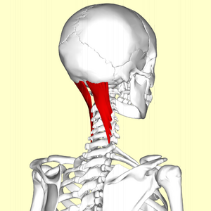
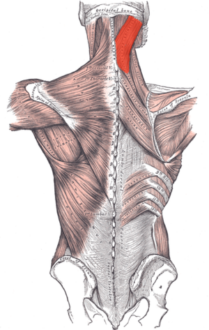
Splenius capitis is a thick, flat muscle at the posterior aspect of the neck arising from the midline and extending superolaterally to the cervical vertebrae and, along with the splenius cervicis, comprise the superficial layer of intrinsic back muscles.[1]
In relation to its surrounding musculature, it sits:
- Deep to the trapezius
- Superficial to the semispinalis capitis and the longissimus capitis
- Forms the floor of the posterior neck triangle, positioned between the sternocleidomastoid and the trapezius
Origin[edit | edit source]
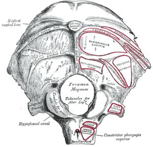
Lower half of ligamentum nuchae (C4-C6) and spinous process of C7-T3[1]
Insertion[edit | edit source]
Superior nuchal line, mastoid process of temporal bone, and rough surface adjoining occipital bone[1]
Nerve and Supply[edit | edit source]
Nerve: Lateral branches of the posterior rami of the middle and lower cervical spinal nerves.[1]Blood: Muscular branches of the occipital artery from the external carotid artery.[1]
Action[edit | edit source]
Splenius capitis assists in supporting the head in the erect position.[1]
Acting bilaterally: extension of the head and cervical spine
Acting unilaterally: lateral flexion of the head and neck and rotation the head to the same side (when working synergistically with sternocleidomastoid). [1]
The muscle tightens with mandibular protrusive movement and in the wide opening movement of the lower jaw.[2]
Synergists: splenius cervicis, semispinalis capitis, semispinalis cervicis, superior portion of trapezius.
Physiotherapy[edit | edit source]
Dysfunction of splenius capitis may be found in those with mechanical chronic neck pain[3] or whiplash disorders. In people with neck pain there may also be overactivity of superficial muscles, splenius capitis and inhibition of Semispinalis Cervicis.[4] Effective management of this condition should include exercises that focus on activating Semispinalis Cervicis, as well as stretching and myofascial release of the splenius capitis.
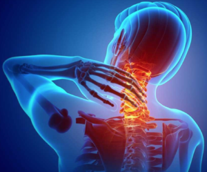
The most common cause of muscle tension headache (MTH) results from inflammatory changes at the site of muscular attachment on the occipital ridge. In the adult, this occurs most often at the attachment of the Splenius Capitis Muscles causing Splenius Capitis Muscle Syndrome. This is a very painful and commonly occurring syndrome. It typically mimics the respective pain reference patterns of temporal tendinitis and migraine headache. The painful headache starts at the lateral margin of the superior nuchal line and medial to the mastoid process. As inflammation develops, entrapment and irritation of Greater Occipital Nerve results. The onset of pain is often caused by eg motor vehicular trauma, blunt trauma, falls. The most frequent cause is postural, from prolonged periods of keeping the head in a downward, rotated and forward position. The muscle tension results in micro trauma to the muscular attachment, swelling ensues, and myalgia/neuralgia result. [2][5].
References[edit | edit source]
- ↑ 1.0 1.1 1.2 1.3 1.4 1.5 1.6 Moore KL. Dalley AF. Agur AMR. Clinically Orientated Anatomy. 7th edition. Philadelphia: Lippincott Williams & Wilkins, 2014.
- ↑ 2.0 2.1 PPM Splenius Capitis Muscle Syndrome Available: https://www.practicalpainmanagement.com/pain/maxillofacial/splenius-capitis-muscle-syndrome (accessed 3.2.2022)
- ↑ Bonilla-Barba L. Lima Florencio L. Rodriguez-Jimenez J. Falla D. Fernandez-de-las-Penas C. Ortega-Santiago R. Women with mechanical neck pain exhibit increased activation of their superficial neck extensors when performing the cranio-cervical flexion test. Musculoskeletal Science and Practice. 2020;49:102222.
- ↑ Schomacher J. Erlenwein J. Dieterich A. Petzke F. Petzkeb F. Falla D. Can neck exercises enhance the activation of the semispinalis cervicis relative to the splenius capitis at specific spinal levels? Manual therapy. 2015;20:694-702.
- ↑ Pain doctor Muscle tension headaches Available:https://paindoctor.typepad.com/martial_arts_injuries/2009/06/muscle-tension-headache-splenius-capitis-syndrome.html (accessed 3.2.2022)
- ↑ Painotopia. Splenius capitis and cervicis pain & trigger points – myofascial release. Avaliable from https://www.youtube.com/watch?v=ZFSn6QEMyPM (accessed 14/11/2021)
- ↑ MSK Neurology. Exercise for the splenius capitis muscle. Avaliable from https://www.youtube.com/watch?v=dEpNRI-DfKw (assessed 14/11/2021)


