Scoliosis
Original Editors - Gregory Maes
Top Contributors - Lucas De Bondt, Amrita Patro, Lydia Xenou, Gregory Maes, Lucinda hampton, Kim Jackson, Nikhil Benhur Abburi, Admin, Selena Horner, Lieselot Longe, Ellen De Boitselier, Evan Thomas, Oyemi Sillo, Liese Bosman, Jason Coldwell, Laure VanderDonckt, 127.0.0.1, WikiSysop, Aminat Abolade, Linde Van Droogenbroeck, Ginika Jemeni, Blessed Denzel Vhudzijena, Ellen Hopmans, Derycker Andries, Vidya Acharya and Scott Buxton </div>
Search Strategy[edit | edit source]
I searched for articles about scoliosis on pubmed and web of knowledge. Searched for basic information and treatments for scoliosis on the internet using google. And I found a lot of information in a chapter about scoliosis in a syllabus of Professor Vaes.
keywords: scoliosis, physical therapy, idiopathic scoliosis, scoliosis treatment, scoliosis diagnosis
Definition/Description[edit | edit source]
Scoliosis is a sideward’s curving of the spine, resulting in one or even two curves, making the spine look like a S. In some cases the spine even shows a rotation component This rotation starts when the scoliosis becomes more pronounced. This is called a torsion-scoliosis, causing a gibbus. Scoliosis can be present from birth. It is then called congenitive scoliosis. Other sorts of scoliosis can be developed during growth, any causes for this are still not found. We then speak of idiopathic scoliosis. There are several types of idiopathic scoliosis. They are classified by location of the (single or double) curve in the spine.
Clinically Relevant Anatomy[edit | edit source]
The spine is a number of vertebra that are connected with muscles and ligaments. Between each vertebra we can find a disc. Each disc content a nucleus pulposis surrounded by an annulus fibrosis. We have 7 cervical vertebra, 12 thoracic vertebra and 5 lumbar vertebra.
For more detailed information I’d like to add next link, which has all the information needed to understand the working of the spine: http://www.spineuniverse.com/anatomy
Epidemiology /Etiology[edit | edit source]
Scoliosis may be structural or non-structural.
I. Non-structural scoliosis[14]
A non-structural curve will usually have no rotational element, being a pure coronal plane deformity. A non-structural scoliosis may be due to:
- Pelvic tilt secondary to leg length inequality
- Pain or irritation (seen with disc prolapsed and other painful conditions, such as osteoid osteoma, typically triggering muscle spasm)
- …
The key feature of non-structural scoliosis is that the curve is reversible. It will spontaneously straighten when the underlying cause is corrected or removed. For example: In the case of pelvic tilt scoliosis, the curve will disappear when the pelvis is leveled and this can be achieved by sitting the patient or by equalizing any leg-length-discrepancy with blocks.
II. Structural scoliosis[14]
A structural curve will usually have a rotational element. The structural scoliosis is irreversible and may be classified according to the underlying etiology:
- Idiopathic scoliosis (70%) = Scoliosis without detectable underlying cause. This is the most common type.
- Congenital scoliosis (15%) = The scoliosis is present from birth. The vertebral disorders that cause congenital scoliosis may be due to either failure of formation or failure of segmentation or a combination of these, leading to a mixed deformity.
- Neuromuscular scoliosis (10%) = Scoliosis due to neuromuscular diseases such as cerebral palsy, poliomyelitis,….
- Trauma, tumour and infection are also possible causes, but are not frequently encountered.
- Rare conditions, including hereditary and mesenchymal abnormalities such as neurofibromatosis, Marfan’s syndrome, EhlerseDanlos syndrome etc. make up the remainder.
Characteristics/Clinical Presentation[edit | edit source]
Differential Diagnosis[edit | edit source]
Symptoms for scoliosis can be (2):
- Sideways curvature of the spine
- Sideways body posture
- One shoulder raised higher than the other
- Clothes not hanging properly
- Local muscular aches
- Local ligament pain
Diagnostic Procedures[edit | edit source]
The most common diagnostic procedures are:
- X-rays of the spine
- Measuring the leg length.
- A bone scan, MRI, or computed tomography (CT) scan may be necessary in difficult diagnostic problems.
Outcome Measures[edit | edit source]
add links to outcome measures here (also see Outcome Measures Database)
Examination[edit | edit source]
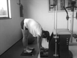
Screening procedures for scoliosis are done either by the Adam forward bend test or using other optical techniques like a scoliometer, while the radiographic measurement of Cobb angle is considered as the golden standard. The Cobb angle is the angle between lines drawn along the upper end plate of the most tilted vertebrae above the curve’s apex and the lower end plate of the most tilted vertebrae below the apex. The Adam forward bend test can be performed in both standing and sitting position. In standing position, the examined person is asked to bend forward looking down, keeping the feet together, the knees straightened, shoulders loose and hands positioned in front of knees or shins with elbows straight and palms opposed. If the scoliosis is present in both standing as bending position, the scoliosis is structural. If the scoliosis is present in standing position but disappears when the examined person is bended forward, the scoliosis is not structural.
In the sitting position, the examined person is seated on a chair with a height of 40 cm. The examined person is asked to bend forward and place his head between the knees, with his shoulders loose, elbows straight and hands positioned between the knees. The scoliometer is an inclinometer designed to measure trunk asymmetry, or axial trunk rotation. It’s used at three areas: at upper thoracic (T3-T4), main thoracic (T5-T12) and at the thoraco-lumbar area (T12-L1 or L2-L3). If the measurement is equal to 0°, there is a symmetry at the particular level of the trunk. An asymmetry at the particular level of the trunk is found, if the scoliometer measurement is equal to any other value[16].
Medical Management
[edit | edit source]
add text here
Physical Therapy Management
[edit | edit source]
Scoliosis is not just a lateral curvature of the spine, it’s a three dimensional condition. To manage scoliosis, we need to work in three planes: the sagittal, frontal and transverse. Different methods have already been studied. [5]
The conservative therapy consists of: physical exercises, bracing, manipulation, electrical stimulation and insoles. There is still discussion about the fact that conservative therapy is effective or not. Some therapists follow the ‘wait and see’ method. This means that at one moment; the Cobb degree threshold will be achieved. Then, the only possibility is a spinal surgery. [4]
The physical therapist has three important tasks: to inform, advice and instruct.
For the treatment of scoliosis, it’s not only important to do the correct exercises but the physical therapist also needs to inform the patient about his/her situation (if the patient is still a child, then he needs to inform the parents too). An educational program makes sure that the therapy accuracy from the patient improves. [1]
Some physical therapists recommend a brace to prevent the worsening of the scoliosis. An often used brace is the Milwaukee brace. Nevertheless the evidence for bracing is controversial. Maruyama T., Nakao Y. and Takeshita T. studied the effect of bracing in a review (2011). They compared brace treatment with no-treatment, other conservative treatments or surgery. The analyzed outcome measures were the radiological progression of the curve, surgery and quality of life. Results demonstrate that brace treatment is better than no-treatment (observation) or electrical stimulation. There is also no negative influence on the quality of life of patients with an idiopathic scoliosis. We can conclude that bracing is recommended as a treatment for female patients with a Cobb angle of 25-35°. The evidence level of some studies in the review was limited, so further research is necessary. [2]
In the literature there is evidence that exercises have beneficial effects on patients with idiopathic scoliosis. [7] [11]
The aims of the physical therapy consist of:[3][7][11]
• Autocorrection 3D
• Coordination
• Equilibrium
• Ergonomy
• Muscular endurance/ strength
• Neuromotorial control of the spine
• Increase of ROM
• Respiratory capacity/ education
• Side-shift
• Stabilisation
In the literature there are different exercise therapies.
An important one is the conservative three-dimensional Schroth method consisting of curve-specific exercises and corrective breathing techniques. The purpose of these exercises is to derotate, deflex and to correct the spine in the sagittal plane while elongating the spine. The patient needs to be focused about re-establishing spinal symmetry. The key to a successful therapy is to work consistently to correct the spine. Another – difficult- part of the therapy is to learn to shorten the muscles on the convex side of the spine and lengthen the muscles on the concave side of the side. This because the muscles become imbalanced on opposite sides. [5]
The exercise position approaches the functional/structural threshold. Schroth therapy takes advantage of the overcorrected positions. Basic corrections are reviewed by using mirrors in front and at the back of the patient. This posture requires concentration and coordination, applying the correct breathing, and well-adapted muscle length and tension. [6]
But there are also other exercises that have been found effective. Scientific Exercises Approach to Scoliosis (SEAS) exercises is for example been found effective in reducing the rate of progression of scoliosis compared with usual care and help to avoid brace prescription. [7] [8] [9]
The SEAS exercises are, according to the Italian Scientific Spine Institute (ISICO), based on a specific form of Active Self-correction (ASC), that is taught individually to each single patient. This is to achieve the maximum possible correction. ASC is then associated with stabilising exercises that include neuromotor control, proprioceptive training and balance. The exercises are also incorporated into their daily living activities. SEAS approach does also involve the parents. They are getting together with the patiënt a cognitive-behavioral approach to maximize compliance to treatment. [7][10]
We mentioned earlier that one of the aims is respiratory capacity/education. The severity of the curvature can cause a pressure on airways and lungs. The patient can experience trouble while breathing. Therefore the therapist must insert breathing exercises to complete his/her management. If the risk of pulmonary dysfunction (as a result of the pressure of the spine) is too high, surgery is indicated. [11][12]
Solache-Carranco and M.G. Sánchez-Bringas (2011) studied the effectiveness of a respiratory rehabilitation program in children with scoliosis. This are the techniques that they used:
- Respiratory education techniques (abdominal-diaphragmatic ventilation, thoracic mobilisation, ventilation at rest and during activities of daily living). This for mobilisation and prevention of stiffness of chest and skeletal muscles.
- Postural drainage and vibration to evacuate mucus and decrease the resistance of the airways.
- Relaxation techniques to make sure that the patients would have better control of respiration (to counteract dyspnea).
They found that the respiratory rehabilitation had a positive effect on increasing pulmonary function of children with scoliosis. [13]
Management of congenital scoliosis
The therapeutic options in cases of congenital scoliosis include conservative or surgical approaches.
I. Conservative treatment
With regard to conservative treatment of patients with congenital scoliosis, it should be noted that there are limited data available in the literature. A review (level of evidence 2) concluded that patients with specific types of segmentation failures, like unilateral unsegmented bars, will not benefit from conservative treatment, while the same applies to formation failures with curves of > 20 degrees in infancy. Nevertheless, there are reports that a conservative approach might be beneficial in mild cases with formation failures in the first three years of life. Furthermore, the review concluded that in patients with formation failures further investigation is needed to document where a conservative approach (bracing treatment) would be necessary. In general, most congenital scoliotic curves are not flexible and therefore are resistant to repair with bracing. For this reason, the use of braces mainly aims to prevent the progression of secondary curves that develop above and below the congenital curve, causing imbalance. In these cases, they may be applied until skeletal maturity[15].
II. Surgical treatment
Spinal surgery in patients with congenital scoliosis is regarded as a safe procedure and many authors claim that surgery should be performed as early as possible to prevent the development of severe local deformities and secondary structural deformities that would require more extensive fusion later. Most of the time Surgery is performed during adolescence, but newer techniques allow good correction to be accomplished into early adulthood. The goals for surgical treatment are to prevent progression and to improve spinal alignment and balance. The hips and shoulders should be level, and the head over the sacrum while maintaining sagittal alignment. The spine is corrected with a combination of rods, hooks, screws and wires while being fused by bone graft – either from the patient, a cadaver or artificially. On the other hand, early and late complications are also described in literature, concerning not only intraoperative and immediate postoperative problems, but also the safety and efficacy of the spinal instrumentation and the possibility of developing neurological disorders and the long-term effect these may have on both lung function and the quality of life. A review (2011, level of evidence 2) concluded that patients with segmentation failures should be treated surgically early, according to the rate of deformity formation and certainly before the pubertal growth spurt to try to avoid cor-pulmonale, even though there is lack of evidence for that in the long-term[15].
Management of non–structural scoliosis[17]
Pilates: This intervention was divided in three parts:
1. Preparation (warming up + stretch)
Warming –up consisted of eight minutes walking on a treadmill or an elliptical machine.
After the warming –up each patient had to do some stretching exercises:
- Spine forward stretching:
- The patient sits on the floor with a straight back and the legs stretched. The patient has to bring her trunk forwards
- Goal: Stretching the posterior muscle chain and mobilizing the vertebral spine
- Upper rolling
- The patient lies supine with the arms besides the body. The patient has to raise both legs till the toes touches the floor. Than this person has to unroll her spine slowly (vertebra by vertebra)
- Goal: stretching the posterior chain, mobilizing the spine and strengthen the abdomen
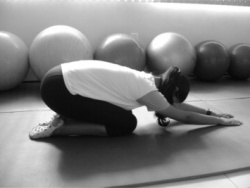
- Child position
- The patient sits in a four support position and has to stretch her spine, her arms are stretched and she has to push her hands against the floor. Then she has to lower her spine.
- Goal: Stretching the thoracic paravertebral, lumbar and gluteal regions and mobilizing the vertebral spine
<span style="line-height: 1.5em;" />- Forward leg pull
- The patient sits in a four support position. Then she has to raise the right arm and leg while the spine stays aligned. Than the same exercise but change arm and leg.
- Goal: Stretching the concavity of the vertebral spine.
2. Specific exercises:
For these exercise they made use of Swiss balls, FlexBall Quarks,.. It’s important that the patients learn to breathe right during exercise.
- Hip movements with a large ball (65 cm diameter)
- Goal: Strengthening the gluteal muscles and developing the equilibrium
- Inverted abdominal skills with a ball (55 cm diameter)
- Goal: Strengthening the infraabdominal region and the ischiotibial muscles.
-Rising into a seated position:
- Goal: Strengthening the M. rectus abdominis
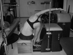
- Lateral spine movement on a step chair with a spring of 0.1410 kgf positioned in the rings to provide major resistance
- Goal: Stretching the lateral muscle chain according to the direction of convexity of the scoliosis
- Lateral spine movement:
- Goal: Stretching the lateral muscle chain according to the direction of convexity of the scoliosis
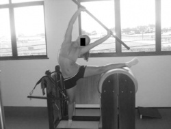
- Flexibility on the step chair with a spring of 0,1410 kgf positioned in the rings to provide major resistance:
- Goal: Mobilize the spine and stretching the paravertebral thorax and lumbar muscles
3. Returning to a relaxed position (relaxation) :
It consist out three movements, the patient has to repeat each exercise three times for five minutes.
All exercises has to be performed rapidly. The purpose of these exercises are metabolic recovery and relaxation of the used muscles.
Prognosis[edit | edit source]
Surgery
The outcome needs to be age matched since the demands of daily life, professional performance, and leisure activities vary substantially in the different age groups. Most of the patients who are still professionally active do not return to their previous work if it was a physically demanding job, but almost all of those had already stopped working before the surgery, because of pain limitations. When analyzed, regarding their overall daily activity by different questionnaires, most of these patients irrespective of age have improved in almost all categories of quality of life, and the use of regular pain medication is reduced substantially in more than 70% of these patients[18].
However for the elderly the outcome is less positive because of the age. The major problem of these patients after surgery, once surgical complications and implant failures have not occurred, is the residual back pain mostly as an expression of muscular spasms and pain due to unbalanced or chronic contractures of the paravertebral muscles[19].
Bracing
Whether brace treatment for scoliosis is successful is often described using the curve progression of more than 5° before skeletal maturity as a benchmark for bracing failure rather than spine surgery[20]. Some use 10° of curve progression or preventing the curve from reaching 45° at skeletal maturity[21].
The outcome data should be determined from the percentage of patients with: less than 5° or greater than 6° of progression at maturity, curves exceeding 45° at maturity, and progression resulting in the recommendation for surgery. Bracing studies should have a minimum of 2 years follow up beyond skeletal maturity. The first study to use these criteria determined a brace should prevent progression in 70% of patients to be considered effective]. Regardless of the recommended standardized parameters, the goal of bracing idiopathic curves remains consistent: control the curve, prevent progression, and avoid surgical intervention[22].
Resources
[edit | edit source]
add text here
Clinical Bottom Line[edit | edit source]
add text here
Recent Related Research (from Pubmed)[edit | edit source]
add text here
References[edit | edit source]
- Dr K. Lau, Scoliosis: Literature Review of Current Treatment Modalities and Exercise Therapy. Level of evidence: 5
- T. Maruyama et al., Effectiveness of brace treatment: a systematic review, 7th International Conference on conservative management of spinal deformities Montreal, 2010. Level of evidence: 3A
- H.R Weiss, S. Negrini, Physical exercises in the treatment of idiopathic scoliosis at risk of brace treatment – SOSORT consensus paper 2005, Scoliosis. 2006. Level of evidence: 2C
- C Fusco, F Zaina et al, Physical exercises in the treatment of adolescent idiopathic scoliosis: An updated systematic review, Informa Healthcare USA, 2011. Level of evidence: 1A
- Scoliosis 3DC- Schroth Method Exercise. Is it Scoliosis? 17 November 2011. Level of evidence: 5
- A. Hennes, D. Turnbull, Rehabilitation in patients with spinal deformities: A description of the Schroth method, Informa Healthcare USA 01/21/11. Level of evidence: 2
- Stefano Negrini, Fabio Zaina, et al., specific exercises reduce brace prescription in adolescent idiopathic scoliosis: a prosprective controlled cohort study with worst-case analysis, Journal of Rehabilitation Medcine, 2008. Level of evidence: 1B
- Negrini S, Negrini A, et al., A controlled prospective study on the efficacy of SEAS.02 exercises in preventing progression and bracing in mild idiopathic scoliosis, Stud Health Technol Inform,2006. Level of evidence: 1B
- Alessandra Negrini, Silvana Parzini, et al., Adult scoliosis can be reduced through specific SEAS exercises: a case report, scoliosis, 2008. Level of evidence: 4
- SOSORT guidelines read on the website of SOSORT, editor is Jean Claude de Mauroy: http://www.sosort2012.org/index.php?option=com_content&view=article&id=88&Itemid=149 : consulted on 10/11/2012
- Paloma Bas, Marco Romagnoli, et al., Beneficial effects of aerobic training in adolescent patients with moderate idiopathic scoliosis, European Spine Journal, August 2011. Level of evidence: 3B
- M. Takaso, T. Nakazawa, Surgical management of severe scoliosis with high risk pulmonary dysfunction in Duchenne muscular dystrophy: patient function, quality of life and satisfaction, Int Orthop. 2010 June. Level of evidence 4
- A. Solache-Carranco, M.G. Sánchez-Bringas, Evaluation of a respiratory rehabilitation program in children with scoliosis, Cirugia y cirujanos, 2010. Level of evidence 3B
- Nigel W. Gummerson, Peter A. Millner. (ii) Scoliosis in children and teenagers. Orthopaedics and trauma. 2011. Vol. 25, num. 6, pp. 403-413. Level of evidence = 5
- Angelos Kaspiris, Theodoros B. Grivas, Hans-Rudolf Weiss and Deborah Turnbull. Surgical and conservative treatment of patients with congenital scoliosis: a search for long-term results. Scoliosis. 2011, Vol. 6, num. 12, pp. 1-17. Level of evidence = 2
- Petros Patias, Theodoros B Grivas, Angelos Kaspiris, Costas Aggouris and Evangelos Drakoutos, ‘A review of the trunk surface metrics used as Scoliosis and other deformities evaluation indices.’, Scoliosis, 2010. Level of evidence = 1
- Alves de Arau’jo M.E., Bezerra da Silva E., Bragade Mello D., Ali Cader S., Shiguemi Inoue Salgado A., Henrique Martin Dantas E., ‘The effectiveness of the Pilates method: Reducing the degree of non-structural scoliosis, and improving flexibility and pain in female college students’, Journal of Bodywork & Movement Therapies, 2012, 16, 191 -198. Level of evidence = 1
- Rinella A, Bridwell K, Kim Y, RudzkiJ, Edwards C, Roh M, Lenke L, Berra. Late complications of adult idiopathic scoliosis primary fusions to L4 and above: the effect of age and distal fusion level. 2004, Spine 29(3):318–325. Level of evidence = 3
- M. Aebi. Adult Scoliosis. Eur Spine J (2005) 14: 925–948. Level of evidence = 3
- Shaughnessy WJ Review Advances in scoliosis brace treatment for adolescent idiopathic scoliosis. Orthop Clin North Am. 2007 Oct; 38(4):469-75, v. Level of Evidence = 1
- Richards BS, Bernstein RM, D'Amato CR, Thompson GH Spine Review Standardization of criteria for adolescent idiopathic scoliosis brace studies: SRS Committee on Bracing and Nonoperative Management. 2005 Sep 15; 30(18):2068-75; discussion 2076-7. Level of evidence: 1
- Weinstein SL. Bracing in Adolescent Idiopathic Scoliosis Trial (BrAIST). University of Iowa, National Institute of Arthritis and Musculoskeletal and Skin Diseases (NIAMS) 2008. Level of Evidence: 1


