Posterior longitudinal ligament: Difference between revisions
Rachael Lowe (talk | contribs) No edit summary |
Evan Thomas (talk | contribs) mNo edit summary |
||
| Line 35: | Line 35: | ||
== References == | == References == | ||
<references /> | <references /><br> | ||
[[Category:Anatomy]][[Category:Cervical_Anatomy]][[Category: | [[Category:Anatomy]] [[Category:Cervical_Anatomy]] [[Category:Thoracic_Anatomy]] [[Category:Lumbar_Anatomy]] [[Category:Ligaments]] | ||
Revision as of 09:07, 26 August 2016
Original Editor - Rachael Lowe
Top Contributors - Rachael Lowe, Adam Vallely Farrell, Kim Jackson, Evan Thomas, WikiSysop and Lucinda hampton
Description[edit | edit source]
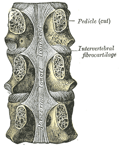
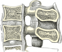
Forming the anterior wall of the vertebral canal, this strong ligament spans from the body of the Axis (C2) to the posterior surface of the sacrum. Like its anterior counterpart the Anterior longitudinal ligament, its deep fibres are intersegmental while the more superficial fibres can span up to four vertebral levels.
In the cervical and thoracic regions it has a uniform width over the bodies and discs, but in the lumbar region the ligament is widest at the levels of the intervertebral discs where it is firmly anchored to the Annulus fibrosus, cartilage of the Vertebral end plates, and the margins of the vertebrae.
Speriorly it blends with the Tectorial membrane.
Attachments[edit | edit source]
Arising at the superior margin of one vertebra they span to the inferior margin of the vertebra that they attach to.
Function[edit | edit source]
Limits flexion of the vertebral column and reinforces the intervertebral disc.
Recent Related Research (from Pubmed)[edit | edit source]
Failed to load RSS feed from http://www.ncbi.nlm.nih.gov/entrez/eutils/erss.cgi?rss_guid=1diOUTbPywntr3hBYmsw0khWvqd3kty2WoRO9r6wFNZWE5esZ5|charset=UTF-8|short|max=10: Error parsing XML for RSS
References[edit | edit source]


