Pectoralis major: Difference between revisions
No edit summary |
No edit summary |
||
| Line 26: | Line 26: | ||
== '''Function''' == | == '''Function''' == | ||
[[ | [[File:Parallel bars.jpeg|right|frameless]] | ||
The function of the pectoralis major is 3-fold and dependent on which heads of muscles are involved<ref name=":2" />. | |||
* With the origin fixed, the pectoralis major adducts, medially rotates, and transversely adducts arm at glenohumeral joint. | * With the origin fixed, the pectoralis major adducts, medially rotates, and transversely adducts arm at glenohumeral joint. | ||
* It assists in flexion of the arm (via its clavicular head) | * It assists in flexion of the arm (via its clavicular head) | ||
* It assists in extension of the arm (via the sternocostal head) at the glenohumeral joint. | * It assists in extension of the arm (via the sternocostal head) at the glenohumeral joint. | ||
* It depresses the shoulder girdle at acrimioclavicular and sternoclavicular joints. | * It depresses the shoulder girdle at acrimioclavicular and sternoclavicular joints. | ||
* With the insertion fixed, it may assist in elevating thorax, as in forced inspiration. It is considered as an accessory muscle of inspiration. | * With the insertion fixed, it may assist in elevating thorax, as in forced inspiration. It is considered as an accessory muscle of inspiration. | ||
* In crutch-walking or parallel-bar work, it will assist in supporting the weight of the body. <ref name=":0" /><ref name=":1">Standring, S. Gray's Anatomy: The Anatomical Basis of Clinical Practice. Gray's Anatomy Series 41st edition. Elsevier (2016).</ref><br> | * In crutch-walking or parallel-bar work, it will assist in supporting the weight of the body. <ref name=":0" /><ref name=":1">Standring, S. Gray's Anatomy: The Anatomical Basis of Clinical Practice. Gray's Anatomy Series 41st edition. Elsevier (2016).</ref><br> | ||
== Clinical relevance | == Clinical relevance == | ||
[[File:Poland Syndrom.jpg|alt=Poland Syndrom|thumb| | [[File:Poland Syndrom.jpg|alt=Poland Syndrom|thumb| | ||
Poland Syndrom | Poland Syndrom | ||
]] | ]] | ||
=== '''List of clinical correlated''' === | ==='''List of clinical correlated'''=== | ||
- Poland's syndrome<ref>US National Library of Medecine. Genetics Home Reference. Available from: https://ghr.nlm.nih.gov/condition/poland-syndrome</ref> | - Poland's syndrome<ref>US National Library of Medecine. Genetics Home Reference. Available from: https://ghr.nlm.nih.gov/condition/poland-syndrome</ref> | ||
- Chondro-epitrochlearis<ref>J Clin Diagn Res. NCBI. Unilateral Existence of Chondro-epitrochlearis: Its Embryological Perspectives and Clinical ImplicationsTitle of electronic publication/webpage. Available from: https://www.ncbi.nlm.nih.gov/pmc/articles/PMC5020218/ (accessed Jul 2016)</ref> | - Chondro-epitrochlearis<ref>J Clin Diagn Res. NCBI. Unilateral Existence of Chondro-epitrochlearis: Its Embryological Perspectives and Clinical ImplicationsTitle of electronic publication/webpage. Available from: https://www.ncbi.nlm.nih.gov/pmc/articles/PMC5020218/ (accessed Jul 2016)</ref> | ||
=== '''Weakness''' === | ==='''Weakness'''=== | ||
'''Upper part of pectoralis major''': | '''Upper part of pectoralis major''': | ||
| Line 52: | Line 53: | ||
Decreases the strength of adduction obliquely toward the opposite hip.From a supine position, if the subjects arm is placed diagonally overhead, it will be difficult to lift arm from a table. The subject will also have difficulty holding any large or heavy object in both hands either at or near waist level.<ref name=":0" /> | Decreases the strength of adduction obliquely toward the opposite hip.From a supine position, if the subjects arm is placed diagonally overhead, it will be difficult to lift arm from a table. The subject will also have difficulty holding any large or heavy object in both hands either at or near waist level.<ref name=":0" /> | ||
=== '''Shortness''' | ==='''Shortness'''=== | ||
'''Upper part of pectoralis major''': | '''Upper part of pectoralis major''': | ||
| Line 67: | Line 68: | ||
{{#ev:youtube|2G1nW7YNz5o}} | {{#ev:youtube|2G1nW7YNz5o}} | ||
== Assessment | == Assessment == | ||
[[Image:Gray410 pectoralis major.png|right|245x245px]] | |||
=== '''Test the pectoralis major''' === | === '''Test the pectoralis major''' === | ||
The two heads of pectoralis major muscle can be tested separately: | The two heads of pectoralis major muscle can be tested separately: | ||
Revision as of 05:44, 7 January 2022
Description[edit | edit source]
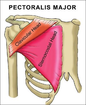
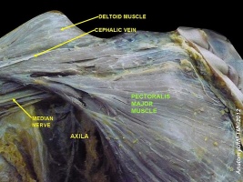
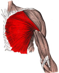
The pectoralis major is the superior most and largest muscle of the anterior chest wall. It is a thick, fan-shaped muscle that lies underneath the breast tissue and forms the anterior wall of the axilla[1].The pectoralis major is the most superficial muscle in the pectoral region.There are 2 heads of the pectoralis major, the clavicular and the sternocostal, which reference their area of origin.[2]
- Pectoralis injury is relatively rare. Tendon tears occurring almost exclusively in males between 20 and 40 years old, and around 50% of these are sustained during weight-bearing exercises eg bench press, when the arm under load is in extension and external rotation.[1]
- The pectoralis major is active in deep or forced inspiration, but not expiration. [3]
Origin[edit | edit source]
The pectoralis makor consists of two heads; the clavicular and sternocostal head :
- Clavicular head – originates from the anterior surface of the medial half of clavicle.
- Sternocostal head – is the larger of the two heads and originates from :
- The anterior surface of the manubrium and body of sternum,
- The anterior surface of the superior six costal cartilages.
- Superior part of the aponeurosis of external oblique muscle.[3]
Image 2: Well defined pectorals.
Insertion[edit | edit source]
The upper and lower fibers of pectoralis major insert to the crest of greater tubercle of the humerus. Upper fibers are more anterior and caudal on the crest, while posterior fibers twist on themselves and are more posterior and cranial than the upper fibers.[3]
Nerve supply and Arterial supply[edit | edit source]
The 2 heads of the pectoralis major have different nervous supplies.
- The clavicular head derives its nerve supply from the lateral pectoral nerve. The lateral pectoral nerve arises directly from the lateral cord of the brachial plexus
- The medial pectoral nerve innervates the sternocostal head. The medial pectoral nerve arises from the medial cord.[1]
Function[edit | edit source]
The function of the pectoralis major is 3-fold and dependent on which heads of muscles are involved[1].
- With the origin fixed, the pectoralis major adducts, medially rotates, and transversely adducts arm at glenohumeral joint.
- It assists in flexion of the arm (via its clavicular head)
- It assists in extension of the arm (via the sternocostal head) at the glenohumeral joint.
- It depresses the shoulder girdle at acrimioclavicular and sternoclavicular joints.
- With the insertion fixed, it may assist in elevating thorax, as in forced inspiration. It is considered as an accessory muscle of inspiration.
- In crutch-walking or parallel-bar work, it will assist in supporting the weight of the body. [3][4]
Clinical relevance[edit | edit source]
[edit | edit source]
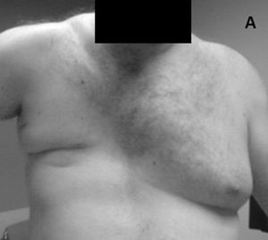
- Poland's syndrome[5]
- Chondro-epitrochlearis[6]
Weakness[edit | edit source]
Upper part of pectoralis major:
Decrease the ability to draw the arm in horizontal adduction across the chest, making it difficult to touch the hand to the opposite shoulder. Decreases the strength of shoulder flexion and medial rotation.
Lower part of pectoralis major:
Decreases the strength of adduction obliquely toward the opposite hip.From a supine position, if the subjects arm is placed diagonally overhead, it will be difficult to lift arm from a table. The subject will also have difficulty holding any large or heavy object in both hands either at or near waist level.[3]
Shortness[edit | edit source]
Upper part of pectoralis major:
The range of motion in horizontal abduction and lateral rotation of the shoulder is decreased. Shortness of the pectoralis holds the humerus in medial rotation and adduction and, secondarily, results in abduction of the scapula from the spine.
Lower part of pectoralis major:
A forward depression of the shoulder girdle from the pull of pectoralis major on the humerus often accompanies the pull of the tight pectoralis minor on the scapula. Flexion and abduction ranges of motion overhead are limited. [3]
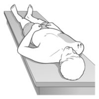
- Observing the patient lies supine with upper arms on the table, hands resting palm down on the lower abdomen. The practitioner observes from the head and notes whether either shoulder is held in an anterior position in relation to the thoracic cage. If one or both shoulders are forward of the thorax, pectoralis muscles are short
Trigger Points
The trigger points in the pectoralis major muscle can produce symptoms that are nearly identical to the pain associated with having a heart attack or angina pectoris. Referred pain from these trigger points is experienced in the chest, front of the shoulder, down the inside of the arm, and along the inside of the elbow. They may also produce tenderness in the breast and nipple hypersensitivity.[7]
Assessment[edit | edit source]
Test the pectoralis major[edit | edit source]
The two heads of pectoralis major muscle can be tested separately:
The clavicular head of pectoralis major can be tested by transversely adducting the arm at the glenohumeral joint against resistance, during which It can be seen and palpated.
The sternocostal head of petoralis major can be tested by adducting the arm ar the glenohumeral joint against resistance, during it can be seen and palpated.[4]
Power[edit | edit source]
Upper (clavicular) part of pectoralis major:
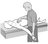
Position: Supine. The examiner holds the opposite shoulder firmly on the table. The triceps maintains the elbow on the extension. Test: Starting with the elbow extended and with the shoulder in 90 degrees flexion and slight medially rotation, the humerus is horizontally adducted towards the sternal end of clavicle. Pressure: Against the forearm in direction of horizontal abduction.
Lower (sternal) part of pectoralis major:
Position: Supine. The examiner places one hand on the opposite iliac crest to hold the pelvis firmly on the table. Test: Starting with the elbow extended and with the shoulder in flexion and slight medially rotation, adduction of the arm obliquely toward the opposite iliac crest. Pressure: Against the forearm obliquely, in a lateral and cranial direction.[3]
Length[edit | edit source]
Upper (clavicular) part of pectoralis major:
Position: Supine with the knees bent and the low back flat on the table. Test: The examiner places the subjects arm in horizontal abduction, with the elbow extended and the shoulder in lateral rotation (palm upward). Normal length: Full horizontal abduction with lateral rotation, the arm flat on the table without trunk rotation.In this position the tendon of pectoralis major at the sternum should not be found to be unduly tense, even with maximum abduction of the arm, unless the muscle is short.Shortness:The extended arm does not drop down to table level. Limitations may be recorded as slight, moderate or marked; measured in degrees using goniometer or measured in inches using a ruler to record the number of inches between the table and lateral epicondyle.
Lower (sternal) part of pectoralis major:
Position:Supine with the knees bent and the low back flat on the table. Test: The examiner places the subjects arm in position of approximately 135 degrees of abduction (in line with the lower fibers), with the elbow extended. The shoulder will be in a lateral rotation. Normal length: Arm drops to table level, with the low back remaining flat on the table. Shortness:The extended arm does not drop down to table level. Limitations may be recorded as slight, moderate or marked; measured in degrees using goniometer or measured in inches using a ruler to record the number of inches between the table and lateral epicondyle.[3]
Treatment[edit | edit source]
Strengthening[edit | edit source]
Stretching[edit | edit source]
Manual therapy[edit | edit source]
Resources[edit | edit source]
See also[edit | edit source]
References[edit | edit source]
- ↑ 1.0 1.1 1.2 1.3 Solari F, Burns B. Anatomy, Thorax, Pectoralis Major Major.Available:https://www.ncbi.nlm.nih.gov/books/NBK525991/ (accessed 7.1.2022)
- ↑ http://teachmeanatomy.info/upper-limb/muscles/pectoral-region/ (accessed 1 July 2018).
- ↑ 3.0 3.1 3.2 3.3 3.4 3.5 3.6 3.7 Kendall F, McCreary E, Provance P,Rodgers M,Romani W. Muscles:Testing and function with posture and pain. 5th ed. Philadelphia: Lippincott Williams & Wilkins, 2005.
- ↑ 4.0 4.1 Standring, S. Gray's Anatomy: The Anatomical Basis of Clinical Practice. Gray's Anatomy Series 41st edition. Elsevier (2016).
- ↑ US National Library of Medecine. Genetics Home Reference. Available from: https://ghr.nlm.nih.gov/condition/poland-syndrome
- ↑ J Clin Diagn Res. NCBI. Unilateral Existence of Chondro-epitrochlearis: Its Embryological Perspectives and Clinical ImplicationsTitle of electronic publication/webpage. Available from: https://www.ncbi.nlm.nih.gov/pmc/articles/PMC5020218/ (accessed Jul 2016)
- ↑ http://www.triggerpointtherapist.com/blog/pectoralis-major-pain/pectoralis-major-trigger-points-cardiac-copycats/ (accessed 1 July 2018).
