Parkinson's - Anatomy, Pathology, Prognosis and Diagnosis
Original Editors - Your name will be added here if you created the original content for this page.
Top Contributors - Kim Jackson, Laura Ritchie, Rachael Lowe, Wendy Walker, Mariam Hashem, Grace Barla, Ewa Jaraczewska, Robin Tacchetti, Lauren Lopez, Lucinda hampton, Jess Bell, Admin, Evan Thomas, Scott Buxton, Naomi O'Reilly, Tarina van der Stockt, 127.0.0.1, Merinda Rodseth, Tony Lowe and WikiSysop
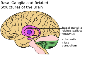
Parkinson’s continues to be considered predominantly a disorder of the basal ganglia.
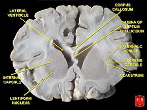
The basal ganglia are a group of nuclei situated at the base of the forebrain.
The striatum, composed of the caudate and putamen, is the largest nuclear complex of the basal ganglia. The striatum receives excitatory input from several areas of the cerebral cortex, as well as inhibitory and excitatory input from the dopaminergic cells of the substantia nigra pars compacta (SNc). These cortical and nigral inputs are received by the spiny projection neurons, which are of 2 types:
- Those that project directly to the internal segment of the globus pallidus (GPi), the major output site of the basal ganglia. The consequence of this pathway is to increase the excitatory drive from thalamus to cortex i.e. as the motor cortex increases firing rates, this results in increased activity in the corticospinal tract and eventually the muscles, so ‘turns up’ the action of the motor system.
- Those that project to the external segment of the globus pallidus (GPe), establishing an indirect pathway to the GPi via the subthalamic nucleus (STN). The consequence of the indirect pathway is to decrease the excitatory drive from thalamus to cortex. The increase in inhibition of the thalamic neurons in effect ‘turns down’ motor activity from the cortex


