Osteochondritis Dissecans of the Knee
Original Editors - Tania Appelmans as part of the Vrije Universiteit Brussel Evidence-based Practice Project
Top Contributors - Tania Appelmans, Tarina van der Stockt, Mats Vandervelde, Charlotte Bellen, Admin, Michelle Lee, Kim Jackson, Daphne Jackson, Wanda van Niekerk, 127.0.0.1, Evan Thomas, WikiSysop and Claire Knott
Definition[edit | edit source]
Osteochondritis dissecans is an idiopathic disease which affects the subchondral bone and its overlying articular cartilage due to loss of blood flow. [1] This may result in separation and instability of a segment of cartilage and free movement of these osteochondral fragments within the joint space.[2] (Level of evidence: 1A) That process can lead to pain, loose body formation and joint effusion.[1] (Level of evidence: 1A)
Clinically Relevant Anatomy [edit | edit source]
The knee (art.genus) is a synovial joint where 3 bones articulate with each other: femur, tibia and patella. It consists of 2 articulations. The first is located between the femur and tibia (art. femorotibialis). The femoral condyles (lateral and medial) which are the distal rounded ends of the femur, articulate with the proximal side of the tibia (tibia plateau). The second joint is the one between the femur and the patella. [3] (Level of evidence: 5)
The articular bones are covered by white, shiny and elastic cartilage. The smooth articular surface of the femur roll and slide on the tibia plateau. Synovial fluid nourishes and lubricates the cartilage.Cite error: Invalid <ref> tag; name cannot be a simple integer. Use a descriptive title In patients with osteochondritis dissecans, the subchondral bone with his articular cartilage doesn’t get any blood supply anymore and degenerates.[4] (Level of evidence: 3B)
Epidemiology /Etiology[edit | edit source]
Osteochondritis dissecans can be split into a juvenile form (JOCD) and an adult form (OCD)[1] [5].
(Level of evidences: 1A, 1B)
There are two main places in the knee joint where osteochondritis dissecans can appear. It mostly affects the femoral condyles, especially the medial condyle on the lateral joint surface (±80%). This area carries the least weight. In 10% of the cases it is located on the patella. OCD is more common in males and bilateral representation is rare (±25%)[1][6][4]
(Level of evidences: 1A, 3B, 2B)
Causes of OCD[edit | edit source]
The cause of OCD is still unknown and mostly multifactorial.[1] It can arise as a result of a direct trauma; when the articular cartilage is damaged (for instance a fall, twist, sprain, tackle, etc.)[8]. The tibial plateau could damage one of the condyles of the femur [9][1].
Repetitive microtrauma due to high levels of participation in sports can also be a factor.[6](Level of evidence: 2B) Other possible etiologies are: chemical changes at the surface located in the subchondral bone, genetic conditions, growth disorders, hereditary factors, ischemia, etc.[10][11]
Stages of OCD[edit | edit source]
There are four distinct stages of OCD [12][13][11][6]:
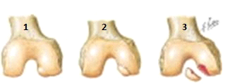
Stage one: ischemic osteonecrose begin to arise in a part of the subchondral bone, because the tissue is not well vascularized.
Stage two: a subchondral osteonecrose.
Stage three: partially detached lesions, a dissecans ‘in situ’.
Stage four: ‘Dissecans’, this is the loosening of the affected bone fragment and the corresponding cartilage of the articular surface. This fragment falls between the moving parts of the knee joint and blocks it. A ‘joint mouse’ is the bone fragment that roams in the joint, because it moves and it is white [8].
|
1) damage at the articular cartilage |
Characteristics/Clinical Presentation[edit | edit source]
- Straining dependent (stabbing) pain [12].
- The knee swells simultaneously with the onset of the pain. The entire knee is irritated because of the loose pieces, and it responds by producing extra synovial fluid in the knee joint [8].
- Having the feeling of knee bends [10][12].
- Stiffness and feeling of instability [10][12].
- When there’s a joint mouse present; the knee cannot be stretched, but still be bent. The knee is ‘locked’, because the bone fragment is located between the bones of the knee joint [9][12].
- OCD can exist for years without symptoms, but suddenly cause discomfort due heavy straining of the joint [12].
Differential Diagnosis[edit | edit source]
If there is no certain radiological determination of osteochondritis dissecans, there can also be alternative causes of the same symptoms that should be sought like:
- Inflammatory arthritites: a group of conditions which affect your own immune system.
- Osteoarthritis: degradation of joints
- Bone cysts: type of cyst in joints
- Septic arthritis: purulent invasion of the knee which produces arthritis
An x-ray, ct scan or MRI scan can be performed to show necrosis of subschondral bone or formation of loose fragments. This can lead to a better diagnosis.[15] (Level of evidence: D)
Diagnostic Procedures
[edit | edit source]
Clinical[edit | edit source]
1 stadium: vague pain at the knee and stiffness after exercising.
2 stadium: mechanical problems, a swollen knee and quadriceps muscle atrophy.
Radiography
[edit | edit source]
Many diagnostic imaging methods (eg, radiography, magnetic resonance imaging (MRI), technetium 99m pyrophosphate joint scintigraphy, bone scans), as well as arthroscopic examination, have been used in an attempt to stage or classify osteochondral lesions. The stages (typically 3 or 4 levels) represent a continuum of tissue degeneration leading to complete disruption and instability of the lesion (loose body). Originally, staging was determined based on radiographic findings. Currently, MRI appears to be the preferred choice for detection of this type of chondral injury and for determination of a lesion's stability.
Outcome Measures[edit | edit source]
add links to outcome measures here (also see Outcome Measures Database)
Examination [edit | edit source]
- The knee feels warmer than the non-injured knee[12].
- There is an intermittent swelling palpable[12][10][8][16].
- Quadriceps muscle atrophy [13][9].
- The passive and active extension of the knee is limited [17].
- Catching or locking of the knee [9][10].
- Tibial external rotation during gait [9].
- Fluid effusion[16](Level of evidence: F5)
- It is possible that both capsular and non-capsular movement restrictions can be found during functional assessment, the severity is dependent on a possible herniation of the knee joint and the degree of joint irritation [12].
- The sensitive location of the abandoned section of the osteochondral fracture can be felt, when the knee is in 90° of flexion [9].
- Wilson's Test: The knee is held in 90° to 30° from full extension while rotating the tibia.[13]
- The test is positive when internal rotation is painful and external rotation relieves symptoms [1]. (Level of evidence: 1A)
- The test is positive when internal rotation is painful and external rotation relieves symptoms [1]. (Level of evidence: 1A)
Medical Management[edit | edit source]
In minor cases rest can be prescribed. The patient has to stop activities for three to six months and the lesion will heal spontaneously, especially with young adolescents.[16]
Normally, immobilization of the knee for a couple of weeks is sufficient in the treatment of growing children. In case immobilization is insufficient, as would normally be the case for adults, a mobilization procedure must be started up. In this procedure, stretching exercises are performed. The range of motion and strengthening ability of the muscles will be gradually increased in the next 3 to 6 months. In the end, in case the knee is not fully recovered, surgery should be necessary. (Level of evidence: C5, F5)
Stages three and four are always treated surgically. Surgery is also required when the conservative treatment in stages one and two was inadequate.[10][17][18] It is recommended to treat surgically when a large part of the femoral condyle has been excavated, because of the risk to develop osteoarthritis.[16]
A variety of surgical methods exist for the management of articular cartilage lesions at the knee, such as OCD. These include the use of arthroscopic lavage or debridement, radio frequency energy, bone drilling, osteochondral autografts or allografts, internal fixation of bone fragments, and autologous chondrocyte implantation .[7] (level of evidence: 3B)
Surgical techniques:
- In stages one and two the articular cartilage is still intact, through retrograde operation trying to tap into to the affected bone ‘from behind’ and clear it. The advantage of this surgical technique is that the articular cartilage stays intact [10][17][9].
- Not yet dissected fragment will be fixed by means of an operation [10][13][11].
- Excision of the fragment and removal of loose bodies [10][13].
- Repair of blood supply by drilling arthroscopic through the cartilage and the hearth of osteochondrosis into the healthy bones[17][13].
- Stabilization of the fragment through pinning or through screw fixation .[9][11][13][17]
- Osteochondral autograft transplantation (OATS).
- Osteochondral allograft transplantation.
- Autologous chondrocyte implantation (ACI) [17][13].
Physical Therapy Management
[edit | edit source]
In stages one and two the condition is localized in the subchondral bone, the cartilage is still intact and gets its nourishment from synovial fluid. In these two stages conservative therapy can be applied[12]. The goals of conservative therapy are: pain reduction, repair the continuity of the surface of the cartilage and to prevent degeneration of the surface of the knee joint.
There is no standard treatment.
1)Resting your joint
Adaption of the strain is needed so the bone can heal. 2 weeks of immobilization and partly support is recommended when having an acute injury. With children whose bones will still grow, the bone defect may heal by taking rest. Long-term immobilization has to be prevented, because joint motion is necessary for the nutrition and strengthening of the cartilage. Sport activities should be stopped temporally [12][17].
2)Therapy
- stretching
- range of motion
-strengthening exercises for the muscles[19](Level of evidence: C5)
First exercises: closed chain exercises, low impact activities like cycle and swim. Using exercises as straight leg raises and ankle band exercises, strength can be maintained. Coactivation or setting of the quadriceps and hamstring can be performed while in an immobilizer or cast. Using neuromuscular electrical stimulation to the quadriceps and hamstrings for coactivation contractions can further augment the strength maintenance program. Following immobilization should be continued, range of motion exercises, as well as progressive quadriceps and hamstring strengthening should be performed. Weight-bearing progression throughout rehabilitation should be to patient tolerance. In facilitating the return to full-weight-bearing status is aquatic therapy very beneficial. To adress any gait deviations that developed during the immobilization and decreased weight-bearing phases of rehabilitation gait training techniques may be used, such as manual facilitation and visual feedback tot the patient via a full length mirror. Additional exercises to restore ankle joint and normal knee proprioception, such as biomechanical ankle platform systems (BAPS board) exercises or unilateral stance, are also beneficial to the athlete planning to return to competition. After this period the sport activities can be partly restart. Next criteria should be managed: the patient is pain free, has a full joint mobility, no swelling, no pressure sensitivity and there’s radiological prove of recovery.
2B) Surgery
An operative treatment is indicated if, after a treatment of three to six months and no recovery has occurred or when the loose fragment is to big[9]. To remove loose fragments or to reattach fragments.[19] [18][20] Immobilization is not necessary before the surgerie. Immediately after the intervention the knee get continuous passive motion for 48 hours. After this therapy is recommend, 8 weeks of rehabilitation exercises on the limb function and recruitment. Between week 6 and 8 weight-bearing is gradually introduced to full weight bearing.[19](Level of evidence: C5)
Key Research[edit | edit source]
add links and reviews of high quality evidence here (case studies should be added on new pages using the case study template)
Resources [edit | edit source]
B. Linden et al,. Osteochondritis dissecans of the femoral condyles: a long-term follow-up study. J Bone Joint Surg Am. 1977;59:769-776.
Level of evidence: B1.
Clinical Bottom Line[edit | edit source]
add text here
Recent Related Research (from Pubmed)[edit | edit source]
Failed to load RSS feed from https://www.ncbi.nlm.nih.gov/entrez/eutils/erss.cgi?rss_guid=1taFIjEhLwO0_vei0GOdUUBQM0e8YR5d_uKUdasEkZ3sCLZgwR|charset=UTF-8|short|max=10: There was a problem during the HTTP request: 422 Unprocessable Entity
References[edit | edit source]
- ↑ 1.0 1.1 1.2 1.3 1.4 1.5 1.6 Erickson BJ, Chalmers PN, Yanke AB, Cole BJ. Surgical management of osteochondritis dissecans of the knee. Curr Rev Musculoskelet Med. 2013 Jun 1;6(2):102-14. http://www.briancolemd.com/wp-content/themes/ypo-theme/pdf/surgical-management-of-osteochondritis-dissecans-of-the-knee.pdf (accessed 11 October 2016) Level of evidence: 1A
- ↑ Pappas AM. Osteochondrosis dissecans. Clinical orthopaedics and related research. 1981 Jul 1;158:59-69. Level of evidence: 1A
- ↑ Prometheus. Bohn Stafleu van Loghum. 2010: p434-444. Level of evidence: 5
- ↑ 4.0 4.1 Jeong JH, Mascarenhas R, Yoon HS. Bilateral osteochondritis dissecans of the femoral condyles in both knees: a report of two sibling cases. 2013 Jun 1;25(2):88-92. https://www.researchgate.net/profile/Randy_Mascarenhas/publication/237061466_Bilateral_Osteochondritis_Dissecans_of_the_Femoral_Condyles_in_Both_Knees_A_Report_of_Two_Sibling_Cases/links/00b7d529818a509cbe000000.pdf (accessed 11 October 2016) Level of evidence: 3B
- ↑ Krause M, Hapfelmeier A, Möller M, Amling M, Bohndorf K, Meenen NM. Healing predictors of stable juvenile osteochondritis dissecans knee lesions after 6 and 12 months of nonoperative treatment. The American journal of sports medicine. 2013 Oct 1;41(10):2384-91. Level of evidence: 1B
- ↑ 6.0 6.1 6.2 Chambers HG, Shea KG, Anderson AF, Brunelle TJ, Carey JL, Ganley TJ, Paterno MV, Weiss JM, Sanders JO, Watters WC, Goldberg MJ. Diagnosis and treatment of osteochondritis dissecans. Journal of the American Academy of Orthopaedic Surgeons. 2011 May 1;19(5):297-306. Level of evidence: 2 B
- ↑ 7.0 7.1 Johnson MP. Physical therapist management of an adult with osteochondritis dissecans of the knee. Physical therapy. 2005 Jul 1;85(7):665-75.fckLRhttp://ptjournal.apta.org/content/85/7/665.short (accessed 11 October 2016) Level of evidence: 3 B Cite error: Invalid
<ref>tag; name "Michael P Johnson" defined multiple times with different content - ↑ 8.0 8.1 8.2 8.3 (1983). Sportinjuries,Brussels, Elsevier. p.478. Cite error: Invalid
<ref>tag; name "Southmayd, W en Hoffman, M." defined multiple times with different content - ↑ 9.0 9.1 9.2 9.3 9.4 9.5 9.6 9.7 9.8 Sailors ME. Recognition and Treatment of Osteochondritis Dissecans of the Femoral Condyles. Journal of athletic training. 1994 Dec;29(4):302. http://www.ncbi.nlm.nih.gov/pmc/articles/PMC1317804/pdf/jathtrain00028-0016.pdf (accessed 11 October 2016) Level of evidence: A2 Cite error: Invalid
<ref>tag; name "Matthew E. et al." defined multiple times with different content - ↑ 10.0 10.1 10.2 10.3 10.4 10.5 10.6 10.7 10.8 Schenk RC, Goodnight JM. Current concept review-Osteochondritis dissecans. J Bone Joint Surg Am. 1996 Mar 1;78(3):439-56. Level of evidence: A1.
- ↑ 11.0 11.1 11.2 11.3 Bohndorf K. Osteochondritis (osteochondrosis) dissecans: a review and new MRI classification. European radiology. 1998 Jan 1;8(1):103-12. Level of evidence: A1
- ↑ 12.00 12.01 12.02 12.03 12.04 12.05 12.06 12.07 12.08 12.09 12.10 (2008). Examination and Treatment of the Knee. Houten: Bohn Stafleu Van Loghum. p.82-87. Level of evidence: A1
- ↑ 13.0 13.1 13.2 13.3 13.4 13.5 13.6 13.7 Defierline AJ, Goldsfein JL, Rue JP, Bach Jr BR. Evaluation and Treatment of Osteochondritis Dissecans Lesions of the Knee. J Knee Surg. 2008;21:106-15.http://www.acldoc.org/Files/Eval%20n%20Treat506.pdf (accessed 12 Oct 2016) Level of evidence: A2.
- ↑ Greene W., Netter's Orthopaedics: The knee and leg. 407 Levels of evidence: E
- ↑ Patient: Osteochondritis dissecans, 2013 fckLR http://www.patient.co.uk/pdf/2549.pdf. Levels of evidence: D
- ↑ 16.0 16.1 16.2 16.3 Outline of Orthopaedics. Churchill Livingstone. 2001: 365-367. Level of evidence: F5
- ↑ 17.0 17.1 17.2 17.3 17.4 17.5 17.6 Pascual-Garrido C, McNickle AG, Cole BJ. Surgical treatment options for osteochondritis dissecans of the knee. American Orthopaedic Society for Sports Medicine. 2009:1-9 2009 Level of evidence: A2.
- ↑ 18.0 18.1 O’Connor MA, Palaniappan M, Khan N, Bruce CE. Osteochondritis dissecans of the knee in children. Bone & Joint Journal.http://www.bjj.boneandjoint.org.uk/content/84-B/2/258.short. 2002 Mar 1;84(2):258-62.
- ↑ 19.0 19.1 19.2 Cite error: Invalid
<ref>tag; no text was provided for refs namedJacobs et al. - ↑ Osteochondritis dissecans, Mayo Clinic, treatment, 2012, http://www.mayoclinic.com/health/osteochondritisdissecans/DS00741/DSECTION=treatments-and-drugsfckLRLevel of evidence: C5


