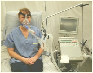
Non Invasive Ventilation
Original Editor - The Open Physio project. Top Contributors - Kim Jackson, George Prudden, Admin, Vidya Acharya, Lucinda hampton, Rachael Lowe, Tomer Yona and WikiSysop
Introduction[edit | edit source]
Non-invasive ventilation (NIV) is the delivery of ventilation support using techniques that do not require an invasive endotracheal airway.[1] NIV achieves comparative physiological benefits to conventional mechanical ventilation by reducing the work of breathing and improving gas exchange.[2]
The intervention is recognised as an effective treatment for respiratory failure in chronic obstructive pulmonary disease, cardiogenic pulmonary oedema and other respiratory conditions without complications such as respiratory muscle weakness, upper airway trauma, ventilator-associated pneumonia, and sinusitis.[1][3]
Aims of NIV[edit | edit source]
- Improve oxygenation
- Improve ventilation
- Relieve hypercapnea
- Reduce the work of breathing
- Avert intubation
- Adjunct for weaning
Uses of NIV[edit | edit source]
- Acute respiratory failure
- Acute/chronic respiratory failure with or without OSA
- Respiratory insufficiency
- Nocturnal Hypoventilation
- End stage cystic fibrosis awaiting transplant
- As an adjunct to airway clearance techniques
Indications[edit | edit source]
- Respiratory distress
- Failure to improve arterial blood gas (ABG) with standard treatment
- Inability to maintain SaO2 > 90%
- pH > 7.28 and pCO2 < 10
- Poor inspiratory effort or tidal volume causing ineffective airway clearance
Contraindications[edit | edit source]
- Facial trauma
- Morbidity
- Cardiovascular collapse
- Uncontrolled arhythmias
- Pneumothorax
- Bronchopleural fistula
- Inability to clear secretions
Equipment check list[edit | edit source]
- Bi-level positive airway pressure (BiPAP) generator
- Anti-bacterial filter
- Smooth bore tubing
- Exhalation port
- Face mask, spacer and headgear
- Oxygen tubing
- Heated humidifier and tubing (if required)
- Oximeter with integral recorder
Setting up the equipment[edit | edit source]
- Measure the patient for the mask
- Connect the headgear and spacer
- Connect all the components from the machine as far as the exhalation port
- Set Mode to "Spontaneous/timed (S/T)"
- Set Intermittent positive airway pressure (IPAP) to 6cm initially
- Set Expiratory positive airway pressure (EPAP) to lowest default setting (2cm or 4cm, depending on the machine being used)
- Have all of this done before bringing the machine to the patient
- Discuss the target settings with the rest of the team, as well as whether supplemental oxygen is required
Explanation to the patient[edit | edit source]
- This will HELP your breathing, it will NOT control your breathing
- It will reduce the effort of breathing
- It will improve your oxygen level
- You will still be able to communicate, drink and cough
- We will try it for a short period of time at first and I will stay with you while you get used to it
- You will feel a rush of air initially but try a few breaths of varying lengths and you will realise that you control the machine and not the other way around
- We will start at low pressure - feel it on your hand
- When you have adjusted to it, you will feel more comfortable and may fall asleep
Sequence[edit | edit source]
- Fit and adjust the nasal mask and headgear - do not overtighten and draw the skin out from under the mask to improve the seal
- Check the oximeter readings and leave the oximeter on the patient
- Turn the machine on and connect it to the patient
- Encourage the patient to adjust breathing as per the instructions given above
- If oxygen saturation improves, ensure that the patient is aware as this will encourage compliance
Parameters[edit | edit source]
- EPAP - range from 2/4cm to 20cm depending on the model. Acts like Positive end expiratory pressure (PEEP), which serves to increase Functional residual capacity (FRC)
- IPAP - range from 2cm to 30 cm. Rarely effective below 12cm. Must be individualised.
- Respiratory rate - range from 4bpm to 30 bpm. On S/T mode, set the baseline to 12bpm
- IPAP- used only with Timed mode (not used at ward level, mainly in Intensive care unit
Settings[edit | edit source]
- Establish target settings in consultation with the rest of the team before going to the bedside.
- When the patient is comfortable at 6cm, start moving the settings in 2cm increments until the target settings are reached.
- Review when the target settings are attained and if the desired gas exchange has not been achieved, discuss supplemental oxygen or respiratory stimulant with the team. If there is still no improvement, the patient may need Intubation.
- In the non-acute patient there should be signs of improvement within 2 hours of initiation of NIV.
- Complete the patient record and leave it at the bedside along with an Instruction sheet.
- Discuss with the nursing staff before leaving.
Troubleshooting[edit | edit source]
- Nasal dryness or rhinorrhoea - add a humidifier
- Bloating or belching - try sips of peppermint water
- Soreness on the bridge of the nose - use a triangle of Granuflex
Facial pressure ulcers[edit | edit source]
Pressure ulcers associated with the use of NIV is a growing clinical problem due to the increased popularity of the intervention. Prevalence of grade I pressure ulcers have been estimated at 5-50% after a couple of hours and 100% after 48 hours[4]. The development of pressure ulcers is associated with poor clinical outcomes, increased complications, and length of hospital stay that compound with the consequences of acute illness. Medical devices such as NIV masks unique risk factors including: the existence of a microclimate individual to the device, the method in which the device is secured, that devices may obscure the skin, and that the areas at risk are not routinely checked[5]. Clinicians’ primary focus has been to attain a mask seal, as air leaks are associated with reduced tolerance to the intervention[6]. The alternating airflow from bi-level positive pressure means that a seal is important to avoid ventilator asynchrony. Therefore strap tension is increased, with the risk of pressure damage a secondary consideration[7]. It is important to consider that the patient may not be able to respond to an uncomfortable mask fit or excessive load delivered to vulnerable areas of skin due to sedation, medication, or neurological disease or injury. Furthermore, the patient may be too weak to reposition the device. Oronasal masks have traditionally been preferred for their comfort and ease of use however other interfaces have been recommended as superior[8]. Prophylactic interventions should also be considered[9].
Resourses[edit | edit source]
References[edit | edit source]
- ↑ 1.0 1.1 Nava S, Hill N. Non-invasive ventilation in acute respiratory failure. Lancet 2009; 374(9685): 250-9.
- ↑ Vitacca M, Ambrosino N, Clini E, et al. Physiological Response to Pressure Support Ventilation Delivered before and after Extubation in Patients Not Capable of Totally Spontaneous Autonomous Breathing. American Journal of Respiratory and Critical Care Medicine 2001; 164: 638-41.
- ↑ Pingleton SK. Complications of acute respiratory failure. Am Rev Respir Dis 1988; 137(6): 1463-93.
- ↑ Carron M, Freo U, BaHammam AS, et al. Complications of non-invasive ventilation techniques: a comprehensive qualitative review of randomized trials. Br J Anaesth 2013; 110(6): 896-914.
- ↑ Black JM, Cuddigan JE, Walko MA, Didier LA, Lander MJ, Kelpe MR. Medical device related pressure ulcers in hospitalized patients. Int Wound J 2010; 7(5): 358-65.
- ↑ Dellweg D, Hochrainer D, Klauke M, Kerl J, Eiger G, Kohler D. Determinants of skin contact pressure formation during non-invasive ventilation. J Biomech 2010; 43(4): 652-7.
- ↑ Yamaguti WP, Moderno EV, Yamashita SY, et al. Treatment-related risk factors for development of skin breakdown in subjects with acute respiratory failure undergoing noninvasive ventilation or CPAP. Respir Care 2014; 59(10): 1530-6.
- ↑ Vaschetto R, De Jong A, Conseil M, et al. Comparative evaluation of three interfaces for non-invasive ventilation: a randomized cross-over design physiologic study on healthy volunteers. Crit Care 2014; 18(1): R2.
- ↑ Weng MH. The effect of protective treatment in reducing pressure ulcers for non-invasive ventilation patients. Intensive Crit Care Nurs 2008; 24(5): 295-9.


