Ligamentum flavum: Difference between revisions
Rachael Lowe (talk | contribs) No edit summary |
Rachael Lowe (talk | contribs) No edit summary |
||
| Line 7: | Line 7: | ||
== Description == | == Description == | ||
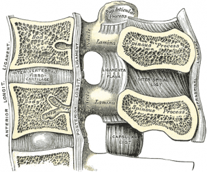
[[Image: | [[Image:Cervical vertebrae lig flavum.png|right|300px]]The ligamenta flavum is a short but thick ligament that connects the laminae of adjacent vertebrae from C2 to S1. It consists of 80% elastin and 20% collagen<ref>Nikolai Bogduk. Chapter 4: Ligaments of the lumbar spine In: Clinical Anatomy of the Lumbar Spine and Sacrum. Elsevier.</ref>. | ||
At each intersegmental level the ligamentum flavum is a paired structure being represented symmetrically on both sides. | At each intersegmental level the ligamentum flavum is a paired structure being represented symmetrically on both sides. | ||
Revision as of 15:56, 19 January 2014
Original Editor - Rachael Lowe
Top Contributors - Rachael Lowe, Kim Jackson, Sehriban Ozmen, Andeela Hafeez, WikiSysop, Ahmed Nasr, Admin and Evan Thomas
Description[edit | edit source]
The ligamenta flavum is a short but thick ligament that connects the laminae of adjacent vertebrae from C2 to S1. It consists of 80% elastin and 20% collagen[1].
At each intersegmental level the ligamentum flavum is a paired structure being represented symmetrically on both sides.
In the neck region the ligaments are thin, but broad and long; they are thicker in the thoracic region, and thickest in the lumbar region.
Attachments[edit | edit source]
Arises from the lower half of the anterior surface of the lamina above and attaches to the posterior surface and upper margin of the lamina below.
On each side the ligament divides into a medial and lateral portion. The medial portion passes to the back of the next lower lamina. The lateral portion passes in front of the fact joint where it attaches to the anterior aspect of the inferior and superior articular processes and forms the anterior capsule. The most lateral fibres extend beyond the superior articular process to the pedicle below.
Function[edit | edit source]
Their marked elasticity serves to preserve the upright posture, and to assist the vertebral column in resuming it after flexion. It resists excess separation of adjacent vertebral lamina and prevents buckling of the ligament into the spinal canal during extension, which would cause canal compression.
The lateral portion prevents the anterior capsule of the facet joint being nipped within the joint cavity during movement.
Pathology[edit | edit source]
Hypertrophy of this ligament may cause spinal stenosis because it lies in the posterior portion of the vertebral canal.
Examination[edit | edit source]
Recent Related Research (from Pubmed)[edit | edit source]
Failed to load RSS feed from http://www.ncbi.nlm.nih.gov/entrez/eutils/erss.cgi?rss_guid=18cpB6wjstduh6c8cE0OHYoNErYbmlFpcDRJHS-90YIdPbYo19|charset=UTF-8|short|max=10: Error parsing XML for RSS
References[edit | edit source]
- ↑ Nikolai Bogduk. Chapter 4: Ligaments of the lumbar spine In: Clinical Anatomy of the Lumbar Spine and Sacrum. Elsevier.


