Lateral Collateral Ligament of the Knee: Difference between revisions
mNo edit summary |
No edit summary |
||
| Line 100: | Line 100: | ||
<references /> | <references /> | ||
[[Category:Knee_Anatomy]] [[Category:Vrije_Universiteit_Brussel_Project]] [[Category:Sports_Injuries]][[Category:Musculoskeletal/ | [[Category:Knee_Anatomy]] [[Category:Vrije_Universiteit_Brussel_Project]] [[Category:Sports_Injuries]][[Category:Musculoskeletal/Orthopaedics]] [[Category:Knee]] [[Category:Assessment]] | ||
Revision as of 23:21, 11 March 2018
Original Editors - Dorien Scheirs, Joris De Pot
Top Contributors - Saimat Lachinova, Admin, Joris De Pot, Kim Jackson, Dorien Scheirs, Rachael Lowe, Leana Louw, WikiSysop, Oyemi Sillo, George Prudden, Kai A. Sigel, Tony Lowe, Derycker Andries and Naomi O'Reilly
Definition/Description[edit | edit source]
On this page you will find some information about the fibular collateral ligament of the knee. Functional anatomy, technique and the different grades of injury from the LCL will be explained. More information about the ligament can be founded at the page of Wouter Claesen. An injury at the Lateral Collateral Ligament is an injury at the lateral side of the knee. There are several tests for testing the Lateral Collateral Ligament. ‘The fibular collateral ligament is the primary varus stabilizer of the knee.’[1]
Clinically Relevant Anatomy[edit | edit source]
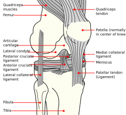
The lateral collateral ligament (LCL) is one of four critical ligaments involved in stabilizing the knee joint. The medial collateral ligament, the anterior cruciate ligament and the posterior cruciate ligament are the other stabilizers of the knee.
The lateral collateral ligament or fibular collateral ligament has its origin on the lateral epicondyle of the femur and runs to the fibular head. [2][3]
When the knee is extended the LCL is stretched and it is loose when the knee is flexed (more than 30°).[4] The fibular collateral ligament is primary restraint to varus rotation from 0-30° of knee flexion, varus rotation is when the distal part of the leg below the knee is deviated inward, resulting in a bowlegged appearance. Secondarily it also acts to resist internal rotation forces of the tibia. The LCL has no direct contact with the joint capsule or lateral meniscus, it’s separated from it by a small fat pad.
LCL Injuries[edit | edit source]
There are different grades for the LCL injuries, they will be classified as follows:[4][4]
Grade 1: Some tenderness and minor pain at the lateral side of the knee, pain when the distal part of the leg below the knee is deviated inward (varus stress). This means there have been small tears in the ligament.
Grade 2: Noticeable looseness in the knee, a joint space opening of about 5-10mm is present when moved by hand. There have been larger tears in the ligament = Joint instability of the knee. Major pain and tenderness at the inner side of the knee and some swelling is a consequence.[5]
Grade 3: Noticeable looseness in the knee, a joint space opening of >10mm is present when moved by hand. This means the ligament is completely torn. Considerable pain and tenderness at the inner side of the knee and some swelling are also noted. There may also be a tear of the anterior cruciate ligament.[5]
For grade 1 and 2 the suggested treatment includes rest, ice, compression, elevation (RICE)
Grade 3 is best treated with surgical intervention
These injuries are much less common than medial collateral ligament (MCL) injuries because the opposite leg usually guards against direct blows to the medial side of the knee.
Symptoms of a tear in the lateral collateral ligament are:[6]
o Knee swelling outside the joint. This symptom occurs also in bursitis, patellar tendonitis and growth plate injury. The swelling may cause the joint to appear larger or abnormally shaped.
o Locking or catching of the knee with movement
o Pain or tenderness along the outside of the knee
o Knee gives way, or feels like it is going to give way, when it is active or stressed in a certain way
Purpose
[edit | edit source]
The lateral collateral ligament stress test (varus stress test) is used to estimate the integrity of the lateral collateral ligament, to see whether it is this ligament that causes the instability in the knee.
The purpose of this test is to determine if there is looseness in the ligament and if an MRI would be necessary. Serious tears or ruptures of the lateral collateral ligament may require surgery.
The varus or adduction stress test evaluates the lateral collateral ligament. To perform this test, the physiotherapist has to place the knee in thirty degrees of flexion. While he is stabilizing the knee, he adducts the ankle. If the knee joint adducts greater than normal (compare with the uninjured leg), the test is positive. This is indicative of a lateral collateral ligament tear.
Technique
[edit | edit source]
From all knee injury’s the Lateral Collateral Ligament Injury only takes 6%.[7]
The Varus stress test:
The Varus stress test shows a lateral joint line gap. It is possible that the ligament is damaged. The patients legg is abducted and the knee is flexed in about 30°, so the knee joint is in the closed packed position.[8] The sensitivity of the test is 25% . the reliability of this test in extension is 68% and in 30° flexion it is only 56%. The test is fairly solid.[9]
How to do the Varus test?
The test can be executed in 0° en 30 ° flexion. The physiotherapist puts one hand on the end of the femur at the medial side of the knee. His other hand is placed on the lateral side on the tibia. The practitioner is trying to stress the lateral collateral ligament by pushing the knee with both hands. It looks like he is going to break the leg. The patient has to rotate the hip maximally.[10] When the therapist feels a soft spot, the ligament is injured.[11]
Resources
[edit | edit source]
Books:
{1}M.shünke, E.S. (2005). Anatomische atlas prometheus, algemene anatomie en bewegingsapparaat. Bohn Stafleu van Loghum
{2}William C.Whiting, R.F. (2008). Biomechanics of musculoskeletal injury. Second Edition
{4}Schoen, D.C.(2000). Adult orthopaedic nursing. Lippincott
Sites:
{3.0}Sherwin SW Ho, MD. Lateral collateral knee ligament injury, Updated: Feb 28, 2010 http://emedicine.medscape.com/article/89819-overview (secondary source)
Primary source: LaPrade RF, Terry GC. Injuries to the posterolateral aspect of the knee. Association of anatomic injury patterns with clinical instability. Am J Sports Med. Jul-Aug 1997;25(4):433-8.
{3.1}Sherwin SW Ho, MD. Lateral collateral knee ligament injury, Updated: Feb 28, 2010 http://emedicine.medscape.com/article/89819-overview (secondary source)
Primary source: Griffin LY. Acute knee injuries. Sports Medicine. New York, NY: John Wiley & Sons, Inc; 1994:2255-60.
{3.2}Sherwin SW Ho, MD. Lateral collateral knee ligament injury, Updated: Feb 28, 2010 http://emedicine.medscape.com/article/89819-overview (secondary source)
Primary source: Snider RK, ed. Essentials of Musculoskeletal Care. Rosemont, Ill: American Academy of Orthopaedic Surgeons; 2000:336-8.
{5}Linda J. Vorvick, MD. Lateral collateral ligament (LCL) injury, Update Date: 6/13/2010 http://www.nlm.nih.gov/medlineplus/ency/article/001079.htm (secondary source)
Primary source: De Carlo M, Armstrong B. Rehabilitation of the knee following sports injury. Clin Sports Med. 2010;29:81-106.
Articles:
- Robert F. LaPrade, Spiridonov SI, Coobs BR, Ruckert PR, Griffith CJ. Fibular collateral ligament anatomical reconstructions: a prospective outcomes study. Am J Sports Med. 2010; 38: 2005-2012
- Benjamin R. Coobs, Robert F. LaPrade, Chad J. Griffith, Bradley J. Nelson. Biomechanical Analysis of an Isolated Fibular (Lateral) Collateral Ligament Reconstruction Using an Autogenous Semitendinosus Graft. Am J Sports Med. 2007; 35: 1521-1527
- DE Cooper. Tests for posterolateral instability of the knee in normal subjects. Results of examination under anesthesia. J bone Joint Surg Am. 1991; 73: 30-36
Clinical Bottom Line[edit | edit source]
The lateral collateral ligament, located on the lateral side of the knee has its origin on the lateral epicondyle of the femur and his insertion on the fibular head. It is the primary varus stabilizer of the knee. This ligament is restraint to varus rotation from 0-30° of knee flexion and it also acts to resist internal rotation forces of the tibia.
Recent Related Research (from Pubmed)[edit | edit source]
References
[edit | edit source]
- ↑ R.F. LAPRADE, Journal of sports medicine, 2005, July the first 2010
- ↑ M.shünke, E.S (2005). Anatomische atlas prometheus, algemene anatomie en bewegingsapparaat. Bohn Stafleu van Loghum
- ↑ William C.Whiting, R.F. (2008). Biomechanics of musculoskeletal injury. Second Edition
- ↑ 4.0 4.1 4.2 eMedicine on Medscape, Sherwin SW Ho, MD. Lateral Collateral Knee Ligament Injury, Updated: Feb 28, 2010 http://emedicine.medscape.com/article/89819-overview
- ↑ 5.0 5.1 Schoen, D.C.(2000). Adult orthopaedic nursing. Lippincott
- ↑ Medlineplus, Linda J. Vorvick, MD. Lateral collateral ligament (LCL) injury, Update Date: 6/13/2010 http://www.nlm.nih.gov/medlineplus/ency/article/001079.htm
- ↑ H.B.TANDETER, P.SHVARTZMAN, ‘Acute knee injuries: Use of decision rules for selective radiograph ordering’, 1999
- ↑ DEMOS MEDICAL PUBLISHING, 2004
- ↑ L. MERRIMAN, W.TURNER, ‘Assessment of the lower limb’, Elsevier health sciences, 2002
- ↑ L. MERRIMAN, W.TURNER, ‘Assessment of the lower limb’, Elsevier health sciences, 2002
- ↑ .P. NOGALSKI, ‘Collateral ligament pathology, knee’, 2009
- ↑ Physiotutors. Varus Stress Test of the Knee⎟Lateral Collateral Ligament. Available from: https://www.youtube.com/watch?v=sg1gk6QKARw


