Joint Line Tenderness of the Knee: Difference between revisions
No edit summary |
No edit summary |
||
| (46 intermediate revisions by 9 users not shown) | |||
| Line 1: | Line 1: | ||
<div class="editorbox"> | <div class="editorbox"> | ||
'''Original Editor '''- [[User:Anne-Laure Vanherwegen|Anne-Laure Vanherwegen]], [[User:Layla Lemaire|Layla Lemaire]], [[User:Sarah Jacobs|Sarah Jacobs]] and Lyn Bruyndockx as part of the [[Vrije Universiteit Brussel Evidence-based Practice Project]] | '''Original Editor '''- [[User:Anne-Laure Vanherwegen|Anne-Laure Vanherwegen]], [[User:Layla Lemaire|Layla Lemaire]], [[User:Sarah Jacobs|Sarah Jacobs]] and Lyn Bruyndockx as part of the [[Vrije Universiteit Brussel Evidence-based Practice Project]] | ||
'''Top Contributors''' - {{Special:Contributors/{{FULLPAGENAME}}}} | |||
</div> | |||
== Description / Purpose == | |||
= | The Joint Line Tenderness (JLT) test is a physical examination test commonly used to screen for sensitivity related to meniscal injuries.<ref name="Akseki 2003">Akseki D, Pinar H, Karaoglan O. [https://pubmed.ncbi.nlm.nih.gov/12845289/ The accuracy of the clinical diagnosis of meniscal tear with or without associated anterior cruciate ligament tears.] Acta Orthop Traumatol Turc. 2003;37:193-198 (2B)</ref> <ref name="Osmon">Osman TE. [https://www.arthroscopyjournal.org/article/S0749-8063(03)00736-9/pdf The accuracy of joint line tenderness by physical examination in the diagnosis of meniscal tears.] Arthroscopy: The Journal of Arthroscopic & Related Surgery 2003; 19(8): 850–854. (1B)</ref> The test can be used if pain is localised to either the medial or lateral aspect of the joint. This is suggested to correlate with degenerative pathology of the articular joint cartilage or compromised integrity of the medial or lateral meniscus. <ref name=":0">Lerew S, Stoker S, Nallamothu S. [https://smrj.scholasticahq.com/article/11765-the-rules-of-four-a-systematic-approach-to-diagnosing-common-musculoskeletal-conditions-of-the-knee The rules of four: a systematic approach to diagnosing common musculoskeletal conditions of the knee.] SMRJ 2020; 4(2). </ref> | ||
A person with JLT has joint pain that increases when pressing on the surface of the joint or moving the joint through its normal range of motion. <ref name="Stephen" /> Symptoms that may coexist with joint tenderness are joint stiffness, joint swelling, joint redness, joint warmth, joint pain and joint deformity.<ref name="Stephen">Stephen J, et al. Joint tenderness overview. DSHI systems 2010(5)</ref> | |||
The test is usually performed with the patient relaxed and lying on the table, but it can also be executed in a sitting position, with the knee in a flexed position of 90°. The test can be executed for the lateral border of the knee (lateral meniscus) and for the medial border of the knee (medial meniscus). <ref name="Wadley">Wadey V, Mohtadi N, Bray R, Frank C. [https://pubmed.ncbi.nlm.nih.gov/17550711/ Positive predictive value of maximal posterior joint-line tenderness in diagnosing meniscal pathology: a pilot study]. Can J Surg. 2007; 50(2): 96–100. (1B)</ref><br><br> | |||
{| cellspacing="1" cellpadding="1" width="80%" border="0" align="center" | |||
|- | |||
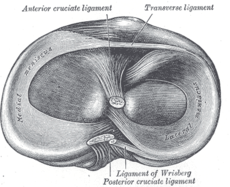
| [[Image:Menisci from above.png|thumb|center|300px|Menisci from Above]] | |||
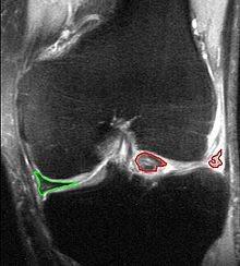
| [[Image:Meniscal tear MRI.jpg|thumb|center|300px|Meniscal Tear on MRI]] | |||
|} | |||
= | == Clinically Relevant Anatomy == | ||
The | The knee is the largest joint of the body, consisting of the patellofermoral and the tibiofemoral joint. <ref>Gupton M, Imonugo O, Terreberry R. [https://www.ncbi.nlm.nih.gov/books/NBK500017/ Anatomy, Bony Pelvis and Lower Limb, Knee.] StatPearls. Available from: https://www.ncbi.nlm.nih.gov/books/NBK500017/<nowiki/>(accessed 23-5-2022). </ref> The latter contains two menisci, a medial and lateral meniscus located between the corresponding femoral condyle and tibial plateau. The meniscus is a shiny-white tissue comprised of specialised extracellular matrix molecules. Each of them has a region-specific innervation and vascularisation. Both menisci are critical for a well-functioning knee joint.<ref name="Elleftherios">Makris E, Hadidi P, Athanasiou K. [https://pubmed.ncbi.nlm.nih.gov/21764438/ The knee meniscus: structure-function, pathophysiology, current repair techniques, and prospects for regeneration.] Biomaterials 2011; 32(30): 7411–7431. (5)</ref> | ||
= | == Technique == | ||
{| class="FCK__ShowTableBorders" cellspacing="1" cellpadding="1" width="40%" border="0" align="right" | |||
|- | |||
| align="right" | | |||
{{#ev:youtube|nuHtTi4mX7M|350}} <ref>Clinically Relevant Technologies. Joint Line Tenderness - Knee (CR). Available from: http://www.youtube.com/watch?v=nuHtTi4mX7M[last accessed 15/09/14]</ref> | |||
|} | |||
Each side of the tibiofemoral joint line is palpated separately to evaluate the maximal joint line sensitivity, i.e. the palpated point from the joint line that gives discomfort and is more tender than the unaffected leg at the same anatomic location.<ref name="Wadley" /><ref name="Elleftherios" /> | |||
The knee needs to be flexed in 90°. The border of the joint line at the sides of the patellar ligament and the soft border between the highness of the femur above and below the tibia should be identified. The joint line palpation of the knee starts from the medial border of the patellar ligament towards the posterior aspect of the knee. Beginning at the lateral border of the patellar ligament, the lateral joint line | The knee needs to be flexed in 90°. The border of the joint line at the sides of the patellar ligament and the soft border between the highness of the femur above and below the tibia should be identified. The joint line palpation of the knee starts from the medial border of the patellar ligament towards the posterior aspect of the knee. Beginning at the lateral border of the patellar ligament, the lateral joint line is palpated in a similar way along the joint line in the posterior direction. The medial and lateral joint lines have to be palpated separately. The borders of the tibial plateau and the femoral condyles are palpated to affirm the presence of isolated posterior/medial joint line tenderness. The borders of the patella are not palpated to assess for any tenderness.<ref name="Wadley" /> | ||
The test is positive if the patient | The test is positive if the patient experiences pain or tenderness during palpation of the medial or lateral joint lines.<ref name="Wadley" /><ref name="Elleftherios" /> | ||
= | == Diagnostic accuracy == | ||
Selecting the most accurate diagnostic tests by evaluating their clinical performance is considered a valuable clinical skill in physiotherapy practice. <ref>Fritz J, Wainner R. [https://pubmed.ncbi.nlm.nih.gov/11688591/ Examining diagnostic tests: an evidence based perspective.] Physical Therapy 2001; 81(9):1546-1564.</ref> A quick reference guide of the most commonly used test statistics and interpretation in physiotherapy practice can be found in [https://www.physio-pedia.com/Test_Diagnostics Test Diagnostics]. | |||
The sensitivity of a test is the proportion of people having the disorder who show a positive test result, and who actually have the disease or dysfunction i.e. A/(A+C) in Table 1.<ref name="Hing">Hing W. [https://www.ncbi.nlm.nih.gov/pmc/articles/PMC2704345/ Validity of the McMurray’s Test and Modified Versions of the Test: A Systematic Literature Review.] J Man Manip Ther 2009; 17(1):22-35. (1A)</ref> For the joint line tenderness test, sensitivity can be described as the ability of the test to correctly identify those individuals with JLT having a meniscal tear.<ref name="Blackburn">Blackburn T, Craig E. [https://pubmed.ncbi.nlm.nih.gov/7454779/ Knee Anatomy: A Brief Review]. Phys Ther 1980; 60(12):1556-1560. (5)</ref> On the other hand, the specificity of a test shows the proportion of people not actually having the disorder and showing a negative test result i.e. D/(B+D) in Table 1.<ref name="Hing" /> In other words, this is the chance of a negative outcome of the JLT in a person who does not have a meniscal pathology.<ref name="Blackburn" /> | |||
{| class="wikitable" | |||
|+ | |||
!Table 1 | |||
!'''Reference standard positive''' | |||
!'''Reference standard negative''' | |||
|- | |||
|Diagnostic test positive | |||
|A | |||
True (+) results | |||
|B | |||
False (+) results | |||
|- | |||
|Diagnostic test negative | |||
|C | |||
False (-) results | |||
|D | |||
True (-) results | |||
|} | |||
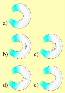
[[Image:Meniscal tears.png|thumb|right|300px|Different Types of Meniscal Tears]] | |||
Sensitivity and specificity values can be combined in the calculation of the likelihood ratio (LR).<ref name="Davidson">Davidson M. The interpretation of diagnostic tests: A primer for physiotherapists. Australian Journal of Physiotherapy 2002; 48(3): 227-232. (5)</ref> For the calculation of the LR, the following formula is usually applied: <ref name="Davidson" /><br>• Likelihood ratio of a positive test = sensitivity/(1-specificity)<br>• Likelihood ratio of a negative test = (1-sensitivity)/specificity | |||
When we simplify the formula of the likelihood ratio, the following applies:<br>LR+ = true positive/false positive<br>LR- = false negative/true negative <br>True positive value: the proportion of people who test positive who have the disorder.<br>False negative value: the proportion of people who test positive who don’t have the disorder.<br>True negative value: the proportion of people who test negative who don’t have the disorder.<br>False negative value: the proportion of people who test negative who have the disorder.<ref name="Davidson" /> | |||
For the interpretation of the LR, there are a few simple rules. | For the interpretation of the LR, there are a few simple rules. Firstly, the LR of a positive test must be larger than 1, and the higher the LR, the more certain you can be that a positive test indicates that the person truly has the specific disorder. Secondly, on the contrary of the positive test, the LR of the negative test must be below 1. The lower it is, the more certain you can be that a negative test indicates that the person does not have the disorder.<ref name="Davidson" /> | ||
The specificity and sensitivity of the JLT test is suggested to be high in general and thus,the diagnostic accuracy of the test is high. However, the scores of the test for the lateral meniscus are suggested to be significantly higher than the scores of the test for the medial meniscus. In a study by Osman,<ref name="Osmon" /> the medial sensitivity was 86% and the specificity 67%, in contrast to the lateral sensitivity 92% and specificity 97%. This has also been confirmed by other authors.<ref name="Akseki 2004">Akseki D, Ozcan O, Boya H, Pinar H. [https://pubmed.ncbi.nlm.nih.gov/15525928/ A new weight-bearing meniscal test and a comparison with McMurray's test and joint line tenderness.] Arthroscopy 2004; 20(9): 951-958.(2B)</ref><ref name="Horn">Horn, A. [https://commons.pacificu.edu/work/ns/e65db3a2-d2bc-480e-bedd-6af078325e1a Diagnostic Accuracy of Orthopedic Special Tests for Meniscal Injury.] Oregon, Pacific University Research Repository, 2011. (5)</ref> In a similar way, the LR for the lateral test scores are higher than the medial test.<ref name="Horn" /> Therefore, a positive correlation has been found between JLT and meniscal lesions with a high sensitivity but a relatively lower specificity i.e. patients with JLT may not exclusively have meniscal tears, especially medial meniscus tears. <ref name="Blackburn" /><ref name="Akseki 2004" /> The accuracy of JLT predicting meniscal pathology also decreases in the presence of an anterior cruciate ligament tear. <ref name="Akseki 2003" /> | |||
== Comparative studies == | |||
The use of the JLT as a stand alone test is not advocated in the clinical decision making process to guide treatment.<ref name="Shelbourne">Shelbourne KD, Benner RW. [https://pubmed.ncbi.nlm.nih.gov/19634720/ Correlation of joint line tenderness and meniscus pathology in patients with subacute and chronic anterior cruciate ligament injuries.] J Knee Surg 2009; 22(3): 187-190. (2B)</ref> <ref>Gupta Y, Mahara D, Lamichhane A. [https://www.ncbi.nlm.nih.gov/pmc/articles/PMC5389077/ McMurray's test and joint line tenderness for medial meniscus tear: are they accurate?] Ethiop J Health Sci 2016; 26(6):567-572.</ref>During a physical examination, when JLT is used along with other tests such as the [https://www.physio-pedia.com/McMurrays_Test McMurrays test] and the jointline fullness, the accuracy of clinically diagnosing of meniscal tears improves.<ref name="Couture">Couture JF., Al-Juhani W., Forsythe ME, Lenczner E., Marien R., Burman M. [https://pubmed.ncbi.nlm.nih.gov/23016068/ Joint line fullness and meniscal pathology.] Sports Health 2012; 4(1): 47-50.(1B)</ref> Physical examination and clinical meniscus tests in addition to a well taken history are considered important when diagnosing a meniscal tear, and may prevent further expensive investigations such as the MRI. <ref>Rose N, Gold S. [https://www.arthroscopyjournal.org/article/S0749-8063(96)90032-8/fulltext A comparison of accuracy between clinical examination and magnetic resonance imaging in the diagnosis of meniscal and anterior cruciate ligament tears.] Arthroscopy 1996; 12(4):398-405.</ref> <ref>Decary S, Fallaha M, Fremont P, Martel -Pelletier J, Pelletier J-P, Feldman D, et al. [https://www.sciencedirect.com/science/article/abs/pii/S1934148217313898 Diagnostic validity of combining hisotry elements and physical examination tests for traumatic and degenerative symptomatic meniscal tears.] </ref>For a well done taken anamnesis, we refer you to ’het gezondheidsprofiel’ of P. Vaes. <ref name="Vaes">P. Vaes, Het gezondheidsprofiel, standaard uitgeverij, 2011, pp 0-160. (1A)</ref> JLT has been reported to be the most sensitive but the least specific meniscus test.<ref name="Akseki 2004" /> This is because it has the highest rate of sensitivity compared with the [[McMurrays Test|McMurray test]], the [[Apley's Test|Apley’s test]], the [[Ege's Test|Ege’s test]] and the [[Thessaly test|Thessaly test]] which reviews pain on forced extension.<ref name="Osmon" /><ref name="Akseki 2004" /><ref name="Kurosaka">Kurosaka M, Yagi M, Yoshiya S, Muratsu H, Mizuno K. [https://pubmed.ncbi.nlm.nih.gov/10653292/ Efficacy of the axially loaded pivot shift test for the diagnosis of a meniscal tear.] International Orthopaedics. 1999, 271–274.(1B)</ref> | |||
JLT has a lower rate of overall accuracy (83%) compared with other tests but is has a higher rate of accuracy in lateral meniscus compared with medial meniscus (93%).<ref name="Akseki 2004" /><ref name="Kurosaka" /> However, all tests, except for the Ege’s test and the Thessally test, are performed in non-weight-bearing positions whereas most of the symptoms of a torn meniscus occur during weight bearing activities.<ref name="Akseki 2004" /> Ege’s test and the Thessaly test, which have compression with weight bearing or clinician-applied axial rotation, were found to have the strongest diagnostic accuracy but with smaller samples in the studies.<ref name="Akseki 2004" /><ref name="Karachalios">Karachalios T, Hantes M, Zibis A. [https://pubmed.ncbi.nlm.nih.gov/15866956/ Diagnostic accuracy of a new clinical test (the Thessaly test) for early detection of meniscal tears]. J Bone Joint Surg Am. 2005; 87: 955-62. (2B)</ref> | |||
= | When the JLT is compared to the Ege’s test and the McMurray test, there is no statistically significant difference between the three tests in detecting meniscal tears. JLT and Ege’s tests are the most accurate tests for medial meniscus tears but the specificity of the Ege’s test is higher.<ref name="Akseki 2004" /> Some studies show a lower sensitivity for the McMurray test than for the joint line tenderness test in diagnosing meniscal tears,<ref name="Osmon" /> but others don't. <ref>Galli M, Marzetti E. [https://www.sciencedirect.com/science/article/abs/pii/S0003999316311595 Accuracy of the McMurray and Joint Line Tenderness Tests in the Diagnosis of Chronic Meniscal Tears:an ad hoc receiver operator characteristic analysis approach.] Arch Phys Med Rehabil 2017; 9:1897-1899.</ref> The JLT has also been compared to the McMurray and the Apley’s test with similar results. JLT has a higher sensitivity but the specificity values were larger with Apley’s test compared to JLT and McMurray’s test. <ref name="Akseki 2004" /><ref name="Karachalios" /> | ||
== Clinical bottom line == | |||
The Joint Line Tenderness (JLT) is a physical examination test commonly applied alongside other tests such as the [https://www.physio-pedia.com/McMurrays_Test McMurrays test] and the jointline fullness to screen for the diagnosis of meniscal injuries. The sensitivity of the JLT test is suggested to be high in general but specificity is relatively lower. In other words, patients with JLT may not exclusively have meniscal tears, especially medial meniscus tears. Furthermore, the value of the JLT predicting meniscal pathology is lower in the presence of an anterior cruciate ligament tear. Physical examination and clinical meniscus tests in addition to a well taken history should be used when diagnosing a meniscal tear, and may prevent further expensive investigations such as the MRI. | |||
== References == | |||
<references /> | |||
<br> | |||
[[Category:Assessment]] | |||
[[Category:Special_Tests]] | |||
[[Category:Knee - Assessment and Examination]] | |||
[[Category:Knee]] | |||
[[Category:Pain]] | |||
[[Category:Musculoskeletal/Orthopaedics]] | |||
[[Category:Vrije_Universiteit_Brussel_Project]] | |||
[[Category:Sports Medicine]] | |||
[[Category:Athlete Assessment]] | |||
[[Category: | |||
Latest revision as of 18:42, 24 May 2022
Original Editor - Anne-Laure Vanherwegen, Layla Lemaire, Sarah Jacobs and Lyn Bruyndockx as part of the Vrije Universiteit Brussel Evidence-based Practice Project
Top Contributors - Angeliki Chorti, Laura Ritchie, Anne-Laure Vanherwegen, Kim Jackson, Evan Thomas, 127.0.0.1, Admin, Rachael Lowe, Kai A. Sigel and Wanda van Niekerk
Description / Purpose[edit | edit source]
The Joint Line Tenderness (JLT) test is a physical examination test commonly used to screen for sensitivity related to meniscal injuries.[1] [2] The test can be used if pain is localised to either the medial or lateral aspect of the joint. This is suggested to correlate with degenerative pathology of the articular joint cartilage or compromised integrity of the medial or lateral meniscus. [3]
A person with JLT has joint pain that increases when pressing on the surface of the joint or moving the joint through its normal range of motion. [4] Symptoms that may coexist with joint tenderness are joint stiffness, joint swelling, joint redness, joint warmth, joint pain and joint deformity.[4]
The test is usually performed with the patient relaxed and lying on the table, but it can also be executed in a sitting position, with the knee in a flexed position of 90°. The test can be executed for the lateral border of the knee (lateral meniscus) and for the medial border of the knee (medial meniscus). [5]
Clinically Relevant Anatomy[edit | edit source]
The knee is the largest joint of the body, consisting of the patellofermoral and the tibiofemoral joint. [6] The latter contains two menisci, a medial and lateral meniscus located between the corresponding femoral condyle and tibial plateau. The meniscus is a shiny-white tissue comprised of specialised extracellular matrix molecules. Each of them has a region-specific innervation and vascularisation. Both menisci are critical for a well-functioning knee joint.[7]
Technique[edit | edit source]
| [8] |
Each side of the tibiofemoral joint line is palpated separately to evaluate the maximal joint line sensitivity, i.e. the palpated point from the joint line that gives discomfort and is more tender than the unaffected leg at the same anatomic location.[5][7]
The knee needs to be flexed in 90°. The border of the joint line at the sides of the patellar ligament and the soft border between the highness of the femur above and below the tibia should be identified. The joint line palpation of the knee starts from the medial border of the patellar ligament towards the posterior aspect of the knee. Beginning at the lateral border of the patellar ligament, the lateral joint line is palpated in a similar way along the joint line in the posterior direction. The medial and lateral joint lines have to be palpated separately. The borders of the tibial plateau and the femoral condyles are palpated to affirm the presence of isolated posterior/medial joint line tenderness. The borders of the patella are not palpated to assess for any tenderness.[5]
The test is positive if the patient experiences pain or tenderness during palpation of the medial or lateral joint lines.[5][7]
Diagnostic accuracy[edit | edit source]
Selecting the most accurate diagnostic tests by evaluating their clinical performance is considered a valuable clinical skill in physiotherapy practice. [9] A quick reference guide of the most commonly used test statistics and interpretation in physiotherapy practice can be found in Test Diagnostics.
The sensitivity of a test is the proportion of people having the disorder who show a positive test result, and who actually have the disease or dysfunction i.e. A/(A+C) in Table 1.[10] For the joint line tenderness test, sensitivity can be described as the ability of the test to correctly identify those individuals with JLT having a meniscal tear.[11] On the other hand, the specificity of a test shows the proportion of people not actually having the disorder and showing a negative test result i.e. D/(B+D) in Table 1.[10] In other words, this is the chance of a negative outcome of the JLT in a person who does not have a meniscal pathology.[11]
| Table 1 | Reference standard positive | Reference standard negative |
|---|---|---|
| Diagnostic test positive | A
True (+) results |
B
False (+) results |
| Diagnostic test negative | C
False (-) results |
D
True (-) results |
Sensitivity and specificity values can be combined in the calculation of the likelihood ratio (LR).[12] For the calculation of the LR, the following formula is usually applied: [12]
• Likelihood ratio of a positive test = sensitivity/(1-specificity)
• Likelihood ratio of a negative test = (1-sensitivity)/specificity
When we simplify the formula of the likelihood ratio, the following applies:
LR+ = true positive/false positive
LR- = false negative/true negative
True positive value: the proportion of people who test positive who have the disorder.
False negative value: the proportion of people who test positive who don’t have the disorder.
True negative value: the proportion of people who test negative who don’t have the disorder.
False negative value: the proportion of people who test negative who have the disorder.[12]
For the interpretation of the LR, there are a few simple rules. Firstly, the LR of a positive test must be larger than 1, and the higher the LR, the more certain you can be that a positive test indicates that the person truly has the specific disorder. Secondly, on the contrary of the positive test, the LR of the negative test must be below 1. The lower it is, the more certain you can be that a negative test indicates that the person does not have the disorder.[12]
The specificity and sensitivity of the JLT test is suggested to be high in general and thus,the diagnostic accuracy of the test is high. However, the scores of the test for the lateral meniscus are suggested to be significantly higher than the scores of the test for the medial meniscus. In a study by Osman,[2] the medial sensitivity was 86% and the specificity 67%, in contrast to the lateral sensitivity 92% and specificity 97%. This has also been confirmed by other authors.[13][14] In a similar way, the LR for the lateral test scores are higher than the medial test.[14] Therefore, a positive correlation has been found between JLT and meniscal lesions with a high sensitivity but a relatively lower specificity i.e. patients with JLT may not exclusively have meniscal tears, especially medial meniscus tears. [11][13] The accuracy of JLT predicting meniscal pathology also decreases in the presence of an anterior cruciate ligament tear. [1]
Comparative studies[edit | edit source]
The use of the JLT as a stand alone test is not advocated in the clinical decision making process to guide treatment.[15] [16]During a physical examination, when JLT is used along with other tests such as the McMurrays test and the jointline fullness, the accuracy of clinically diagnosing of meniscal tears improves.[17] Physical examination and clinical meniscus tests in addition to a well taken history are considered important when diagnosing a meniscal tear, and may prevent further expensive investigations such as the MRI. [18] [19]For a well done taken anamnesis, we refer you to ’het gezondheidsprofiel’ of P. Vaes. [20] JLT has been reported to be the most sensitive but the least specific meniscus test.[13] This is because it has the highest rate of sensitivity compared with the McMurray test, the Apley’s test, the Ege’s test and the Thessaly test which reviews pain on forced extension.[2][13][21]
JLT has a lower rate of overall accuracy (83%) compared with other tests but is has a higher rate of accuracy in lateral meniscus compared with medial meniscus (93%).[13][21] However, all tests, except for the Ege’s test and the Thessally test, are performed in non-weight-bearing positions whereas most of the symptoms of a torn meniscus occur during weight bearing activities.[13] Ege’s test and the Thessaly test, which have compression with weight bearing or clinician-applied axial rotation, were found to have the strongest diagnostic accuracy but with smaller samples in the studies.[13][22]
When the JLT is compared to the Ege’s test and the McMurray test, there is no statistically significant difference between the three tests in detecting meniscal tears. JLT and Ege’s tests are the most accurate tests for medial meniscus tears but the specificity of the Ege’s test is higher.[13] Some studies show a lower sensitivity for the McMurray test than for the joint line tenderness test in diagnosing meniscal tears,[2] but others don't. [23] The JLT has also been compared to the McMurray and the Apley’s test with similar results. JLT has a higher sensitivity but the specificity values were larger with Apley’s test compared to JLT and McMurray’s test. [13][22]
Clinical bottom line[edit | edit source]
The Joint Line Tenderness (JLT) is a physical examination test commonly applied alongside other tests such as the McMurrays test and the jointline fullness to screen for the diagnosis of meniscal injuries. The sensitivity of the JLT test is suggested to be high in general but specificity is relatively lower. In other words, patients with JLT may not exclusively have meniscal tears, especially medial meniscus tears. Furthermore, the value of the JLT predicting meniscal pathology is lower in the presence of an anterior cruciate ligament tear. Physical examination and clinical meniscus tests in addition to a well taken history should be used when diagnosing a meniscal tear, and may prevent further expensive investigations such as the MRI.
References[edit | edit source]
- ↑ 1.0 1.1 Akseki D, Pinar H, Karaoglan O. The accuracy of the clinical diagnosis of meniscal tear with or without associated anterior cruciate ligament tears. Acta Orthop Traumatol Turc. 2003;37:193-198 (2B)
- ↑ 2.0 2.1 2.2 2.3 Osman TE. The accuracy of joint line tenderness by physical examination in the diagnosis of meniscal tears. Arthroscopy: The Journal of Arthroscopic & Related Surgery 2003; 19(8): 850–854. (1B)
- ↑ Lerew S, Stoker S, Nallamothu S. The rules of four: a systematic approach to diagnosing common musculoskeletal conditions of the knee. SMRJ 2020; 4(2).
- ↑ 4.0 4.1 Stephen J, et al. Joint tenderness overview. DSHI systems 2010(5)
- ↑ 5.0 5.1 5.2 5.3 Wadey V, Mohtadi N, Bray R, Frank C. Positive predictive value of maximal posterior joint-line tenderness in diagnosing meniscal pathology: a pilot study. Can J Surg. 2007; 50(2): 96–100. (1B)
- ↑ Gupton M, Imonugo O, Terreberry R. Anatomy, Bony Pelvis and Lower Limb, Knee. StatPearls. Available from: https://www.ncbi.nlm.nih.gov/books/NBK500017/(accessed 23-5-2022).
- ↑ 7.0 7.1 7.2 Makris E, Hadidi P, Athanasiou K. The knee meniscus: structure-function, pathophysiology, current repair techniques, and prospects for regeneration. Biomaterials 2011; 32(30): 7411–7431. (5)
- ↑ Clinically Relevant Technologies. Joint Line Tenderness - Knee (CR). Available from: http://www.youtube.com/watch?v=nuHtTi4mX7M[last accessed 15/09/14]
- ↑ Fritz J, Wainner R. Examining diagnostic tests: an evidence based perspective. Physical Therapy 2001; 81(9):1546-1564.
- ↑ 10.0 10.1 Hing W. Validity of the McMurray’s Test and Modified Versions of the Test: A Systematic Literature Review. J Man Manip Ther 2009; 17(1):22-35. (1A)
- ↑ 11.0 11.1 11.2 Blackburn T, Craig E. Knee Anatomy: A Brief Review. Phys Ther 1980; 60(12):1556-1560. (5)
- ↑ 12.0 12.1 12.2 12.3 Davidson M. The interpretation of diagnostic tests: A primer for physiotherapists. Australian Journal of Physiotherapy 2002; 48(3): 227-232. (5)
- ↑ 13.0 13.1 13.2 13.3 13.4 13.5 13.6 13.7 13.8 Akseki D, Ozcan O, Boya H, Pinar H. A new weight-bearing meniscal test and a comparison with McMurray's test and joint line tenderness. Arthroscopy 2004; 20(9): 951-958.(2B)
- ↑ 14.0 14.1 Horn, A. Diagnostic Accuracy of Orthopedic Special Tests for Meniscal Injury. Oregon, Pacific University Research Repository, 2011. (5)
- ↑ Shelbourne KD, Benner RW. Correlation of joint line tenderness and meniscus pathology in patients with subacute and chronic anterior cruciate ligament injuries. J Knee Surg 2009; 22(3): 187-190. (2B)
- ↑ Gupta Y, Mahara D, Lamichhane A. McMurray's test and joint line tenderness for medial meniscus tear: are they accurate? Ethiop J Health Sci 2016; 26(6):567-572.
- ↑ Couture JF., Al-Juhani W., Forsythe ME, Lenczner E., Marien R., Burman M. Joint line fullness and meniscal pathology. Sports Health 2012; 4(1): 47-50.(1B)
- ↑ Rose N, Gold S. A comparison of accuracy between clinical examination and magnetic resonance imaging in the diagnosis of meniscal and anterior cruciate ligament tears. Arthroscopy 1996; 12(4):398-405.
- ↑ Decary S, Fallaha M, Fremont P, Martel -Pelletier J, Pelletier J-P, Feldman D, et al. Diagnostic validity of combining hisotry elements and physical examination tests for traumatic and degenerative symptomatic meniscal tears.
- ↑ P. Vaes, Het gezondheidsprofiel, standaard uitgeverij, 2011, pp 0-160. (1A)
- ↑ 21.0 21.1 Kurosaka M, Yagi M, Yoshiya S, Muratsu H, Mizuno K. Efficacy of the axially loaded pivot shift test for the diagnosis of a meniscal tear. International Orthopaedics. 1999, 271–274.(1B)
- ↑ 22.0 22.1 Karachalios T, Hantes M, Zibis A. Diagnostic accuracy of a new clinical test (the Thessaly test) for early detection of meniscal tears. J Bone Joint Surg Am. 2005; 87: 955-62. (2B)
- ↑ Galli M, Marzetti E. Accuracy of the McMurray and Joint Line Tenderness Tests in the Diagnosis of Chronic Meniscal Tears:an ad hoc receiver operator characteristic analysis approach. Arch Phys Med Rehabil 2017; 9:1897-1899.


