Introduction to Neuroanatomy
Original Editor - Your name will be added here if you created the original content for this page.
Top Contributors - Joanne Garvey, Lucinda hampton, Naomi O'Reilly, Laura Ritchie, Kim Jackson, Kate Sampson, Rachael Lowe, Tarina van der Stockt, Admin, WikiSysop, Simisola Ajeyalemi, Adam Vallely Farrell, Rucha Gadgil, Rewan Elsayed Elkanafany, Jess Bell, Scott Buxton, George Prudden and Mande Jooste
Overview of the Nervous System[edit | edit source]
The nervous system can be divided into 3 significant parts.
These are
- Autonomic Nervous System (ANS)
- Peripheral Nervous System (PNS)
- Central Nervous System (CNS)

1. Autonomic Nervous System
The ANS's main role is to innervate the internal and glandular organs. The peripheral component is defined in terms of the enteric, sympathetic, and parasympathetic systems. Efferent fibres of the ANS originate from the intermediate zone (lateral column) of the spinal cord or specific cranial nerve. and sacral nuclei, and synapse in a ganglion. These are are different for sympathetic or parasympathetic systems. Afferent fibres from the organs innervated by the ANS travel via the dorsal root to the spinal cord.
2. Peripheral Nervous System
The PNS consists of nerve trunks that are formed from both afferent axons which conduct sensory information to the spinal cord, and efferent fibres which transmit impulses primarily to muscles. If these a particular nerve is damaged, then there is resulting weakness to the muscle it supplies as well as sensory loss from the region it conveys information from.
The peripheral nerves connect with the spinal cord through foramina in the vertebra of the spine, or with the brain through foramina in the skull.
3. Central Nervous System
The CNS consists of the spinal cord and brain. The spinal cord connect to the brain via the brainstem which is situated at the base of the brain. This is composed of the medulla, pons, and mid-brain. It is in the brainstem that discrete collections of nuclei are situated for the formation of 10 of the 12 cranial nerves. The brainstem and the cerebellum
Cerebral Hemispheres of the Brain The cerebral hemispheres are composed of 4 major lobes
- Occipital
- Parietal
- Temporal (medial part of which are a series of structures including the Hippocampus)
- Frontal
The outer layer of the cerebral hemisphere is termed the cerebral cortex. This is inter-connected via pathways that run sub-cortically. It is these connections as well as the connections from the cerebral cortex to the brainstem, spinal cord and nuclei deep within the cerebral hemisphere that form the white matter of the cerebral hemishere.
The deep nuclei include structures such as thebasal ganglia and the thalamus.


Meninges
The CNS is enclosed within the skull and vertebral column. These structures are separated by a series of membranes known as the Meninges. The Pia Mater is separated from the delicate arachnoid membrane by the subarachnoid space, which is then in turn separated from theDura mater by the Sub-dural space.
Neurons[edit | edit source]
- The Cell body contains the nucleus and surrounding cytoplasm. It is the focus of the cells metabolic processes, housing the mitochondria, golgi apparatus and peroxisomes.
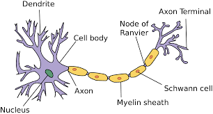
- The Dendrites are neuronal processes that taper from the cell body outwards. They produce many branches and transmit information towards the cell body from synapses on the dendritic tree. their primary role is to increase the surface area for synapse formation, allowing a great number of synapses to integrate together.
- There is only one Axon per neuron. originates at the axon hillock conducting information away from the cell body towards the nerve terminal and synapses. It can however, branch to produce several processes. The initial segment of the axon, as it emerges from the cell body, is the most excitable part of the neuron because it has a high density of sodium channels at this point. Therefore, it is at this point that the action potential is generated.
- A Lipid bilayer (cell membrane) encloses the neuron. It is within this that proteins are located. Some of these form ion channels, others form receptors to certain chemicals that are released by neurons. Others act as ion pumps, moving ions across the membrane. e.g Na+ - K+ exchange pump.
- The Axolemma is the axonal surface membrane while the axoplasm is contained within it.
- Many axons are surrounded by a Myelin sheath. This alters the conducting properties of the axon, allowing for fast action potential propagation, while the strength of the signal is maintained. This is able to happen due to the gaps in the sheath called the Nodes of Ranvier which contain many ion channels. The myelin sheath surrounds the axon from the origin near the cell body along the length of the axon to the terminal, before the axon branches.
- The myelin sheath is formed by Schwann cells in the PNS and Oligodendrocytes in the CNS.
- The synapse is the junction where the neuron meets another cell. in the CNS this will be another neuron. however in the PNS this may be a muscle cell, glandular cell or other organs. The physiology of synapses will be dealt with on the physiopedia page Introduction to Neuro-physiology.

Anatomy of the Spinal Cord and Associated Pathways[edit | edit source]
- The spinal cord lies within the vertebral canal, extending from the foramen magnum to the lowest border of the first lumbar vertebra.
- It is enlarged at 2 sites, the cervical and lumbar region.
- The lower part of the spinal canal contains the lower lumbar and sacral nerves known as the Cauda Equina.
- Sensory nerve fibres enter the spinal cord via the Dorsal (posterior) roots. The cell bodies for these neurons are situated in the dorsal root ganglia.
- Motor and preganglionic autonomic fibres exit via the ventral (anterior) root


This short video clip gives an overview of spinal cord anatomy
Ascending Sensory Pathways
- Spinothalamic tract: From Dorsal horn lam. I,III,IV,V. crosses midline in spinal cord, projects to brain stem and contr-lateral thalamus. Conveys pain and temperature.
- Dorsal column Medial lemniscal pathway: Afferents from mechanoreceptors, muscle and joint receptors. terminates in dorsal column nuclei of medulla. Forms medial lemniscus at this level and synapses in ventroposterior nucleus of thalamus. Conveys proprioception, light touch and vibration.
- Spinocerebellar tract: From spinal cord interneurons. It has 2 tracts a) Dorsal SCT relays via inferior cerebellar peduncle and b)VCT relays via superior cerebellar peduncle to the cerebellum. it conveys proprioceptive information and on-going activity in the spinal cord interneurons.
Descending Motor Tracts
- Corticospinal (pyramidal) tract: From the motor cortex, premotor cortex, and somatosensory cortex. Has a role in sensory processing and fractionated finger movements.
- Rubrospinal tract: Originates from the magnocellular part of the red nucleus in the brain. It projects towards common structures with the CoST, particularly those involved with distal motor control. There is debate as to how significant this tract is.
- Vestibulospinal Tract: Originates from Deiters nucleus in the medulla and innervates the extensor and axial muscles. It is involved in balance control and posture.
- Reticulospinal Tract: This tract begins in the caudal reticular formation in the pons and medulla. Provides both excitable and inhibitory effects on the interneurons in the spinal cord, and to a lesser extent, it also acts on the motor neurons. Its main action is to dampen down activity in the spinal cord. without this pathway, there is increased extensor tone observed.
Spinal Motorneurons.
Alpha and Gamma motorneurons (MNs) are both found in the ventral (anterior) horn.
Alpha motorneurons are the largest motor neurons in the nervous system. They innervate skeletal muscle.
Gamma Motorneurons innervate intrafusal muscle fibres of the muscle spindle.
Motor neurons are arranged somatotopically across the ventral horn. The more medially placed MNs innervate proximal muscles while laterally placed MNs innervate distal muscles.
Brainstem[edit | edit source]
Recent Related Research (from Pubmed)[edit | edit source]
Extension:RSS -- Error: Not a valid URL: Feed goes here!!|charset=UTF-8|short|max=10
References[edit | edit source]
References will automatically be added here, see adding references tutorial.

