Introduction to Neuroanatomy: Difference between revisions
No edit summary |
No edit summary |
||
| Line 77: | Line 77: | ||
== The Hypothalamus == | == The Hypothalamus == | ||
The hypothalamus lies on either side of the 3rd ventricle, below the thalamus and between the optic chiasm and the midbrain. It receives a large input from limbic structures. It has a significantly large efferent output to the ANS and has a highly significant role in the control of pituitary endocrine function. | The hypothalamus lies on either side of the 3rd ventricle, below the [[thalamus]] and between the optic chiasm and the midbrain. It receives a large input from limbic structures. It has a significantly large efferent output to the ANS and has a highly significant role in the control of pituitary endocrine function. | ||
The hypothalamus as well as inputting the ANS also has a large part to play in the homeostasis of many physiological systems such as hunger, thirst, water and sodium balance and temperature regulation. it also plays a role in memory and emotional responses, providing autonomic and endocrine responses. | The hypothalamus as well as inputting the ANS also has a large part to play in the homeostasis of many physiological systems such as hunger, thirst, water and sodium balance and temperature regulation. it also plays a role in memory and emotional responses, providing autonomic and endocrine responses. | ||
| Line 85: | Line 85: | ||
{{#ev:youtube|TVhm2rBGhB0|400}}<ref>Neuroscientifically Challenged. 2-Minute Neuroscience: Hypothalamus & Pituitary Gland. Available from: http://www.youtube.com/watch?v=TVhm2rBGhB0 [last accessed 19/10/2019] </ref> | {{#ev:youtube|TVhm2rBGhB0|400}}<ref>Neuroscientifically Challenged. 2-Minute Neuroscience: Hypothalamus & Pituitary Gland. Available from: http://www.youtube.com/watch?v=TVhm2rBGhB0 [last accessed 19/10/2019] </ref> | ||
== Limbic System == | == [[Limbic System]] == | ||
This refers to a number of areas within the brain lying mainly on the medial side of the temporal lobe. It includes a number of structures as seen in the diagram. | This refers to a number of areas within the brain lying mainly on the medial side of the temporal lobe. It includes a number of structures as seen in the diagram. | ||
Revision as of 06:42, 29 December 2020
Original Editor - Joanne Garvey and Naomi O'Reilly
Top Contributors - Joanne Garvey, Lucinda hampton, Naomi O'Reilly, Laura Ritchie, Kim Jackson, Rachael Lowe, Kate Sampson, Admin, Tarina van der Stockt, WikiSysop, Simisola Ajeyalemi, Adam Vallely Farrell, Rucha Gadgil, Rewan Elsayed Elkanafany, Jess Bell, Mande Jooste, Scott Buxton and George Prudden
Overview of the Nervous System[edit | edit source]

The nervous system is made up of vast neural networks; signalling within these circuits enables thinking, language, feeling, learning, memory, and all function and sensation. It is well-established that through plasticity of existing cells our nervous systems can adapt to situations not previously encountered, but it also has been shown that cells (NSCs) are plastic and involved in creating new connections in adaptation and response to injury.[1]
The Nervous System has three specific functions:
- Sensory Input - Sensory receptors present in the skin and organs respond to external & internal stimuli by generating nerve impulses that to the central nervous system
- Integration - The brain and spinal cord of the Central Nervous System combine and sum up all the data received from the body and send out nerve impulses.
- Motor Output - The nerve impulses from the Central Nervous System go to the effectors (muscles and glands). Muscle contractions and gland secretions are responses to stimuli received by sensory receptors.
The Nervous System is divided into two main divisions.[2]
- Central Nervous System (CNS)
- Peripheral Nervous System (PNS)
Central Nervous System[edit | edit source]
The CNS 2 parts
The Brain is divided into four main parts[1]:
- Brain stem, consisting of the medulla, pons, and midbrain
- Cerebellum
- Diencephalon, with the thalamus and hypothalamus
- Cerebral hemispheres (comprised of the cerebral cortex, basal ganglia, white matter, hippocampi and amygdalae). The right and left hemisphere are connected by the corpus callosum which facilitates communication between both sides of the brain. The Hemispheres are then further divided into four lobes.
2. The Spinal Cord (the caudal extension of the CNS).
Clinical Significance
Many neurological conditions affect the CNS. They range dramatically in scope, impact, and nature of the effect. Some conditions lead to progressively impaired movement eg Parkinson disease. Huntington chorea. The demyelination in multiple sclerosis can cause acute attacks, and over time, chronic degradation of function. Others may impact cognition such as the various dementias. Epilepsy can cause uncontrolled excitation. Headaches often impair the daily function of patients. Traumatic injuries can cause plegia or paresis and may result a wide range of deficits depending on the location and extent of the lesion[1].
Cerebral Cortex[edit | edit source]

The cerebrum consists of two cerebral hemispheres, the right and left hemisphere are connected by the corpus callosum which facilitates communication between both sides of the brain, with each hemisphere in the main connection to the contralateral side of the body i.e. the left hemisphere of the cerebrum receives information from the right side of the body resulting in motor control of the right side of the body and vice versa.
The hemispheres are then further divided into four lobes;
- Occipital
- Parietal
- Temporal (medial part of which are a series of structures including the Hippocampus)
- Frontal
The outer layer of the cerebral hemisphere is termed the cerebral cortex. This is inter-connected via pathways that run sub-cortically. It is these connections as well as the connections from the cerebral cortex to the brainstem, spinal cord and nuclei deep within the cerebral hemisphere that form the white matter of the cerebral hemisphere. The deep nuclei include structures such as the basal ganglia and the thalamus.
Meninges[edit | edit source]
The CNS is enclosed within the skull and vertebral column. These structures are separated by a series of membranes known as the Meninges. The Pia Mater is separated from the delicate arachnoid membrane by the subarachnoid space, which is then in turn separated from the Dura mater by the Sub-dural space.
Neurons[edit | edit source]

The cell body contains the nucleus and surrounding cytoplasm. It is the focus of the cells metabolic processes, housing the mitochondria, golgi apparatus and peroxisomes.
- The Dendrites are neuronal processes that taper from the cell body outwards. They produce many branches and transmit information towards the cell body from synapses on the dendritic tree. their primary role is to increase the surface area for synapse formation, allowing a great number of synapses to integrate together.
- There is only one Axon per neuron. originates at the axon hillock conducting information away from the cell body towards the nerve terminal and synapses. It can however, branch to produce several processes. The initial segment of the axon, as it emerges from the cell body, is the most excitable part of the neuron because it has a high density of sodium channels at this point. Therefore, it is at this point that the action potential is generated.
- A Lipid bilayer (cell membrane) encloses the neuron. It is within this membrane that proteins are located. Some of these form ion channels, others form receptors to certain chemicals that are released by neurons. Others act as ion pumps, moving ions across the membrane. e.g Na+ - K+ exchange pump.
- The Axolemma is the axonal surface membrane while the axoplasm is contained within it.
- Many axons are surrounded by a Myelin sheath. This alters the conducting properties of the axon, allowing for fast action potential propagation, while the strength of the signal is maintained. This is able to happen due to the gaps in the sheath called the Nodes of Ranvier which contain many ion channels. The myelin sheath surrounds the axon from the origin near the cell body along the length of the axon to the terminal, before the axon branches.
- The myelin sheath is formed by Schwann cells in the PNS and Oligodendrocytes in the CNS.
- The synapse is the junction where the neuron meets another cell. in the CNS this will be another neuron. however in the PNS this may be a muscle cell, glandular cell or other organs. The physiology of synapses will be dealt with on the Physiopedia page Introduction to Neuro-physiology.
Neuroglial Cells[edit | edit source]
There are four main classes of neuroglial cells within the CNS.
- Astrocytes: Small stellate cells. Found throughout CNS. Form structural and supporting framework for neuronal cells and capillaries. They maintain integrity of blood brain barrier (BBB). They store and release some neurotransmitters. Disperse excess ions. Important role in development of NS and may have a role in injury recovery. May have a role in presenting antigen to the immune system when CNS and BBB damaged.
- Oligodendrocytes: Responsible for myelination of CNS neurons. Large numbers in the white matter. Each Olig. forms myelin for 3-50 fibres, and many others surround fibres without forming sheaths. Clinical disorders of these cells cause central demyelination in conditions such as multiple sclerosis.
- Ependymal cells: Important for enabling movement of cerebrospinal fluid (CSF) as well as interacting with with Astrocytes to form a barrier separating the ventricles and CSF from neuronal environment. They line the central canal in the spinal cord.
- Microglial Cells: found throughout white and grey matter of the CNS. They are phagocytic in nature. Mediate immune responses within the CNS.
And in the PNS:
- Schwann Cell: Found only in the PNS. Responsible for the myelination of the peripheral nerves by wrapping the cell around the axon. There are multiple layers of scwann cell membrane wrapped around the nerve. One schwann cell wraps around one axon and provides myelin for one internode. They are important for regeneration of damaged peripheral axons.
Basal Ganglia[edit | edit source]
See link
The Hypothalamus[edit | edit source]
The hypothalamus lies on either side of the 3rd ventricle, below the thalamus and between the optic chiasm and the midbrain. It receives a large input from limbic structures. It has a significantly large efferent output to the ANS and has a highly significant role in the control of pituitary endocrine function.
The hypothalamus as well as inputting the ANS also has a large part to play in the homeostasis of many physiological systems such as hunger, thirst, water and sodium balance and temperature regulation. it also plays a role in memory and emotional responses, providing autonomic and endocrine responses.
It also plays a role in the control of circadian rhythms via retinal input to suprachiasmatic nucleus. There may also be hypothalamus input into sexual and emotional behaviour independent of its endocrine role.
Limbic System[edit | edit source]
This refers to a number of areas within the brain lying mainly on the medial side of the temporal lobe. It includes a number of structures as seen in the diagram.

- The limbic system provides high level processing of sensory information. The main outflow of the limbic system is to the prefrontal cortex and the hypothalamus as well as to cortical areas. It appears to have a role in attaching behavioural significance and response to a given stimulus.
- Damage to this area has profound effects on emotional responses.
- Long term potentiation (LTP) is the increase in the strength of a synaptic transmission with repetitive use, it can be seen to be effected in the hippocampus (primarily is involved with memory) and is thought to be important for memory acquisition.
Brainstem[edit | edit source]
See link
CNS Blood Supply[edit | edit source]
- Arterial blood supply to the brain comes from four vessels;
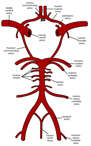

- Right and Left Internal Carotid
- Right and Left Vertebral Arteries
Basilar Artery:
This has a number of branches: anterior and inferior cerebellar artery, artery to the labyrinth, pontine branches and superior cerebellar artery.
Posterior Cerebral Arteries:
These supply blood to the posterior parietal cortex, occipital lobe and inferior temporal lobe. There are several branches of this artery that supply the midbrain, thalamus, subthalamus, posterior internal capsule, optic radiation and cerebral peduncle.
| [5] | [6] |
Venous Drainage
Venous drainage of the brainstem and cerebellum is straight into the dural venous sinuses which are adjacent to the posterior cranial fossa.
The cerebral hemispheres have external and internal veins.
- External veins drain the cortex and drain into the transverse sinus. This then drains into the lateral sinus and empties into the internal jugular vein.
- Internal cerebral veins drain the deep structures of the cerebral hemisphere into the great vein of Galen, and then into the sinus.
If occlusion of either of these venous systems then raised intracranial pressure can develop.
Blood Supply to the Spinal Cord
- The spinal cord is supplied by a single anterior spinal artery and paired posterior spinal arteries.
- Anterior spinal artery: arises from the vertebral arteries and extends from the level of the lower brainstem to the tip of the conus medullaris.
- It supplies the ventral medial surface of the medulla and anterior 2/3 of the spinal cord.
- The posterior spinal arteries supply the dorsal 1/3 of the cord.
- There are reinforcing branches from other arteries along the length of the cord.
- If occlusion occurs, it is normally of the anterior spinal artery, producing loss of power and spinothalamic sensory deficit, but dorsal column sensory capabilities are maintained.
The Cerebellum.[edit | edit source]

See link
Spinal Cord[edit | edit source]

See link
Peripheral Nervous System[edit | edit source]
The PNS consists of nerve trunks that are formed from both afferent axons which conduct sensory information to the spinal cord, and efferent fibres which transmit impulses primarily to muscles. If a particular nerve is damaged, there is resulting weakness to the muscle it supplies as well as sensory loss from the region it conveys information from.
The peripheral nerves connect with the spinal cord through foramina in the vertebra of the spine or with the brain through foramina in the skull.
Autonomic Nervous System[edit | edit source]
The main role of the ANS is to innervate the internal and glandular organs. Functions of the ANS include the regulation of “circulation, respiration, metabolism, secretion, body temperature, and reproduction.” The peripheral component is defined in terms of the sympathetic, and parasympathetic systems. Efferent fibres of the ANS originate from the intermediate zone (lateral column) of the spinal cord or specific cranial nerve and sacral nuclei before synapsing in a ganglion. These are different for sympathetic or parasympathetic systems. Afferent fibres from the organs innervated by the ANS travel via the dorsal root to the spinal cord.
The ANS includes nerve cells and fibres which innervate internal and glandular organs.There are always two successive neurons; preganglionic with cell body in the CNS and postganglionic.
The ANS is divided into two Divisions:
Sympathetic[edit | edit source]
Preganglionic neurons found in lateral horn of spinal cord from upper thoracic to mid-lumbar cord (T1-L3). Postganglionic cell bodies found in vertebral and prevertebral ganglia. Uses Noradrenalin as postganglionic transmitter.
Parasympathetic[edit | edit source]
Preganglionic neurons have cell bodies in the brainstem and sacrum. Postganglionic cell bodies are found adjacent to or within the walls of the organ they supply. Uses acetylcholine (Ach) as postganglionic transmitter.
Both Parasympathetic and Sympathetic systems use Ach at the level of the ganglia.
Somatic Nervous System[edit | edit source]
The Somatic Nervous System (SNS) is made up of nerves that are connected to skin, muscles and sensory organs (the eyes, ears, nose, skin, etc.). The SNS or voluntary nervous system is concerned with reactions to external stimulation. This system is under conscious control and is responsible for skeletal muscle contraction by way of the 31 pairs of spinal nerves. This system enables our voluntary control of muscles, as well as our reception of sights, sounds, sensations, tastes and smells.
| [7] | [8] |
Sensory Systems[edit | edit source]

The sensory system is where information is transmitted to the spinal cord and brain from peripheral sensory receptors. These are specialised neurons or nerve endings.
The sensory receptor, afferent axon and cell body are known as the primary afferent. The process by which the signals are transmitted through this system is known as sensory transduction. The signal produced is sent to the CNS via peripheral or cranial nerves via several synapses, eventually terminating in the cortex where it is analysed.
There are five main sensory systems in mammals.
- touch/pressure
- vision
- hearing and balance
- taste
- smell/olfaction
Sensory receptors[edit | edit source]
A sensory receptor's adequate stimulus is the stimulus modality for which it possesses the adequate sensory transduction apparatus. Adequate stimulus can be used to classify sensory receptors:
- Baroreceptors respond to pressure in blood vessels
- Chemoreceptors respond to chemical stimuli
- Electromagnetic radiation receptors respond to electromagnetic radiation[1] Infrared receptors respond to infrared radiation
- Photoreceptors respond to visible light
- Ultraviolet receptors respond to ultraviolet radiation
- Electroreceptors respond to electric fields Ampullae of Lorenzini respond to electric fields, salinity, and to temperature, but function primarily as electroreceptors
- Hydroreceptors respond to changes in humidity
- Magnetoreceptors respond to magnetic fields
- Mechanoreceptors respond to mechanical stress or mechanical strain
- Nociceptors respond to damage, or threat of damage, to body tissues, leading (often but not always) to pain perception
- Osmoreceptors respond to the osmolarity of fluids (such as in the hypothalamus)
- Proprioceptors provide the sense of position
- Thermoreceptors respond to temperature, either heat, cold or both
Pain Systems[edit | edit source]
Pain is defined as an unpleasant sensory or emotional experience, associated with potential or actual tissue damage. Nociception defines the processing of information about damaging stimuli by the nervous system up to the level of the cortex.
Potentially damaging mechanical, thermal, and chemical stimuli are detected by nerve endings called nociceptors, which are found in the skin, on internal surfaces such as the periosteum, joint surfaces, and in some internal organs. The concentration of nociceptors varies throughout the body; they are found in greater numbers in the skin than in deep internal surfaces. The nociceptors are unspecialized free nerve endings that have their cell bodies outside the spinal column in the dorsal root ganglia. Nociceptors are categorized according to the axons which travel from the receptors to the spinal cord or brain.
Nociceptors have a certain threshold; that is, they require a minimum intensity of stimulation before they trigger a signal. Once this threshold is reached a signal is passed along the axon of the neuron into the spinal cord.
In some conditions, excitation of pain fibers becomes greater as the pain stimulus continues, leading to a condition called hyperalgesia.
There are two types of nociceptor:
- A delta fibres: activated by high threshold mechanoreceptors. thinly myelinated.
- Unmyelinated C-fibres: activated by polymodal nociceptors(PMN) and respond to intense mechanical stimulation, high temperatures and irritant chemicals.
There are three main pathways that transmit nociceptive signals to the brain:
- Spinothalamic tract
- Spino reticular tract.
- Spino mesencephalic
Motor Systems[edit | edit source]

Motor systems are the areas of the nervous system responsible for controlling movement. The movement can either be guided by input from the sensory systems (closed-loop) or triggered by a sensory cue, or an internal desire to move (open-loop). Most movements involve both types of control. Closed loop movements tend to involve the axial muscles (posture and balance) while open-loop movements involve the peripheral muscles (locomotion and fine skilled movements).
- Level 1 - Highest level - concerned with initiation, planning, and programming of the movement. Response to internal desire to move (limbic system and post. parietal cortex.
- Level 2 - Cerebellum - responsible for balance and coordination of the movement.
- Level 3 - Control of lower descending neurons via supraspinal descending motor pathways (corticospinal/pyramidal tract and tecto and rubrospinal, vestibulo spinal, extrapyramidal).
- Level 4 - Low level motor organisation in the spinal cord. Descending motor paths, and interneurons. Mediation of spinal cord reflexes. This is where the central pattern generators are situated.
- Level 5 - Lowest level. output neuron of the CNS to the muscle (motorneuron). Receives important input from sensory organs in the periphery - the muscle spindle and golgi tendon organ.
References[edit | edit source]
- ↑ 1.0 1.1 1.2 Parker E. Ludwig; Matthew Varacallo Neuroanatomy, Central Nervous System (CNS) Feb 2019 Available from: ☀https://www.ncbi.nlm.nih.gov/books/NBK442010/ (last accessed 4.1.2020)
- ↑ Barker; Barasi; Neal. Neuroscience at a glance; Blackwell science Ltd; 1999
- ↑ khanacademymedicine. Cerebral cortex. Available from: http://www.youtube.com/watch?v=mGxomKWfJXs [last accessed 19/10/2019]
- ↑ Neuroscientifically Challenged. 2-Minute Neuroscience: Hypothalamus & Pituitary Gland. Available from: http://www.youtube.com/watch?v=TVhm2rBGhB0 [last accessed 19/10/2019]
- ↑ khanacademymedicine. Cerebral blood supply - Part 1. Available from: http://www.youtube.com/watch?v=hfG8J_X1D5Q [last accessed 19/10/2019]
- ↑ khanacademymedicine. Cerebral blood supply - Part 2. Available from: http://www.youtube.com/watch?v=kVulo3qDcUo [last accessed 19/10/2019]
- ↑ khanacademymedicine. Autonomic Nervous System. Available from: http://www.youtube.com/watch?v=TVhm2rBGhB0 [last accessed 19/10/2019]
- ↑ khanacademymedicine. Autonomic vs somatic nervous system. Available from: http://www.youtube.com/watch?v=TVhm2rBGhB0 [last accessed 19/10/2019]
- ↑ UCL Centre for Anaesthesia. An Introduction to Pain Pathways and Mechanisms. Available from: http://www.youtube.com/watch?v=i5V_q7XqQN8 [last accessed 19/10/2019]
- ↑ khanacademymedicine. Motor unit. Available from: http://www.youtube.com/watch?v=vXb0ZvkFkS8 [last accessed 19/10/2019]

