Humerus: Difference between revisions
No edit summary |
No edit summary |
||
| Line 18: | Line 18: | ||
The lesser tuberosity is located inferior to the head, on the anterior part of the humerus, Its very prominent and palpable. | The lesser tuberosity is located inferior to the head, on the anterior part of the humerus, Its very prominent and palpable. | ||
Bicipital (intertubercular) groove is located between the | Bicipital (intertubercular) groove is located between the two tuberosities. The Biceps tendon is placed here. | ||
=== Body Features === | === Body Features === | ||
| Line 34: | Line 34: | ||
== Function == | == Function == | ||
The humerus serves as an attachment to 13 muscles which contribute to the movements of the hand and elbow, and therefore function of the upper limb. | The humerus serves as an attachment to 13 muscles which contribute to the movements of the hand and elbow, and therefore the function of the upper limb. | ||
==Video== | ==Video== | ||
| Line 41: | Line 41: | ||
== Articulations == | == Articulations == | ||
[[Glenohumeral Joint|Glenohumeral joint]] | |||
Elbow joint | Elbow joint | ||
| Line 63: | Line 63: | ||
|Lesser Tubercle | |Lesser Tubercle | ||
|- | |- | ||
|[[ | |[[Pectoralis major|Pectoralis Major]] | ||
|Upper Part of the Anterior Border | |Upper Part of the Anterior Border | ||
|- | |- | ||
|[[Triceps Brachii]] | |[[Triceps brachii|Triceps Brachii]] | ||
|Lower Part of the Lateral Border | |Lower Part of the Lateral Border | ||
Lateral Supracondylar Ridge | Lateral Supracondylar Ridge | ||
| Line 94: | Line 94: | ||
== Clinical Relevance == | == Clinical Relevance == | ||
Proximal end or Head-Surgical Neck Fracture<ref>https://teachmeanatomy.info/upper-limb/bones/humerus/#Clinical_Relevance_Surgical_Neck_Fracture</ref> | Proximal end or Head-Surgical Neck Fracture<ref>https://teachmeanatomy.info/upper-limb/bones/humerus/#Clinical_Relevance_Surgical_Neck_Fracture</ref> | ||
* It is caused by a direct blow on the area or fall on outstretched hand. | * It is caused by a direct blow on the area or fall on an outstretched hand. | ||
* It results in damage to Axillary nerve and Posterior circumflex artery. | * It results in damage to the Axillary nerve and Posterior circumflex artery. | ||
* Axillary nerve damage results in paralysis of deltoid and teres minor muscles. | * Axillary nerve damage results in paralysis of deltoid and teres minor muscles. | ||
Shaft-Mid-shaft fracture<ref>https://teachmeanatomy.info/upper-limb/bones/humerus/#Clinical_Relevance_Mid-Shaft_Fracture</ref> | Shaft-Mid-shaft fracture<ref>https://teachmeanatomy.info/upper-limb/bones/humerus/#Clinical_Relevance_Mid-Shaft_Fracture</ref> | ||
* This fracture causes damage to radial nerve and Profunda brachii artery. | * This fracture causes damage to radial nerve and Profunda brachii artery. | ||
* Sensory loss can be seen over the dorsal surface of hand,proximal parts of lateral 3 and half fingers dorsally. | * Sensory loss can be seen over the dorsal surface of the hand, proximal parts of lateral 3 and a half fingers dorsally. | ||
* Radial nerve palsy results in wrist drop. | * Radial nerve palsy results in wrist drop. | ||
Distal end-Supracondylar fracture<ref>https://teachmeanatomy.info/upper-limb/bones/humerus/</ref> | Distal end-Supracondylar fracture<ref>https://teachmeanatomy.info/upper-limb/bones/humerus/</ref> | ||
* It is a fracture of distal | * It is a fracture of the distal humerus just above the Elbow joint. | ||
* It results in damage to brachial artery and anterior interosseous nerve,the resulting ischemia causes Volkman's ischemic contracture. | * It results in damage to the brachial artery and anterior interosseous nerve, the resulting ischemia causes Volkman's ischemic contracture. | ||
Other conditions | Other conditions | ||
* | * Haematologic, infectious, genetic and neurological disorders cause humerus varus.<ref>https://www.ncbi.nlm.nih.gov/books/NBK534821/#</ref> | ||
* Charcot arthropathy is a rare disorder characterised by debilitating joint destruction.<ref>https://www.ncbi.nlm.nih.gov/books/NBK534821/#</ref> | * Charcot arthropathy is a rare disorder characterised by debilitating joint destruction.<ref>https://www.ncbi.nlm.nih.gov/books/NBK534821/#</ref> | ||
Revision as of 14:23, 23 March 2020
Description[edit | edit source]
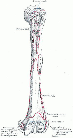
Humerus is a long bone which consists of a shaft (diaphysis) and two extremities (epiphysis). It is the longest bone of the upper extremity.
Structure[edit | edit source]
Upper Extremity Features[edit | edit source]
The head of the humerus is the articular surface of the upper extremity, which is an irregular hemisphere.
The anatomical neck is the part between the head and the tuberosities.
The surgical neck is the part between the tuberosities and the shaft.
The greater tuberosity it is located lateral to the head.
The lesser tuberosity is located inferior to the head, on the anterior part of the humerus, Its very prominent and palpable.
Bicipital (intertubercular) groove is located between the two tuberosities. The Biceps tendon is placed here.
Body Features[edit | edit source]
The body of the humerus has three borders and three surfaces.
Borders[edit | edit source]
- Anterior
- Lateral
- Medial
Surfaces[edit | edit source]
- Antero-lateral
- Antero-medial
- Posterior
Function[edit | edit source]
The humerus serves as an attachment to 13 muscles which contribute to the movements of the hand and elbow, and therefore the function of the upper limb.
Video[edit | edit source]
Articulations[edit | edit source]
Elbow joint
Muscle Attachments[edit | edit source]
| Muscle | Attachment |
|---|---|
| Supraspinatus | Greater Tubercle |
| Infraspinatus | Greater Tubercle |
| Teres Minor | Greater Tubercle
Upper Part of the Lateral Border |
| Subscapularis | Lesser Tubercle |
| Pectoralis Major | Upper Part of the Anterior Border |
| Triceps Brachii | Lower Part of the Lateral Border
Lateral Supracondylar Ridge |
| Brachioradialis | Lateral Supracondylar Ridge |
| Extensor Carpi Radialis Longus | Lateral Supracondylar Ridge |
| Teres Major | Crest of the Lesser Tubercle |
| Coracobrachialis | Crest of the Lesser Tubercle |
| Brachialis | Medial Supracondylar Ridge |
| Pronator Teres | Medial Supracondylar Ridge |
| Latissimus Dorsi | Bicipital Groove |
Clinical Relevance[edit | edit source]
Proximal end or Head-Surgical Neck Fracture[2]
- It is caused by a direct blow on the area or fall on an outstretched hand.
- It results in damage to the Axillary nerve and Posterior circumflex artery.
- Axillary nerve damage results in paralysis of deltoid and teres minor muscles.
Shaft-Mid-shaft fracture[3]
- This fracture causes damage to radial nerve and Profunda brachii artery.
- Sensory loss can be seen over the dorsal surface of the hand, proximal parts of lateral 3 and a half fingers dorsally.
- Radial nerve palsy results in wrist drop.
Distal end-Supracondylar fracture[4]
- It is a fracture of the distal humerus just above the Elbow joint.
- It results in damage to the brachial artery and anterior interosseous nerve, the resulting ischemia causes Volkman's ischemic contracture.
Other conditions
- Haematologic, infectious, genetic and neurological disorders cause humerus varus.[5]
- Charcot arthropathy is a rare disorder characterised by debilitating joint destruction.[6]
References[edit | edit source]
- ↑ http://www.bartleby.com/107/illus207.html
- ↑ https://teachmeanatomy.info/upper-limb/bones/humerus/#Clinical_Relevance_Surgical_Neck_Fracture
- ↑ https://teachmeanatomy.info/upper-limb/bones/humerus/#Clinical_Relevance_Mid-Shaft_Fracture
- ↑ https://teachmeanatomy.info/upper-limb/bones/humerus/
- ↑ https://www.ncbi.nlm.nih.gov/books/NBK534821/#
- ↑ https://www.ncbi.nlm.nih.gov/books/NBK534821/#
Gray H., 2000, Anatomy of Human Body, Twentieth Edition, New York, Bartleby.com


