Facial Nerve: Difference between revisions
Scott Buxton (talk | contribs) No edit summary |
Scott Buxton (talk | contribs) No edit summary |
||
| Line 112: | Line 112: | ||
== References == | == References == | ||
<references /><br> | |||
< | <br><br> | ||
[[Category:Neurology]] [[Category:Anatomy]] [[Category:Nerves]] | |||
[[Category:Neurology]][[Category:Anatomy]][[Category:Nerves]] | |||
Revision as of 20:14, 20 May 2015
Original Editor - Wendy Walker.
Top Contributors - Wendy Walker, Kim Jackson, Scott Buxton, Lucinda hampton, Rishika Babburu, Merinda Rodseth, Admin, Evan Thomas, Samson Chengetanai, Tarina van der Stockt, Priyanka Chugh, WikiSysop and Jess Bell
Topic Expert - Wendy Walker
Description[edit | edit source]
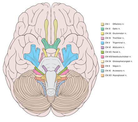
The Facial Nerve is the seventh Cranial Nerve.
It is composed of approximately 10,000 neurons, 7,000 of which are myelinated and innervate the nerves of facial expression.
The remaining 3,000 fibres are somatosensory and secretomotor, and are known as the Nervus Intermedius.
Movements Produced[edit | edit source]
All movements of facial expression, including:
Smile, close eyes, pucker lips, wrinkle nose, raise eyebrows, frown.
General Course of Nerve[edit | edit source]
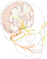
The facial nerve has six named segments [1]:
- intracranial (cisternal) segment
- meatal segment (internal auditory canal) - 8mm - zero branches
- labyrinthine segment (IAC to geniculate ganglion) - 3-4mm - 3 branches (from geniculate ganglion)
- tympanic segment (from geniculate ganglion to pyramidal eminence) - 8-11mm - zero branches
- mastoid segment (from pyramidal eminence to stylomastoid foramen) - 8-14mm - 3 branches
- extratemporal segment (from stylomastoid foramen to division into major branches) 15-20mm - 9 branches
Intracranial (cisternal) segment
The nerve emerges immediately beneath the pons, lateral to the abducens nerve and medial to the vestibulocochlear nerve and is joined by the nervus intermedius, which has emerged lateral to the main trunk. Together the two travel through the cerebellopontine angle to the internal acoustic meatus
Meatal segment
Having been joined by the nervus intermedius, they are located in the superior upper quadrant, above the falciform crest and anterior to Bill's bar.
Labyrinthine segment
As the facial nerve and nervus intermedius pass through the anterior superior quadrant of theinternal acoustic meatus it enters the Fallopian canal, passing anterolaterally between and superior to the cochlea (anterior) and vestibule (posterior), and then runs back posteriorly at thegeniculate ganglion (where the nervus intermedius joins the facial nerve and where fibers for taste synapse). It is here that three branches originate:
- greater superficial petrosal nerve
- lesser petrosal nerve
- external petrosal nerve
The labyrinthine segment is the shortest only measuring 3-4 mm. It is also the narrowest and the most susceptible to vascular compromise.
Tympanic segment
As the nerve passes posteriorly from the geniculate ganglion it becomes the tympanic segment (8-11 mm in length) and is immediately beneath the lateral semicircular canal in the medial wall of the middle ear cavity. The nerve passes posterior to the cochleariform process, tensor tympani and oval window. Just distal to the pyramidal eminence the nerve makes a second turn (second genu) passing vertically downwards as the mastoid segment.
The tympanic segment has no branches.
Mastoid segment
The mastoid segment, measuring 8-14mm in length, extends from the second genu to the stylomastoid foramen, through what is confusingly referred to as the fallopian canal. It gives off three branches:
- nerve to stapedius
- chorda tympani - terminal branch of the nervus intermedius carrying both secretomotor fibres to the submandibular gland and sublingual gland and taste to the anterior two thirds of the tongue
- nerve from the auricular branch of the vagus nerve (CN X) - pain fibers to the posterior part of the external acoustic meatus
Extratemporal segment
As the nerve exits the stylomastoid foramen, it gives off a sensory branch that supplies part of the external acoustic meatus and tympanic membrane.
It then passes between the posterior belly of the digastric muscle and the stylohyoid muscle and enters the parotid gland. Lying between the deep and superficial lobes of the gland the nerve divides into to main branches at the pes anserinus (Latin = "duck foot") - a superior temporofacial and and inferior cervicofacial branches.
From the anterior border of the gland, five branches emerge: temporal, zygomatic, buccal, mandibular (marginal) and cervical.
There is another detailed diagram of the course of the Facial Nerve on the Facial Palsy page.
5 Distal Branches[edit | edit source]
The facial nerve innervates 14 of the 17 paired muscle groups of the face on their deep side[2]. The 3 facial muscles are innervated from other sources: buccinator, levator anguli oris, and mentalis.
- Temporal (Frontal)
- Zygomatic
- Buccal
- Marginal mandibular
- Cervical
The temporal trunk innervates the following muscles:
- Frontalis
- Orbicularis oculi
- Corrugator supercilii
- Pyramidalis
The zygomatic division innervates the following muscles:
- Zygomaticus major
- Zygomaticus minor
- Elevator ala nasi
- Levator labii superioris
- Caninus
Depressor septi - Compressor nasi
- Dilatator naris muscles
The buccal division gives off fibers to innervate the buccinator and superior part of the orbicularis oris muscle.
Mandibular division innervations are found in the following muscles:
- Risorius
- Quadratus labii inferioris
- Triangularis
- Mentalis
- Lower parts of the orbicularis oris
The cervical division provides platysma innervation.
Embryology[edit | edit source]
By the third week of gestation, the fascioacoustic primordium gives rise to CN VII and VIII. During the fourth week, the chorda tympani can be discerned from the main branch. The former courses ventrally into the first branchial arch and terminates near a branch of the trigeminal nerve that eventually becomes the lingual nerve. The main trunk courses into the mesenchyme, approaching the epibranchial placode.
The geniculate ganglion, nervus intermedius, and greater superficial petrosal nerve are visible by the fifth week. The second branchial arch gives rise to the muscles of facial expression in the seventh and eighth week. To innervate these muscles, the facial nerve courses across the region that eventually becomes the middle ear. By the eleventh week, the facial nerve has arborized extensively. In the newborn, the facial nerve anatomy approximates that of an adult, except for its location in the mastoid, which is more superficial.
Imaging[edit | edit source]
Computed tomography (CT) scanning and magnetic resonance imaging (MRI) are useful in the diagnosis of injury to intratemporal and/or intracranial affections of the facial nerve, as they may reveal temporal fracture patterns (vertical, transversal, mixed) and edema formation. Under certain circumstances, the facial nerve can be viewed, and swelling or disruption may be seen[3].
Recent Related Research (from Pubmed)[edit | edit source]
Failed to load RSS feed from http://www.ncbi.nlm.nih.gov/entrez/eutils/erss.cgi?rss_guid=1nySOMllQ3n2CiHBc3BYbuME_p6vfoPeCy8XODjpmyNdbOZEls|charset=UTF-8|short|max=10: Error parsing XML for RSS
References[edit | edit source]
- ↑ May M, Schaitkin B. May M, Schaitkin B, eds. The Facial Nerve, 2nd Edition. New York, NY: Thieme; 2000.
- ↑ Davis RA, Anson BJ, Budinger JM, et al. Surgical anatomy of the facial nerve and the parotid gland based on a study of 350 cervicofacial halves. Surg Gynecol Obstet. 1956;102:358.
- ↑ Kumar A, Mafee MF, Mason T. Value of imaging in disorders of the facial nerve. Top Magn Reson Imaging. Feb 2000;11(1):38-51. [Medline].


