Distal Radial Fractures
Original Editors
Top Contributors - Laura Ritchie, Diane Hodges Popps, Jocelyn Fu, Kim Jackson, Admin, Katherine Knight, Nupur Smit Shah, WikiSysop, Ulrike Lambrix, Jess Bell, Evan Thomas, Elien Lebuf, Tarina van der Stockt, Karen Wilson, Lauren Lopez, Tony Lowe, Jorge Rodríguez Palomino, Felicia Daigle and Jeremy Brady - Your name will be added here if you are a lead editor on this page.
Search Strategy[edit | edit source]
Search Datebases:Cochrane Library, Medline with full text, pubmed, CINAHL, Search dates: 9/16/10, 9/21/10, 10/26/10.</span>
</span></span>Search terms: Distal radial fracture, distal radius fracture, distal radial treatment, distal radius treatment, radius physical therapy, RCT, Handoll</span>
Definition/Description
[edit | edit source]
Fractures occurring in the distal radius often occur in both children and adults and can be referred to as “wrist fractures.” They are defined as occurring in the distal radius within three centimeters from the radiocarpal joint, where the radius interfaces with the lunate and scaphoid bone of the wrist. The majority of distal radial fractures are closed injuries in which the overlying skin remains intact.[1][2]
Epidemiology /Etiology[edit | edit source]
Distal radial fractures in adults is one of the most common fractures, accounting for one-sixth of all fractures in the emergency department and can be seen predominantly in the white and older population.[1][2][3][4][5] In women, the incidence of occurrence increases with age starting at 40 years old, whereas before this age the incidence in men is much higher. Occurrences in younger adults are usually the result of a high-energy trauma such as a motor vehicle accident. In older adults, such fractures are often the result of a low-energy or moderate trauma such as falling from standing height. This may reflect the greater fragility of the bone due to osteoporosis in the older adult.[1][6]
Distal radial fractures account for up to 72% of all forearm fractures and 8-17% of all extremity fractures.[7] Historically, literature has focused on restoring the anatomic radiocarpal alignment, however the distal radioulnar joint (DRUJ) is also important in restoring hand function.[8]
Multiplanar wrist motion is based on three articulations: radioscaphoid, radiolunate, and distal radioulnar joints. The distal end of the radius forms a “platform” to support the functional demands of the wrist. The medial distal radius ligament and ulnar-syloid based triangular fibrocartilage complex (TFCC) help to stabilize the wrist.[7]The TFCC is the primary intrinsic stabilizer of the DRUJ and is critical for normal DRUJ biomechanics.[8] Bony anatomy can result in malunions with radiographic assessment by measuring the radial inclination, radial length, ulnar variance, and radial tilt as shown in Figure 1. Slight changes may result in malunion and can cause considerable pain and disability. For example, increased dorsal angulation can alter the loading force and result in decreased congruency at the DRUJ thus tightening the interosseous membrane and decreasing pronation-supination.[7] A concomminant injury due to stress on the TFCC is an avulsion fracture of the ulnar styloid process and can result in significant malalignment.[8]
A correlation between the severity of the primary displacement and the expectant loss of reduction over a time period is assumed when treating distal radial fractures with immobilization. Predictive factors of instability at an early (1 week) and a late (6 weeks) time period can be established by radial shortening and dorsal tilt in the early phases and radial shortening, dorsal tilt, and decreased radial inclination in the late phases. Instability is defined as dorsal tilt >15°, volar tilt >20°, ulnar variance >4mm and a radial inclincation <10°.[7][9]
Characteristics/Clinical Presentation[edit | edit source]
Distal radial fractures can be classified based on their clinical appearance and typical deformity. Dorsal displacement, dorsal angulation, dorsal comminution, and radial shortening may all be used to describe the presentation of the fracture. Classification based on fracture patterns such as intra-articular (articular surfaces disrupted) or extra-articular (articular surface of radius intact) may also be used.[1][7] A Colles fracture is typically due to a fall on an outstretched hand and results in a dorsal displacement of the fractured radius. A “Smith’s” fracture is a reverse Colles with a volar displacement. A “Barton’s” fracture is an intra-articular fracture with subluxation or dislocation of the carpus bone. Of the 8 grades for distal radial fractures in the Frykman classification system, half include ulnar syloid involvement.
Complications are common and diverse. They may be the result of injury or treatment, and are associated with poorer outcomes. These can include upper extremity stiffness, carpal tunnel syndrome or median nerve involvement, malunion, carpal instability, DRUJ dysfunction, Dupuytren’s disease, radiocarpal arthritis, tendon/ligament injuries, and complex regional pain syndrome.[4][8][10][11]Residual pain and stiffness at the wrist cannot be compensated for by shoulder or elbow kinematics.
- Malunion--Distal radius malunion is the most common complication, affecting up to 17% of patients. Physical therapists may assess the effect of malunion to determine if surgery is appropriate by performing a detailed physical exam that includes a preoperative history, location and severity of pain, and functional loss.[7][11]
- Compartment Syndrome - Incidence of this complication affects only 1% of patients. Elevate, observe, and loosen cast emergently if compartment syndrome is suspected.[11]
- Complex Regional Pain Syndrome (CRPS) - This complication is observed in 8-35% of patients.[11] CRPS should be suspected when pain, decreased ROM, skin temperature, color, and swelling are out of proportion to the injury. In order to obtain a good functional outcome for this patient population, early recognition and a multidisciplinary treatment approach to address pain and function requires psychiatric and physical/occupational therapy interventions.
- Dupeytren’s Disease - Patients develop mild contractures in the palm along the fourth and fifth rays within six months of a distal radial fracture. The severity of the contractures determines the treatment course.[11]
- Nerve Pathology – Neuropathy may present acutely or throughout treatment. The median nerve is most common (4%), however 1% of patients have ulnar or radial involvement.[4] Physical therapists may need to refer the patient to an orthopedic surgeon.[11]
- Acute Carpal Tunnel Syndrome – Physical therapists must be able to identify acute carpal tunnel syndrome, as delayed treatment is associated with poor outcomes, incomplete recovery, or a prolonged functional recovery time.[4][11]
- Tendon Complications - Physical therapists should be prepared to refer patients to surgery in the event of tendon complications secondary to irritation with inflammation or rupture from impingement.[11]
- Capsule Contracture - Even after physical therapy treatment, some patients do not regain full forearm rotation due to contracture of the distal radioulnar joint capsule. Dorsal contracture limits pronation, volar contracture limits supination, and both may occur together. A DRUJ capsulectomy may be considered if functional ROM is not regained.[8]
Differential Diagnosis[edit | edit source]
Because the mechanism of injury for a distal radial fracture is usually a high energy traumatic incident, radiographs should be taken to confirm the diagnosis and ensure that the surrounding tissues are still intact. Other injuries causing radial sided pain may include TFCC wear or preforation, Galeazzi fracture (fracture to the distal 2/3 of the radius), scaphoid fracture, or radiocarpal ligament injury.
Examination[edit | edit source]
Physical therapists must conduct a thorough physical exam including subjective and objective information.
- Subjective exam includes any information given by the patient about pain experienced, limitations of ROM of the wrist, and activity limitations.[4]
- Objective exam includes assessment of wrist and digit ROM, grip and forearm strength, bony and soft-tissue abnormalities, skin integrity, and nerve involvement.[4]
Health care professionals should always evaluate the ligamentous integrity in the presence of carpal instability and persistent pain as early as possible in order to avoid poor functional outcomes and prolonged recovery. Specific fracture patterns and high energy injuries are strongly indicative of intercarpal ligament involvement.[11]
Medical Management (current best evidence)[edit | edit source]
Orthopedic surgeons typically recommend surgical repair of displaced articular fractures of the distal radius for active, healthy people.[12] The sheer variety of reduction and fixation options is noted based on a series of five Cochrane reviews focusing on this topic alone. Methods include: closed reduction and percutaneous pinning, either extra-focal or intra-focal; bridging external fixation with or without supplemental Kirschner-wire fixation; dorsal plating; fragment-specific fixation; open reduction and internal fixation with a volar plate through a classic Henry approach; or a combination of these methods.[1][2][7][12][13][14][15] Surgical “complications include edema, hematoma, stiffness, infection, neurovascular injury, loss of fixation, recurrent malunion, nonunion or delayed union, instability, tendon irritation or ruptures, osteoarthritis, residual ulnar-side pain, median neuropathy, complex regional pain syndrome, and problems with the bone-graft harvest site."[7]
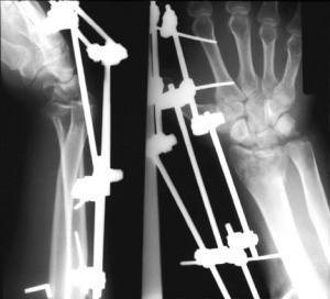
- External Fixation – External fixation is typically a closed, minimally invasive method in which metal pins or screws are driven into the bone via small incisions in the skin. These pins can then be fixed externally by either a plaster cast or securing them into an external fixator frame.[1][2] In comparison to a standard immobilization procedure, external fixation of distal radius fractures reduces redisplacement and yields better anatomical results. However, current evidence for better functional outcomes from external fixation is weak and is also associated with high risk for complications such as pin site infections and radial nerve injuries. A radiographic example is shown in Figure 2.
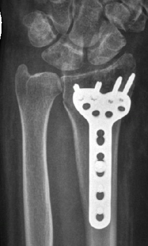
- Internal fixation – Internal fixation involves open surgery where the fractured bone is exposed. Dorsal, volar or T-plates with screws may be used.[7] However, due to the invasive and demanding nature of open surgery, there is an increased risk of infection and soft-tissue damage and therefore this type of fixation is usually reserved for more severe injuries.[13] Figure 3 is a radiographic image of internal fixation.
- Bone Grafts - Upon reduction of distal radial fractures, bony voids are common and can be reduced by inserting bone grafts or bone graft substitutes. Autogenous bone material obtained from the patient themselves or allogenous bone material obtained from cadaver or live donors can be used as filler for reducing bony voids. However, current research describes risk of complications including infection, nerve injury, or donor site pain, and limited evidence that bone scaffolding may improve anatomical or functional outcomes.[16] Bone grafting is required by most procedures except closing wedge.[7]
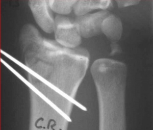
- Percutaneous Pinning - Another strategy in reducing and stabilizing the fractures is percutaneous pinning, which involves insertion of pins, threads or wires through the skin and into the bone.[15][7] This procedure is typically less invasive and reduction of the fracture is closed upon which the pins placed in the bone are used to fix the distal radial fragment. Current indications for the best technique of pinning, the extent and duration of immobilization are uncertain, in which the excess of complications are likely to outweigh therapeutic benefits of pinning. Figure 4 is a radiograph of pinning.
- Closed Reduction - In closed reduction, displaced radial fragments are repositioned using different maneuvers while the arm is in traction. Different methods include manual reduction in which two people pull in the opposite directions to produce and maintain longitudinal traction and mechanical methods of reduction including the use of “finger traps.” However there is insufficient evidence establishing the effectiveness of different methods of closed reduction used in treating distal radial fractures.[14]
- Arthroscopic-assisted reduction has many advantages over open reduction. In addition to being less invasive, arthroscopic-assisted reduction allows for direct visualization and reduction of articular displacement, opportunity to diagnose and treat associated ligamentous injuries, removal of articular cartilage debris, and lavage of radiocarpal joint. The primary limitations for arthroscopic reduction are due the limited number of surgeons with experience, a longer, more difficult procedure, and the potential for compartment syndrome or acute carpal tunnel syndrome with fluid extravasation.[12]
Physical Therapy Management (current best evidence)[edit | edit source]
Current physical therapy management for distal radius fractures covers post-surgical or post-immobilization treatment. The protocols that have been studied range from a single treatment session with a physical therapist at the time of cast removal to a formal course of outpatient therapy. Some patients are not referred for therapeutic intervention at all.[5] Handall et al [17]concluded in a systematic review of rehabilitation for distal radial fractures primarily following immobilization without surgical intervention, that there is insufficient data to establish the effectiveness of various rehabilitation interventions. Modalities such as ice, whirlpool immersion, ultrasound, and intermittent pneumatic compression where insignificant.[5] This may be due to poor study design and the heterogeneity of distal radial fractures themselves.
Distal radial fractures tend to follow a normal course of recovery for pain and disability for up to one year.[18] This recovery pattern is clinically significant for a physical therapist to develop an appropriate plan of care and prognosis for treatment. During the first two months, patients report severe pain during movement and severe disability during activities of daily living as assessed with valid and reliable outcome measures such as the Patient Rated Wrist Evaluation (PRWE) and the Disabilities of the Arm, Shoulder, and Hand Questionnaire (DASH).[18] These deficits are reflected in decreased range of motion and decreased grip strength measurements with strength being more highly correlated with functional ability. However, most patients achieve the majority of recovery within the first six months. A small minority of patients will experience persistent pain and disability at one year post injury regardless of treatment protocol,[4][18][19] especially when pushing up from sit to stand and carrying weight. Patients expressing atypical recovery from distal radial fractures need modified treatment programs with goals toward increasing workability.
The most important goal of conservative physical therapy treatment is to maintain anatomical alignment and surgical reduction following a distal radial fracture in order to avoid common complications and achieve optimal functional outcomes.[6] Table 1 details the recommended physical therapy treatment for the first twelve weeks following fracture reduction.[20] The goals of the initial six weeks are to reduce swelling, minimize stiffness, support the reduced fracture, and promote range of motion. During weeks six through eight, the wrist and forearm range of motion is increased, with a focus on supination.[8] During the late phase of weeks eight through twelve, treatment focuses on maximizing motion and promoting strength of the entire upper extremity. Typical return to full activity is in three to four months.
Key Research[edit | edit source]
Handoll et al conducted eight Cochrane Reviews targeting distal radial fractures. There was insufficient data from which to draw conclusions. This may be due to poor study design and the heterogeneity of distal radial fractures themselves.
• Different methods of external fixation for treating distal radial fractures in adults[2]
• External Fixation versus conservative treatment for distal radial fractures in adults[1]
• Internal fixation and comparisons of different fixation methods for treating distal radial fractures in adults[13]
• Closed reduction methods for treating distal radial fractures in adults[14]
• Percutaneous pinning for treating distal radial fractures in adults[15]
• Bone grafts and bone substitutes for treating distal radial fractures in adults[16]
• Conservative interventions for treating distal radial fractures in adults[10]
• Rehabilitation for distal radial fractures in adults[17]
A 2008 randomized control trial, Kay et al[5] was supportive of physical therapy intervention, though limitations in the study abound. While no significant differences in grip strength and wrist extension were found in the experimental group, it is important to note that some of the secondary measures showed significant improvement as compared to the control group. These include benefits in activity, pain, and satisfaction.
Resources
[edit | edit source]
Clinical Bottom Line[edit | edit source]
Due to the fact that distal radial fractures are one of the most common injuries in orthopedics, it is important for physical therapists to understand the risk factors and treatment options. Although further research is needed to ascertain proper post surgical management, it is recommended based on current evidence that patients should be routinely referred to a physical therapist for education and an exercise program.
Recent Related Research (from Pubmed)[edit | edit source]
see tutorial on Adding PubMed Feed
Extension:RSS -- Error: Not a valid URL: Feed goes here!!|charset=UTF-8|short|max=10
References
[edit | edit source]
- ↑ 1.0 1.1 1.2 1.3 1.4 1.5 1.6 Handoll HHG, Huntley JS, Madhok R. External Fixation versus conservative treatment for distal radial fractures in adults (Review). The Cochrane Library. 2008;4:1-78.
- ↑ 2.0 2.1 2.2 2.3 2.4 Handoll HHG, Huntley JS, Madhok R. Different methods of external fixation for treating distal radial fractures in adults (Review). The Cochrane Library. 2008;4:1-67.
- ↑ Abramo A, Kopylov P, Tagil M. Evaluation of a treatment protocol in distal radius fractures: a prospective study in 581 patients using DASH as outcome. Acta Orthopaedica. 2008;79(3):376-385
- ↑ 4.0 4.1 4.2 4.3 4.4 4.5 4.6 Bienek T, Kusz D, Cielinski L. Peripheral nerve compression neuropathy after fractures of the distal radius. J Hand Surg. (British and European Volume). 2006;31B(3):256-260.
- ↑ 5.0 5.1 5.2 5.3 Kay S, McMahon M, Stiller K. An advice and exercise program has some benefits over natural recovery after distal radius fracture: a randomized trial. Aust J Physiother. 2008;54:253-259.
- ↑ 6.0 6.1 Leung F, Ozkan M, Chow SP. Conservative treatment of intra-articular fractures of the distal radius – factors affecting functional outcomes. Hand Surg. 2000;5(2):145-153.
- ↑ 7.00 7.01 7.02 7.03 7.04 7.05 7.06 7.07 7.08 7.09 7.10 Bushnell BD, Bynum DK. Malunion of the distal radius. J Am Acad Orthop Surg. 2007;15:27-40.
- ↑ 8.0 8.1 8.2 8.3 8.4 8.5 Kleinman WB. Distal radius instability and stiffness; common complications of distal radius fractures. Hand Clin. 2010;26:245-264.
- ↑ Leone J, Bhandari M, Adili A, McKenzie S, Moro JK, Dunlop RB. Predictors of early and late instability following conservative treatment of exta-articular distal radius fractures. Arch Orthop Trauma Surg. 2004;124:38-41.
- ↑ 10.0 10.1 Handoll HHG, Madhok R. Conservative interventions for treating distal radial fractures in adults (Review). The Cochrane Library. 2008;4:1-112.
- ↑ 11.0 11.1 11.2 11.3 11.4 11.5 11.6 11.7 11.8 Patel VP, Paksima N. Complications of distal radius fracture fixation. Bulletin of NYU Hospital for Joint Diseases. 2010;68(2):112-8.
- ↑ 12.0 12.1 12.2 Herzberg G. Intra-articular fracture of the distal radius: arthroscopic-assisted reduction. J Hand Surg. 2010;35A:1517-1519.
- ↑ 13.0 13.1 13.2 Handoll HHG, Watts AC. Internal fixation and comparisons of different fixation methods for treating distal radial fractures in adults (Protocol). The Cochrane Library. 2008;4:1-14.
- ↑ 14.0 14.1 14.2 Handoll HHG, Huntley JS, Madhok R. Closed reduction methods for treating distal radial fractures in adults (Review). The Cochrane Library. 2008;4:1-29.
- ↑ 15.0 15.1 15.2 Handoll HHG, Vaghela MV, Madhok R. Percutaneous pinning for treating distal radial fractures in adults (Review). The Cochrane Library. 2008;4:1-70.
- ↑ 16.0 16.1 Handoll HHG, Huntley JS. Bone grafts and bone substitutes for treating distal radial fractures in adults (Review). The Cochrane Library. 2009;3:1-87.
- ↑ 17.0 17.1 Handoll HHG, Madhok R, Howe TE. Rehabilitation for distal radial fractures in adults (Review). The Cochrane Library. 2008;4:1-78.
- ↑ 18.0 18.1 18.2 MacDermid JC, Roth JH, Richards RS. Pain and disability reported in the year following a distal radius fracture: A cohort study. BMC Musculoskelet Disord. 2003;4:24-36.
- ↑ Moore CM, Leonardi-Bee J. The prevalence of pain and disability one year post fracture of the distal radius in a UK population: A cross sectional survey. BMC Musculoskeletl Disord. 2008;9:129-138.
- ↑ Brotzman BS, Wilk KE. Handbook of Orthopaedic Rehabilitation, 2nd ed. Philadelphia: Mosby.2007.


