Diaphragm Anatomy and Differential Diagnosis: Difference between revisions
Kim Jackson (talk | contribs) m (Text replacement - "[[HIV" to "[[Human Immunodeficiency Virus (HIV)") |
No edit summary |
||
| (17 intermediate revisions by 5 users not shown) | |||
| Line 4: | Line 4: | ||
== Anatomy of the Diaphragm == | == Anatomy of the Diaphragm == | ||
The diaphragm is a major muscle of respiration.<ref>Fayssoil A, Behin A, Ogna A, Mompoint D, Amthor H, Clair B, Laforet P, Mansart A, Prigent H, Orlikowski D, Stojkovic T, Vinit S, Carlier R, Eymard B, Lofaso F, Annane D. [https://content.iospress.com/articles/journal-of-neuromuscular-diseases/jnd170276 Diaphragm: Pathophysiology and Ultrasound Imaging in Neuromuscular Disorders.] J Neuromuscul Dis. 2018;5(1):1-10</ref> | The diaphragm is a major muscle of respiration.<ref>Fayssoil A, Behin A, Ogna A, Mompoint D, Amthor H, Clair B, Laforet P, Mansart A, Prigent H, Orlikowski D, Stojkovic T, Vinit S, Carlier R, Eymard B, Lofaso F, Annane D. [https://content.iospress.com/articles/journal-of-neuromuscular-diseases/jnd170276 Diaphragm: Pathophysiology and Ultrasound Imaging in Neuromuscular Disorders.] J Neuromuscul Dis. 2018;5(1):1-10</ref> | ||
* It is a dome-shaped, "fibromuscular sheet" that separates the thorax from the abdomen<ref name=":0">Bains KN, Kashyap S, Lappin SL. [https://www.ncbi.nlm.nih.gov/books/NBK519558/ Anatomy, Thorax, Diaphragm.] StatPearls [Internet]. 2020 Aug 15.</ref> | * It is a dome-shaped, "fibromuscular sheet" that separates the thorax from the abdomen.<ref name=":0">Bains KN, Kashyap S, Lappin SL. [https://www.ncbi.nlm.nih.gov/books/NBK519558/ Anatomy, Thorax, Diaphragm.] StatPearls [Internet]. 2020 Aug 15.</ref> | ||
* It forms the floor of thorax and roof of abdomen<ref name=":0" /><ref>Kurian J. [https://link.springer.com/chapter/10.1007/978-3-030-31989-2_6 Chest Wall and Diaphragm]. InPediatric Body MRI 2020 (pp. 159-192). Springer, Cham.</ref> | * It forms the floor of the thorax and the roof of the abdomen.<ref name=":0" /><ref>Kurian J. [https://link.springer.com/chapter/10.1007/978-3-030-31989-2_6 Chest Wall and Diaphragm]. InPediatric Body MRI 2020 (pp. 159-192). Springer, Cham.</ref> | ||
* The left side is lower than the right | * The left side is lower than the right - this is because the liver is situated on the right side.<ref name=":0" /> | ||
* The left side may also be located partially inferiorly due to the "push" by the heart<ref name=":0" /><ref>Bordoni B, Purgol S, Bizzarri A, Modica M, Morabito B. [https://www.ncbi.nlm.nih.gov/pmc/articles/PMC6070065/ The influence of breathing on the central nervous system.] Cureus. 2018 Jun;10(6).</ref><ref>Oliver KA, Ashurst JV. [https://pubmed.ncbi.nlm.nih.gov/30020697/ Anatomy, Thorax, Phrenic Nerves.] InStatPearls [Internet] 2020 Jul 27. StatPearls Publishing.</ref> | * The left side may also be located partially inferiorly due to the "push" by the [[Anatomy of the Human Heart|heart]].<ref name=":0" /><ref>Bordoni B, Purgol S, Bizzarri A, Modica M, Morabito B. [https://www.ncbi.nlm.nih.gov/pmc/articles/PMC6070065/ The influence of breathing on the central nervous system.] Cureus. 2018 Jun;10(6).</ref><ref>Oliver KA, Ashurst JV. [https://pubmed.ncbi.nlm.nih.gov/30020697/ Anatomy, Thorax, Phrenic Nerves.] InStatPearls [Internet] 2020 Jul 27. StatPearls Publishing.</ref> | ||
* The peripheral portion of the diaphragm is muscular and is composed of three distinct muscle groups: | |||
** The [[Sternum|sternal]] group originates from the xiphoid process as two fleshy slips.<ref name=":0" /> | |||
** The costal group originates from the inner surfaces of the cartilages and adjacent parts of the six lower [[ribs]]. It "interdigitates with transversus abdominis".<ref name=":0" /> | |||
** The [[lumbar]] group originates from the two crura and the arcuate ligaments, which are in turn inserted into L1 and L2, and sometimes L3 as well.<ref name=":2" /> | |||
* The central portion of the diaphragm is made up of very strong [[Aponeurosis|aponeurotic]] tendinous ligaments - these ligaments do not have any bony attachments.<ref name=":3" />[[File:Henry Vandyke Carter, Public domain, via Wikimedia Commons.png|thumb|Figure 1. Diaphragm anatomy.]] | |||
== Major Openings in the Diaphragm == | == Major Openings in the Diaphragm == | ||
The diaphragm has three major openings (see Figure 1): | |||
# '''Caval hiatus:''' at the level of the T8 vertebra in the central tendon. | # '''Caval hiatus:''' situated at the level of the T8 vertebra in the central tendon. The inferior [[Vena Cava|vena cava]] and some right [[Phrenic Nerve|phrenic nerve]] branches pass through this hiatus.<ref name=":0" /> | ||
# '''Oesophageal hiatus:''' at the level of the T10. | # '''Oesophageal hiatus:''' situated at the level of the T10 vertebra. The oesophagus, the right and left [[Vagus Nerve|vagus]] trunks, the oesophageal branches of the left gastric vessels, and the [[Lymphatic System|lymph]] vessels pass through this hiatus.<ref name=":0" /> | ||
# '''Aortic hiatus:''' anterior to the T12 vertebral body between the crura. | # '''Aortic hiatus:''' located anterior to the T12 vertebral body between the crura. The [[aorta]], thoracic duct, and azygos vein pass through this hiatus.<ref name=":0" /> | ||
== Nerve Supply == | == Nerve Supply == | ||
[[File:Cervical plexus phrenic nerve Primal.png|thumb|Cervical plexus phrenic nerve]] | [[File:Cervical plexus phrenic nerve Primal.png|thumb|Figure 2. Cervical plexus phrenic nerve.]] | ||
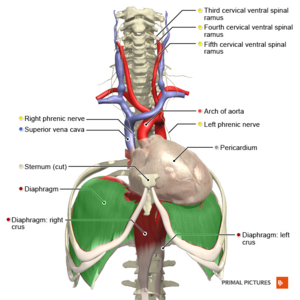
The diaphragm is supplied by the [[Phrenic Nerve]] | The diaphragm is supplied by the right and left [[Phrenic Nerve|phrenic nerves]],<ref name=":0" /> which originate from the ventral rami of C3, C4, C5 and sometimes C6 (see Figure 2). Each phrenic nerve divides into four trunks.<ref name=":3" /> | ||
* '''Motor nerve supply:'''<ref name=":1">Patel PR, Bechmann S. Elevated Hemidiaphragm. 2021 Aug 9. In: StatPearls [Internet]. Treasure Island (FL): StatPearls Publishing; 2021 | * '''Motor nerve supply:'''<ref name=":1">Patel PR, Bechmann S. Elevated Hemidiaphragm. [Updated 2021 Aug 9]. In: StatPearls [Internet]. Treasure Island (FL): StatPearls Publishing; 2021 Jan-. Available from: https://www.ncbi.nlm.nih.gov/books/NBK559255/</ref> | ||
# | # The left hemidiaphragm is supplied by the left phrenic nerve | ||
# | # The right hemidiaphragm is supplied by the right phrenic nerve | ||
* '''Sensory nerve supply:''' | * '''Sensory nerve supply:''' | ||
*# The phrenic nerve innervates the parietal pleura and the peritoneum which covers the | *# The phrenic nerve innervates the parietal pleura and the peritoneum, which covers the central surfaces of the diaphragm.<ref name=":0" /> | ||
*# The phrenic nerve is made up of large-diameter myelinated, small-diameter myelinated, and unmyelinated fibres. The large diameter fibres fire when the diaphragm contracts | *# The bottom six intercostal nerves innervate the periphery of the diaphragm<ref name=":0" /> | ||
*#* Activation of the phrenic nerve modulates the sympathetic motor outflow.<ref name=":0" /> | *# The phrenic nerve is made up of large-diameter myelinated, small-diameter myelinated, and unmyelinated fibres. The large-diameter fibres fire when the diaphragm contracts, while the small-diameter fibers fire throughout respiration.<ref>Nair J, Streeter KA, Turner SM, Sunshine MD, Bolser DC, Fox EJ, Davenport PW, Fuller DD. [https://journals.physiology.org/doi/full/10.1152/jn.00484.2017 Anatomy and physiology of phrenic afferent neurons.] Journal of neurophysiology. 2017 Dec 1;118(6):2975-90.</ref> | ||
*#* Phrenic afferents are also involved in the somatosensation of the diaphragm and they make individuals aware of their breathing while they are awake.<ref name=":0" /> | *#* Activation of the phrenic nerve modulates the [[Sympathetic Nervous System|sympathetic]] motor outflow.<ref name=":0" /> | ||
*#* Phrenic afferents are also involved in the [[somatosensation]] of the diaphragm, and they make individuals aware of their breathing while they are awake.<ref name=":0" /> | |||
== Vascular Supply == | == Vascular Supply == | ||
'''Arterial supply:''' | '''Arterial supply:'''<ref name=":5">Whitley A, Křeček J, Kachlik D. The inferior phrenic arteries: A systematic review and meta-analysis. Annals of Anatomy-Anatomischer Anzeiger. 2021 May 1;235:151679.</ref> | ||
* | * Bilateral phrenic arteries, which are the branches of the thoracic aorta | ||
* Pericardiophrenic, musculophrenic arteries | * Pericardiophrenic, musculophrenic arteries | ||
'''Venous supply:''' | * Tributaries from the internal mammary arteries | ||
* Inferior phrenic | '''Venous supply:'''<ref name=":5" /> | ||
* Inferior phrenic veins (drain into the inferior vena cava) | |||
== Fascial Attachments == | == Fascial Attachments == | ||
=== | === Vertebrae === | ||
* Medial lumbocostal arch<ref name=":0" /> | * Medial lumbocostal arch<ref name=":0" /> | ||
** A tendinous arch in the fascia which covers [[Psoas Major]] | ** A tendinous arch in the [[fascia]] which covers [[Psoas Major|psoas major]] | ||
** Medially | ** Medially: attaches to the side of the L1 vertebral body | ||
** Laterally | ** Laterally: attaches to the front of the L1 transverse process | ||
* Lateral lumbocostal arch<ref name=":0" /> | * Lateral lumbocostal arch<ref name=":0" /> | ||
** A tendinous arch in the fascia which covers the upper | ** A tendinous arch in the fascia which covers the upper part of [[Quadratus Lumborum|quadratus lumborum]] | ||
** Medially | ** Medially: attaches to the front of the L1 transverse process | ||
** Laterally | ** Laterally: attaches to the lower border of rib 12 | ||
=== | === Muscles === | ||
* Quadratus Lumborum (QL) | * [[Quadratus Lumborum|Quadratus lumborum]] (QL) originates at the iliac crest and iliolumbar ligament and it inserts into the inferior border of the 12th rib, and the transverse processes of L1-L4 vertebrae. Part of the diaphragm also attaches to the superior portion of the 12th rib. The fascia is continuous between these attachments.<ref name=":2">Pandya R. [https://members.physio-pedia.com/diaphragm-anatomy-and-differential-diagnosis-course/ Diaphragm Anatomy and Differential Diagnosis Course]. Plus , 2021.</ref> | ||
* | * [[Psoas Major|Pssoas major]] is lateral to the lumbar vertebrae and medial to quadratus lumborum.<ref>KenHub. Psoas major muscle. Available from: https://www.kenhub.com/en/library/anatomy/psoas-major-muscle (last accessed 23 October 2023).</ref> It originates at the vertebral bodies of T12-L4, intervertebral discs between T12-L4 and transverse processes of L1-L5 vertebrae. It inserts into the lesser trochanter of femur.<ref>Physiopedia. [[Functional Anatomy of the Lumbar Spine and Abdominal Wall]]. </ref> | ||
<blockquote>When thinking about the diaphragm, we need to remember that the diaphragm and all these correlated muscles "they form a continuous chain of movements [...] the activity in one muscle group contributes to the efficiency in the other. They should all work together like a smooth machine, a well-oiled machine, whereas discrepancy, deficiency in one of these ends up compromising posture, movement, gait, cardiovascular issues, as well as digestive and oesophageal consequences." -- Rina Pandya<ref name=":2" /></blockquote> | |||
== Aetiology of an Elevated Diaphragm == | == Aetiology of an Elevated Diaphragm == | ||
An elevated hemidiaphragm may | An elevated hemidiaphragm may have both direct and indirect causes. These causes can be grouped into three categories based on the location of the cause, including:<ref name=":1" /><ref name=":2" /> | ||
# Above the diaphragm: | |||
#* decreased [[Lung Volumes|lung volume]] | |||
# Above the diaphragm | #* [[atelectasis]]/collapse | ||
#* | #* prior lobectomy or pneumonectomy | ||
#* [[ | #* pulmonary hypoplasia | ||
#* | |||
#* | |||
# At the level of the diaphragm | # At the level of the diaphragm | ||
#* | #* phrenic nerve palsy | ||
#* | #* diaphragmatic eventration:<ref>Columbia Surgery Diaphragm Eventration Available:https://columbiasurgery.org/conditions-and-treatments/diaphragm-eventration (accessed 9.5.2022)</ref> | ||
#* | #** this is an abnormal placement of the diaphragm - i.e. the diaphragm is located too high in the body | ||
#** this abnormal placement can be due to dysfunction in the nerves that supply the diaphragm or dysfunction of the diaphragm itself | |||
#** in severe cases, diaphragmatic eventration can compress the lungs and affect respiration | |||
#* contralateral [[stroke]]: usually middle cerebral artery (MCA) distribution | |||
# Below the diaphragm | # Below the diaphragm | ||
#* | #* abdominal tumour, e.g. liver metastases or primary malignancy | ||
#* | #* subphrenic abscess | ||
#* | #* distended stomach or colon, including Chilaiditi sign/syndrome | ||
#** Chilaiditi sign is "a radiological finding that occurs when a segment of a large bowel loop or small intestine is interposed between the liver and a diaphragm."<ref name=":6">Kumar A, Mehta D. Chilaiditi Syndrome. [Updated 2023 Apr 10]. In: StatPearls [Internet]. Treasure Island (FL): StatPearls Publishing; 2023 Jan-. Available from: https://www.ncbi.nlm.nih.gov/books/NBK554565/</ref> | |||
#** Chilaiditi syndrome occurs when these changes cause gastrointestinal symptoms<ref name=":6" /> | |||
=== Differential Diagnosis for Elevated Diaphragm === | === Differential Diagnosis for Elevated Diaphragm === | ||
| Line 83: | Line 94: | ||
== Aetiology for Paralysis of the Diaphragm == | == Aetiology for Paralysis of the Diaphragm == | ||
Diaphragmatic paralysis occurs when the nerve supply is interrupted. This interruption might occur in the phrenic nerve | Diaphragmatic paralysis occurs when the nerve supply is interrupted. This interruption might occur in the phrenic nerve, at the cervical spinal cord, or in the brainstem. It is most commonly caused by a phrenic nerve lesion:<ref name=":3">Kokatnur L, Vashisht R, Rudrappa M. Diaphragm Disorders. [Updated 2021 Aug 9]. In: StatPearls [Internet]. Treasure Island (FL): StatPearls Publishing; 2021 Jan-. Available from: https://www.ncbi.nlm.nih.gov/books/NBK470172/</ref><ref name=":4">TeachMe Anatomy. The diaphragm. Available from: https://teachmeanatomy.info/thorax/muscles/diaphragm/ (accessed 30 November 2021).</ref><ref>Rizeq YK, Many BT, Vacek JC, Reiter AJ, Raval MV, Abdullah F, Goldstein SD. Diaphragmatic paralysis after phrenic nerve injury in newborns. Journal of pediatric surgery. 2020 Feb 1;55(2):240-4.</ref> | ||
*'''Mechanical trauma:''' such as nerve damage / ligation during surgery | *'''Mechanical trauma:''' such as nerve damage / ligation during surgery | ||
*'''Compression:''' due to a chest cavity tumour | *'''Compression:''' due to a chest cavity tumour | ||
*'''Myopathies:''' including myasthenia gravis | *'''Myopathies:''' including [[Myasthenia Gravis|myasthenia gravis]] (an [[Autoimmune Disorders|autoimmune disorder]] that affects the neuromuscular junction<ref>Physiopeda. [[Myasthenia Gravis]].</ref>) | ||
*'''Neuropathic:''' including conditions such as diabetic neuropathy, inclusion body myositis, [[dermatomyositis]], [[ | *'''Neuropathic:''' including conditions such as [[Diabetic Neuropathy|diabetic neuropathy]], inclusion body [[Myositis Ossificans|myositis]], [[dermatomyositis]], [[Multiple Sclerosis (MS)|multiple sclerosis]], anterior horn cell disease, chronic demyelinating disease, and neuralgic [[Myopathies|myopathy]] | ||
*''' Inflammation:''' a number of systemic diseases can cause inflammation in the phrenic nerve / diaphragm, which results in diaphragmatic palsy. Examples include: | *''' Inflammation:''' a number of systemic diseases can cause inflammation in the phrenic nerve / diaphragm, which results in diaphragmatic palsy. Examples include: | ||
** Viral infections (e.g. [[Human Immunodeficiency Virus (HIV) | **[[Viral Infections|viral]] infections (e.g. [[Human Immunodeficiency Virus (HIV)|HIV]], West Nile virus and [[poliomyelitis]] virus) | ||
** Bacterial infections (e.g. [[Lyme Disease|Lyme disease]]) | ** [[Bacterial Infections|bacterial]] infections (e.g. [[Lyme Disease|Lyme disease]]) | ||
** Non-infectious causes (e.g. [[sarcoidosis]] and amyloidosis) | ** [[Non-Communicable Diseases|non-infectious causes]] (e.g. [[sarcoidosis]] and [[amyloidosis]]) | ||
*''' Idiopathic:''' around 20 percent of cases have no obvious cause | *''' Idiopathic:''' around 20 percent of cases have no obvious cause | ||
=== Differential Diagnosis for Paralysis of the Diaphragm === | === Differential Diagnosis for Paralysis of the Diaphragm === | ||
* Alveolar hypoventilation | * [[Alveoli|Alveolar]] hypoventilation | ||
* Anterior horn cell or neuromuscular junction disease | * Anterior horn cell or neuromuscular junction disease | ||
* Cerebral haemorrhage | * Cerebral haemorrhage | ||
| Line 107: | Line 118: | ||
== Symptoms of Diaphragmatic Weakness == | == Symptoms of Diaphragmatic Weakness == | ||
# '''Unilateral weakness:''' Often asymptomatic and detected incidentally. Patients show limitations in exercise capacity | # '''Unilateral weakness:''' Often asymptomatic and detected incidentally. Patients show limitations in exercise capacity and lower oxygen saturation levels:<ref name=":3" /> | ||
#* | #* One-third of patients complain of exertional breathlessness | ||
#* | #* Individuals who have "coexisting debilitating cardiopulmonary conditions"<ref name=":3" /> might have dyspnoea at rest | ||
# '''Bilateral weakness:''' dyspnoea | # '''Bilateral weakness:''' patients report varying levels of dyspnoea (i.e. from breathlessness with mild exertion to dyspnoea at rest.<ref name=":3" /> When diaphragm function is further compromised, patients tend to have orthopnoea (i.e. breathlessness when lying supine.<ref name=":2" /><ref name=":3" /> | ||
Progressive hypoventilation can lead to hypercapnia and right heart failure. Hypoxaemia and hypercapnia will be worse when a patient is sleeping.<ref name=":3" /> | |||
== Paradoxical Breathing == | == Paradoxical Breathing == | ||
[[File:Paradoxical Breathing Image - recreated with labels.jpg|thumb|400x400px|Paradoxical | [[File:Paradoxical Breathing Image - recreated with labels.jpg|thumb|400x400px|Figure 3. Paradoxical breathing.]] | ||
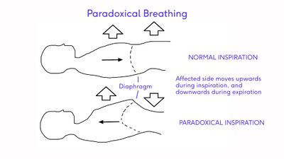
Paralysis of the diaphragm results in a "paradoxical movement". The | Paralysis of the diaphragm results in a "paradoxical movement" (see Figure 3). Typically, when we inhale, the diaphragm lowers and flattens, which causes bulging / blowing / elevation of the stomach. During expiration, the diaphragm relaxes, so it can return to its original position (i.e. dome-like shape), and there is a passive drop of the belly.<ref name=":2" /> | ||
In paradoxical breathing, this process happens in the reverse order.<ref name=":2" /> The diaphragm moves up during inspiration and down during expiration.<ref name=":4" /> | |||
* Unilateral diaphragmatic paralysis is frequently asymptomatic and is often found incidentally on x-ray.<ref name=":4" /> | * Unilateral diaphragmatic paralysis is frequently asymptomatic and is often found incidentally on x-ray.<ref name=":4" /> | ||
* Bilateral paralysis can result in poor exercise tolerance, orthopnoea and fatigue. There will also be a restrictive deficit on lung function tests.<ref name=":4" /> | * Bilateral paralysis can result in poor exercise tolerance, orthopnoea and fatigue. There will also be a restrictive deficit on lung function tests.<ref name=":4" /> | ||
{{#ev:youtube|8TnrNrrEjuE}} | The following optional video provides a demonstration of paradoxical breathing.{{#ev:youtube|8TnrNrrEjuE}} | ||
== References == | == References == | ||
[[Category:Course Pages]] | [[Category:Course Pages]] | ||
[[Category: | [[Category:Plus Content]] | ||
[[Category:Rehabilitation]] | [[Category:Rehabilitation]] | ||
<references /> | <references /> | ||
[[Category:Respiratory]] | [[Category:Respiratory]] | ||
Latest revision as of 11:10, 23 October 2023
Top Contributors - Carin Hunter, Jess Bell, Kim Jackson, Wanda van Niekerk, Lucinda hampton, Merinda Rodseth, Tarina van der Stockt and Ewa Jaraczewska
Anatomy of the Diaphragm[edit | edit source]
The diaphragm is a major muscle of respiration.[1]
- It is a dome-shaped, "fibromuscular sheet" that separates the thorax from the abdomen.[2]
- It forms the floor of the thorax and the roof of the abdomen.[2][3]
- The left side is lower than the right - this is because the liver is situated on the right side.[2]
- The left side may also be located partially inferiorly due to the "push" by the heart.[2][4][5]
- The peripheral portion of the diaphragm is muscular and is composed of three distinct muscle groups:
- The sternal group originates from the xiphoid process as two fleshy slips.[2]
- The costal group originates from the inner surfaces of the cartilages and adjacent parts of the six lower ribs. It "interdigitates with transversus abdominis".[2]
- The lumbar group originates from the two crura and the arcuate ligaments, which are in turn inserted into L1 and L2, and sometimes L3 as well.[6]
- The central portion of the diaphragm is made up of very strong aponeurotic tendinous ligaments - these ligaments do not have any bony attachments.[7]
Major Openings in the Diaphragm[edit | edit source]
The diaphragm has three major openings (see Figure 1):
- Caval hiatus: situated at the level of the T8 vertebra in the central tendon. The inferior vena cava and some right phrenic nerve branches pass through this hiatus.[2]
- Oesophageal hiatus: situated at the level of the T10 vertebra. The oesophagus, the right and left vagus trunks, the oesophageal branches of the left gastric vessels, and the lymph vessels pass through this hiatus.[2]
- Aortic hiatus: located anterior to the T12 vertebral body between the crura. The aorta, thoracic duct, and azygos vein pass through this hiatus.[2]
Nerve Supply[edit | edit source]
The diaphragm is supplied by the right and left phrenic nerves,[2] which originate from the ventral rami of C3, C4, C5 and sometimes C6 (see Figure 2). Each phrenic nerve divides into four trunks.[7]
- Motor nerve supply:[8]
- The left hemidiaphragm is supplied by the left phrenic nerve
- The right hemidiaphragm is supplied by the right phrenic nerve
- Sensory nerve supply:
- The phrenic nerve innervates the parietal pleura and the peritoneum, which covers the central surfaces of the diaphragm.[2]
- The bottom six intercostal nerves innervate the periphery of the diaphragm[2]
- The phrenic nerve is made up of large-diameter myelinated, small-diameter myelinated, and unmyelinated fibres. The large-diameter fibres fire when the diaphragm contracts, while the small-diameter fibers fire throughout respiration.[9]
- Activation of the phrenic nerve modulates the sympathetic motor outflow.[2]
- Phrenic afferents are also involved in the somatosensation of the diaphragm, and they make individuals aware of their breathing while they are awake.[2]
Vascular Supply[edit | edit source]
Arterial supply:[10]
- Bilateral phrenic arteries, which are the branches of the thoracic aorta
- Pericardiophrenic, musculophrenic arteries
- Tributaries from the internal mammary arteries
Venous supply:[10]
- Inferior phrenic veins (drain into the inferior vena cava)
Fascial Attachments[edit | edit source]
Vertebrae[edit | edit source]
- Medial lumbocostal arch[2]
- A tendinous arch in the fascia which covers psoas major
- Medially: attaches to the side of the L1 vertebral body
- Laterally: attaches to the front of the L1 transverse process
- Lateral lumbocostal arch[2]
- A tendinous arch in the fascia which covers the upper part of quadratus lumborum
- Medially: attaches to the front of the L1 transverse process
- Laterally: attaches to the lower border of rib 12
Muscles[edit | edit source]
- Quadratus lumborum (QL) originates at the iliac crest and iliolumbar ligament and it inserts into the inferior border of the 12th rib, and the transverse processes of L1-L4 vertebrae. Part of the diaphragm also attaches to the superior portion of the 12th rib. The fascia is continuous between these attachments.[6]
- Pssoas major is lateral to the lumbar vertebrae and medial to quadratus lumborum.[11] It originates at the vertebral bodies of T12-L4, intervertebral discs between T12-L4 and transverse processes of L1-L5 vertebrae. It inserts into the lesser trochanter of femur.[12]
When thinking about the diaphragm, we need to remember that the diaphragm and all these correlated muscles "they form a continuous chain of movements [...] the activity in one muscle group contributes to the efficiency in the other. They should all work together like a smooth machine, a well-oiled machine, whereas discrepancy, deficiency in one of these ends up compromising posture, movement, gait, cardiovascular issues, as well as digestive and oesophageal consequences." -- Rina Pandya[6]
Aetiology of an Elevated Diaphragm[edit | edit source]
An elevated hemidiaphragm may have both direct and indirect causes. These causes can be grouped into three categories based on the location of the cause, including:[8][6]
- Above the diaphragm:
- decreased lung volume
- atelectasis/collapse
- prior lobectomy or pneumonectomy
- pulmonary hypoplasia
- At the level of the diaphragm
- phrenic nerve palsy
- diaphragmatic eventration:[13]
- this is an abnormal placement of the diaphragm - i.e. the diaphragm is located too high in the body
- this abnormal placement can be due to dysfunction in the nerves that supply the diaphragm or dysfunction of the diaphragm itself
- in severe cases, diaphragmatic eventration can compress the lungs and affect respiration
- contralateral stroke: usually middle cerebral artery (MCA) distribution
- Below the diaphragm
- abdominal tumour, e.g. liver metastases or primary malignancy
- subphrenic abscess
- distended stomach or colon, including Chilaiditi sign/syndrome
Differential Diagnosis for Elevated Diaphragm[edit | edit source]
Other situations which may mimic an elevated hemidiaphragm include:[8]
- Subpulmonic effusion
- Diaphragmatic hernia
- Diaphragmatic rupture
- Tumour of the pleura or diaphragm
Aetiology for Paralysis of the Diaphragm[edit | edit source]
Diaphragmatic paralysis occurs when the nerve supply is interrupted. This interruption might occur in the phrenic nerve, at the cervical spinal cord, or in the brainstem. It is most commonly caused by a phrenic nerve lesion:[7][15][16]
- Mechanical trauma: such as nerve damage / ligation during surgery
- Compression: due to a chest cavity tumour
- Myopathies: including myasthenia gravis (an autoimmune disorder that affects the neuromuscular junction[17])
- Neuropathic: including conditions such as diabetic neuropathy, inclusion body myositis, dermatomyositis, multiple sclerosis, anterior horn cell disease, chronic demyelinating disease, and neuralgic myopathy
- Inflammation: a number of systemic diseases can cause inflammation in the phrenic nerve / diaphragm, which results in diaphragmatic palsy. Examples include:
- viral infections (e.g. HIV, West Nile virus and poliomyelitis virus)
- bacterial infections (e.g. Lyme disease)
- non-infectious causes (e.g. sarcoidosis and amyloidosis)
- Idiopathic: around 20 percent of cases have no obvious cause
Differential Diagnosis for Paralysis of the Diaphragm[edit | edit source]
- Alveolar hypoventilation
- Anterior horn cell or neuromuscular junction disease
- Cerebral haemorrhage
- Cervical fracture
- Decreased pulmonary compliance
- Guillain-Barre syndrome
- Myasthenia gravis
- Peripheral neuropathies
- Pleural adhesions[7]
Symptoms of Diaphragmatic Weakness[edit | edit source]
- Unilateral weakness: Often asymptomatic and detected incidentally. Patients show limitations in exercise capacity and lower oxygen saturation levels:[7]
- One-third of patients complain of exertional breathlessness
- Individuals who have "coexisting debilitating cardiopulmonary conditions"[7] might have dyspnoea at rest
- Bilateral weakness: patients report varying levels of dyspnoea (i.e. from breathlessness with mild exertion to dyspnoea at rest.[7] When diaphragm function is further compromised, patients tend to have orthopnoea (i.e. breathlessness when lying supine.[6][7]
Progressive hypoventilation can lead to hypercapnia and right heart failure. Hypoxaemia and hypercapnia will be worse when a patient is sleeping.[7]
Paradoxical Breathing[edit | edit source]
Paralysis of the diaphragm results in a "paradoxical movement" (see Figure 3). Typically, when we inhale, the diaphragm lowers and flattens, which causes bulging / blowing / elevation of the stomach. During expiration, the diaphragm relaxes, so it can return to its original position (i.e. dome-like shape), and there is a passive drop of the belly.[6]
In paradoxical breathing, this process happens in the reverse order.[6] The diaphragm moves up during inspiration and down during expiration.[15]
- Unilateral diaphragmatic paralysis is frequently asymptomatic and is often found incidentally on x-ray.[15]
- Bilateral paralysis can result in poor exercise tolerance, orthopnoea and fatigue. There will also be a restrictive deficit on lung function tests.[15]
The following optional video provides a demonstration of paradoxical breathing.
References[edit | edit source]
- ↑ Fayssoil A, Behin A, Ogna A, Mompoint D, Amthor H, Clair B, Laforet P, Mansart A, Prigent H, Orlikowski D, Stojkovic T, Vinit S, Carlier R, Eymard B, Lofaso F, Annane D. Diaphragm: Pathophysiology and Ultrasound Imaging in Neuromuscular Disorders. J Neuromuscul Dis. 2018;5(1):1-10
- ↑ 2.00 2.01 2.02 2.03 2.04 2.05 2.06 2.07 2.08 2.09 2.10 2.11 2.12 2.13 2.14 2.15 Bains KN, Kashyap S, Lappin SL. Anatomy, Thorax, Diaphragm. StatPearls [Internet]. 2020 Aug 15.
- ↑ Kurian J. Chest Wall and Diaphragm. InPediatric Body MRI 2020 (pp. 159-192). Springer, Cham.
- ↑ Bordoni B, Purgol S, Bizzarri A, Modica M, Morabito B. The influence of breathing on the central nervous system. Cureus. 2018 Jun;10(6).
- ↑ Oliver KA, Ashurst JV. Anatomy, Thorax, Phrenic Nerves. InStatPearls [Internet] 2020 Jul 27. StatPearls Publishing.
- ↑ 6.0 6.1 6.2 6.3 6.4 6.5 6.6 Pandya R. Diaphragm Anatomy and Differential Diagnosis Course. Plus , 2021.
- ↑ 7.0 7.1 7.2 7.3 7.4 7.5 7.6 7.7 7.8 Kokatnur L, Vashisht R, Rudrappa M. Diaphragm Disorders. [Updated 2021 Aug 9]. In: StatPearls [Internet]. Treasure Island (FL): StatPearls Publishing; 2021 Jan-. Available from: https://www.ncbi.nlm.nih.gov/books/NBK470172/
- ↑ 8.0 8.1 8.2 Patel PR, Bechmann S. Elevated Hemidiaphragm. [Updated 2021 Aug 9]. In: StatPearls [Internet]. Treasure Island (FL): StatPearls Publishing; 2021 Jan-. Available from: https://www.ncbi.nlm.nih.gov/books/NBK559255/
- ↑ Nair J, Streeter KA, Turner SM, Sunshine MD, Bolser DC, Fox EJ, Davenport PW, Fuller DD. Anatomy and physiology of phrenic afferent neurons. Journal of neurophysiology. 2017 Dec 1;118(6):2975-90.
- ↑ 10.0 10.1 Whitley A, Křeček J, Kachlik D. The inferior phrenic arteries: A systematic review and meta-analysis. Annals of Anatomy-Anatomischer Anzeiger. 2021 May 1;235:151679.
- ↑ KenHub. Psoas major muscle. Available from: https://www.kenhub.com/en/library/anatomy/psoas-major-muscle (last accessed 23 October 2023).
- ↑ Physiopedia. Functional Anatomy of the Lumbar Spine and Abdominal Wall.
- ↑ Columbia Surgery Diaphragm Eventration Available:https://columbiasurgery.org/conditions-and-treatments/diaphragm-eventration (accessed 9.5.2022)
- ↑ 14.0 14.1 Kumar A, Mehta D. Chilaiditi Syndrome. [Updated 2023 Apr 10]. In: StatPearls [Internet]. Treasure Island (FL): StatPearls Publishing; 2023 Jan-. Available from: https://www.ncbi.nlm.nih.gov/books/NBK554565/
- ↑ 15.0 15.1 15.2 15.3 TeachMe Anatomy. The diaphragm. Available from: https://teachmeanatomy.info/thorax/muscles/diaphragm/ (accessed 30 November 2021).
- ↑ Rizeq YK, Many BT, Vacek JC, Reiter AJ, Raval MV, Abdullah F, Goldstein SD. Diaphragmatic paralysis after phrenic nerve injury in newborns. Journal of pediatric surgery. 2020 Feb 1;55(2):240-4.
- ↑ Physiopeda. Myasthenia Gravis.


