Cruciate Ligament of Atlas: Difference between revisions
Sally Fahmy (talk | contribs) (Created page with "== Description == The cruciform ligament of atlas (cruciate may substitute for cruciform) is a cruciate ligament in the neck forming part of the atlanto-axial joint. The ligam...") |
Sally Fahmy (talk | contribs) No edit summary |
||
| Line 1: | Line 1: | ||
== Description == | == Description == | ||
The cruciform ligament of atlas (cruciate may substitute for cruciform) is a cruciate ligament in the neck forming part of the atlanto-axial joint. The ligament is named as such because it is in the shape of a cross. | The cruciform ligament of atlas (cruciate may substitute for cruciform) is a cruciate ligament in the neck forming part of the atlanto-axial joint. The ligament is named as such because it is in the shape of a cross. | ||
It consists of two bands: | It consists of two bands: | ||
* Longitudinal band. | |||
* Transverse band. | |||
== Anatomy == | == Anatomy == | ||
'''Atlas osteology''' | '''Atlas osteology''' | ||
* Atlas (C1) is a ring containing two articular lateral masses | |||
* It lacks a vertebral body or a spinous process | |||
* Embryology: ''Forms from 3 ossification centers'' | |||
'' | * Anatomic variation: ''Incomplete formation of the posterior arch is a relatively common anatomic variant and does not represent a traumatic injury'' | ||
'' | |||
'' | |||
== Function == | == Function == | ||
It is an important ligament that holds the posterior dens of C2 in articulation at the atlanto-axial joint. It lies behind a large synovial bursa (surrounded by loose fibrous capsule) | It is an important ligament that holds the posterior dens of C2 in articulation at the atlanto-axial joint. It lies behind a large synovial bursa (surrounded by loose fibrous capsule) and consists of two bands: | ||
consists of two bands: | * Longitudinal band: joins the body of the C2 (axis) to the foramen magnum | ||
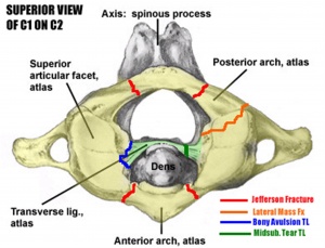
* Transverse band: attaches to the inner margin of the C1 (atlas) lateral masses on both sides. | |||
== Clinial relevance == | |||
[[File:Atlasview.jpg|thumb]] | |||
== Atlas Fracture == | === Atlas Fracture === | ||
Epidemiology | |||
==== Epidemiology ==== | |||
* Makes upto ~7% of cervical spine fractures | |||
* Risk of neurologic injury is low | |||
Pathophysiology | * Commonly missed due to inadequate imaging of occipitocervical junction | ||
includes hyperextension, lateral compression, and axial compression | ==== Pathophysiology ==== | ||
Associated conditions | * Mechanism: includes hyperextension, lateral compression, and axial compression | ||
spine fracture | |||
50% have an associated spine injury | * Associated conditions: | ||
40% associated with axis fx | # spine fracture | ||
Prognosis | # 50% have an associated spine injury | ||
# 40% associated with axis fx | |||
==== Prognosis: ==== | |||
stability dependent on degree of injury and healing potential of transverse ligament. | stability dependent on degree of injury and healing potential of transverse ligament. | ||
== Assessment: == | |||
{| class="wikitable" | |||
| colspan="2" |'''Landells Atlas Fractures Classification''' | |||
|- | |||
|Type I | |||
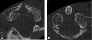
|Isolated anterior or posterior arch fracture. A "plough fracture is an isolated anterior arch fracture caused by a force driving the odontoid through the anterior arch. Stable. Treat with hard collar. | |||
|- | |||
|Type II | |||
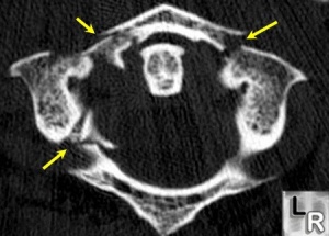
|Jefferson burst fracture with bilateral fractures of anterior and posterior arch resulting from axial load. Stability determined by integrity of transverse ligament. If intact, hard collar. If disrupted, halo vest (for bony avulsion) or C1-2 fusion (for intrasubstance tear).. | |||
|- | |||
|Type III | |||
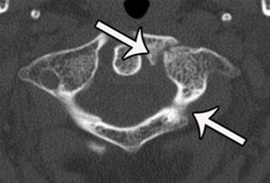
|Unilateral lateral mass fx. Stability determined by integrity of transverse ligament. If stable, treat with hard collar. If unstable, halo vest. | |||
|} | |||
[[File:Atlastype1.jpg|thumb]] | |||
[[File:Typeatlas.jpg|thumb]] | |||
[[File:Type3atlas.jpg|thumb]] | |||
== Treatment: == | |||
* Nonoperative | |||
** '''hard collar vs. halo immobilization for 6-12 weeks''' | |||
*** indications | |||
**** stable Type I fx (intact transverse ligament) | |||
**** stable Jefferson fx (Type II) (intact transverse ligament) | |||
**** stable Type III (intact transverse ligament) | |||
*** technique | |||
**** controversy exists around optimal form of immobilization | |||
* Operative | |||
** posterior C1-C2 fusion vs. occipitocervical fusion | |||
*** indications | |||
**** unstable Type II (controversial) | |||
**** unstable Type III (controversial) | |||
*** technique | |||
**** may consider preoperative traction to reduce displaced lateral masses | |||
== References == | == References == | ||
This article incorporates text in the public domain from the 20th edition of Gray's Anatomy (1918) | This article incorporates text in the public domain from the 20th edition of Gray's Anatomy (1918) | ||
| Line 49: | Line 83: | ||
http://www.orthobullets.com/spine/2015/atlas-fracture-and-transverse-ligament-injuries | http://www.orthobullets.com/spine/2015/atlas-fracture-and-transverse-ligament-injuries | ||
<div class="editorbox"><div class="editorbox">'''Original Editor '''- [[User:Sally Fahmy|Sally Fahmy]]'''Top Contributors''' - {{Special:Contributors/{{FULLPAGENAME}}}}</div></div> | |||
<div class="editorbox"></div> | |||
Revision as of 22:31, 24 August 2017
Description[edit | edit source]
The cruciform ligament of atlas (cruciate may substitute for cruciform) is a cruciate ligament in the neck forming part of the atlanto-axial joint. The ligament is named as such because it is in the shape of a cross.
It consists of two bands:
- Longitudinal band.
- Transverse band.
Anatomy[edit | edit source]
Atlas osteology
- Atlas (C1) is a ring containing two articular lateral masses
- It lacks a vertebral body or a spinous process
- Embryology: Forms from 3 ossification centers
- Anatomic variation: Incomplete formation of the posterior arch is a relatively common anatomic variant and does not represent a traumatic injury
Function[edit | edit source]
It is an important ligament that holds the posterior dens of C2 in articulation at the atlanto-axial joint. It lies behind a large synovial bursa (surrounded by loose fibrous capsule) and consists of two bands:
- Longitudinal band: joins the body of the C2 (axis) to the foramen magnum
- Transverse band: attaches to the inner margin of the C1 (atlas) lateral masses on both sides.
Clinial relevance[edit | edit source]
Atlas Fracture[edit | edit source]
Epidemiology[edit | edit source]
- Makes upto ~7% of cervical spine fractures
- Risk of neurologic injury is low
- Commonly missed due to inadequate imaging of occipitocervical junction
Pathophysiology[edit | edit source]
- Mechanism: includes hyperextension, lateral compression, and axial compression
- Associated conditions:
- spine fracture
- 50% have an associated spine injury
- 40% associated with axis fx
Prognosis:[edit | edit source]
stability dependent on degree of injury and healing potential of transverse ligament.
Assessment:[edit | edit source]
| Landells Atlas Fractures Classification | |
| Type I | Isolated anterior or posterior arch fracture. A "plough fracture is an isolated anterior arch fracture caused by a force driving the odontoid through the anterior arch. Stable. Treat with hard collar. |
| Type II | Jefferson burst fracture with bilateral fractures of anterior and posterior arch resulting from axial load. Stability determined by integrity of transverse ligament. If intact, hard collar. If disrupted, halo vest (for bony avulsion) or C1-2 fusion (for intrasubstance tear).. |
| Type III | Unilateral lateral mass fx. Stability determined by integrity of transverse ligament. If stable, treat with hard collar. If unstable, halo vest. |
Treatment:[edit | edit source]
- Nonoperative
- hard collar vs. halo immobilization for 6-12 weeks
- indications
- stable Type I fx (intact transverse ligament)
- stable Jefferson fx (Type II) (intact transverse ligament)
- stable Type III (intact transverse ligament)
- technique
- controversy exists around optimal form of immobilization
- indications
- hard collar vs. halo immobilization for 6-12 weeks
- Operative
- posterior C1-C2 fusion vs. occipitocervical fusion
- indications
- unstable Type II (controversial)
- unstable Type III (controversial)
- technique
- may consider preoperative traction to reduce displaced lateral masses
- indications
- posterior C1-C2 fusion vs. occipitocervical fusion
References[edit | edit source]
This article incorporates text in the public domain from the 20th edition of Gray's Anatomy (1918)
Anatomy of Spinal Vertebrae Tutorial Archived 2007-10-10 at the Wayback Machine.
Federative Committee on Anatomical Terminology (1998). Terminologia anatomica: international anatomical terminology. Thieme. pp. 27–. ISBN 978-3-13-114361-7. Retrieved 17 June 2010.
http://www.orthobullets.com/spine/2015/atlas-fracture-and-transverse-ligament-injuries


