Costovertebral Joints: Difference between revisions

(category)
No edit summary
 
(9 intermediate revisions by 2 users not shown)
Line 6: Line 6:
</div>  
</div>  


===Introduction===
==Introduction==
[[File:Gray312.png|thumb|Costovertebral articulations. Anterior view.]]
The costovertebral joints are the articulations that connect the heads of the ribs with the bodies of the thoracic vertebrae. Joining of ribs to the vertebrae occurs at two places, the head and the tubercle of the rib. Two convex facets from the head attach to two adjacent vertebrae. This forms a synovial planar (gliding) joint, which is strengthened by the ligament of the head and the intercapital ligament. Articulation of the tubercle is to the transverse process of the adjacent vertebrae. This articulation is reinforced by the dorsal costotransverse ligament<ref>Gray.H,Gray's anatomy 20th edition,page 299</ref>


===Anatomy<ref>thoracic spine. Teach me anatomy.http://teachmeanatomy.info/thorax/bones/thoracic-spine/ last access 29/1/2018</ref> ===
The costovertebral joints describe two groups of synovial plane joints which connect the proximal end of the [[ribs]] with their corresponding [[Thoracic Vertebrae|thoracic vertebrae]], enclosing the thoracic cage from the posterior side.[[File:Gray312.png|Costovertebral articulations. Anterior view.|right|frameless|369x369px]]
The joints of the thoracic spine can be divided into two groups – those that are present throughout the vertebral column, and those unique to the thoracic spine.
Joining of ribs to the vertebrae occurs at two places
# Head - Two convex facets from the head attach to two adjacent vertebrae. This forms a [[Joint Classification|synovial planar (gliding) joint]], which is strengthened by the ligament of the head and the intercapital ligament.
# Tubercle of the rib - Articulation of the tubercle is to the transverse process of the adjacent vertebrae. This articulation is reinforced by the dorsal costotransverse ligament<ref>Gray.H,Gray's anatomy 20th edition,page 299</ref>


'''Present throughout Vertebral Column'''
==Anatomy ==


There are two types of joints present throughout the vertebral column:
=== Costovertebral Joint ===
* '''Between vertebral bodies''' – adjacent vertebral bodies are joined by intervertebral discs, made of fibrocartilage. This is a type of cartilaginous joint, known as a symphysis.
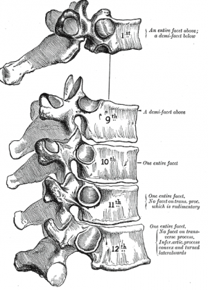
Costovertebral joint consists of the head of the rib (the head of a typical rib has two facets - each facet with a separate synovial joint separated by a ridge.  The head of each rib articulates with:
* The lower rib facet articulates with the upper costal facet of its own vertebra
* The upper facet articulates with the lower facet of the vertebral body above. 
* The first rib articulates with the T1 vertebra only and the lowest three ribs articulate only with their own vertebral body.
There are two types of [[Ligament|ligaments]]:
# intra-articular ligament - attaches the intervertebral disc to the ridge in between the two facets of the head of the rib.
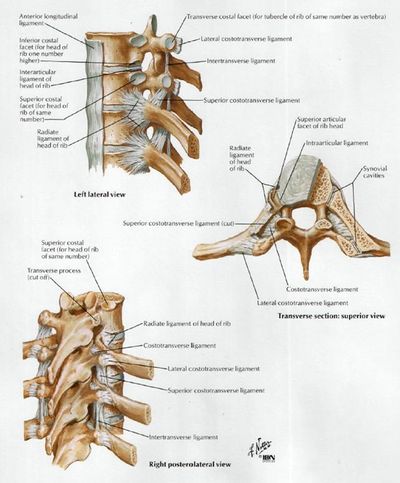
# radiate ligament - formed by three bands which connect the rib head to the vertebral bodies. A superior band runs to the vertebral body above and an inferior band runs to the vertebral body below. A central band runs deep to the anterior longitudinal ligament and blends into the intervertebral disc to join the ligament on the opposite side. In the first rib and the last three ribs, only two bands exist as they only articulate with their own vertebra<ref name=":1">Radiopedia [https://radiopaedia.org/articles/costovertebral-joint Costrovert. Joint] Available from:https://radiopaedia.org/articles/costovertebral-joint (last accessed 10.5.2020)</ref>.
[[File:Afbeelding 3.png|right|frameless]]


* '''Between vertebral arches''' – formed by the articulation of superior and inferior articular processes from adjacent vertebrae. It is a synovial type joint.
=== Costotransverse Joint ===
There are two facets of a tubercle of a rib, the medial and lateral.
# The hyaline cartilage-lined medial facet forms a plane synovial joint with the tip of the transverse process which is reinforced by a capsule.
# The lateral facet is attached to the transverse process through three ligaments:
* Lateral costotransverse ligament - attaches the lateral facet to the tip of the transverse process of the vertebral body.
* Costotransverse ligament - attaches the back of the neck of the rib to the front of the transverse process
* Superior costotransverse ligaments - attaches the neck of the rib to the underside of the transverse process of the vertebra above
The lower two ribs are only attached by ligaments and do not form synovial joints with the transverse process<ref name=":1" />.


Each '''costovertebral joint''' consists of the head of the rib articulating with:
===Movements===
* Superior costal facet of the corresponding vertebra
The movements on these joints are called ‘pump-handle’ or ‘bucket-handle’ movements, and are limited to a small degree of gliding and rotation of the rib head.
* Inferior costal facet of the superior vertebra
* The function of these movements is to enable lifting of the ribs upwards and outwards during breathing.
* Intervertebral disc separating the two vertebrae
* The end result is the increase of the lateral diameter of the thorax and subsequent expansion of the lung parenchyma as the air is being inhaled<ref name=":0">Kenhub [https://www.kenhub.com/en/library/anatomy/costovertebral-joints CV joints] Available from:https://www.kenhub.com/en/library/anatomy/costovertebral-joints (last accessed 10.5.2020)</ref>.
Within this joint, the intra-articular ligament of head of rib attaches the rib head to the intervertebral disc. Only slight gliding movements can occur at these joints, due to the close articulation of their components.
The costovertebral complex is an essential component of the biomechanics of chest wall movement. The costovertebral ligaments make the actions of the costovertebral joints and intervertebral movement possible.  The ligaments function to:
* Affix, stabilize and allow some motion of the ribs on the thoracic vertebra at the costovertebral joint. Their presence helps with the load-bearing, protection, posture and scaffolding roles that the thoracic cage provides with their stabilization properties .
* Allow and limit movement of the ribs at the transverse joint to allow for maximum expansion of the thoracic cavity as needed for respiratory demand. Their actions on both the costovertebral and intervertebral complexes allow lateral bending and axial rotation.


===Ligaments===
=== Muscles That Act on the Costovertebral Joints ===
The thoracic spine is strengthened by the presence of numerous ligaments.
[[File:Image017.jpg|right|frameless|483x483px]]
The prime movers of the costovertebral joints are the muscles of respiration.ie
* [[Muscles of Respiration|Respiratory diaphragm]]
* [[Muscles of Respiration|Intercostal muscles.]]
However, all the muscles that attach to the ribs and which are sorted as accessory respiratory muscles can cause the movements on these joints;


'''Present Throughout Vertebral Column'''
[[sternocleidomastoid]], [[scalene]], [[Serratus Anterior|serratus anterior]], [[pectoralis major]], [[Pectoralis Minor|pectoralis minor]], [[Latissimus Dorsi Muscle|latissimus dorsi]] and [[Serratus Posterior|serratus posterior]] superior.
* '''[[Anterior longitudinal ligament|Anterior]]''' '''and [[Posterior longitudinal ligament|posterior longitudinal ligaments]]''': Long ligaments that run the length of the vertebral column, covering the vertebral bodies and intervertebral discs.


* '''[[Ligamentum flavum]]:''' Connects the laminae of adjacent vertebrae.
===Innervation of the Costovertebral Ligaments ===
Both types of costovertebral joints are innervated by the lateral branches of the posterior rami of spinal nerves C8-T11.
* The innervation has a segmental pattern
* Each joint receives the fibers from a spinal nerve of its numerically equivalent level and from the level above.<ref name=":0" />


* '''[[Interspinous ligament]]:''' Connects the spinous processes of adjacent vertebrae.
==Clinical Significance==
 
Costotransverse disorders are disorders affecting or involving the costotransverse and costovertebral joints and ligaments. Important considerations:
* '''[[Supraspinous ligament]]''': Connects the tips of adjacent spinous processes.
* Often overlooked yet it can be responsible for pain in the thoracic region or functional impairments of the thoracic spine.  
A number of small ligaments also support the costovertebral joints:
* May occur due to trauma, degenerative changes, tumours, deformities or muscular spasm.  
* '''Radiate ligament of head of rib''' – Fans outwards from the head of the rib to the bodies of the two vertebrae and intervertebral disc.
* A diagnosis is usually made through a clinical examination with treatment consisting of mobilisation, exercise therapy, injections and/or fixation of unstable joints<ref>B A Young, H E Gill, R S Wainner, T W Flynn. Thoracic costotransverse joint pain patterns: a study in normal volunteers. BMC Musculoskelet disord. 2008; 9: 140 </ref>.
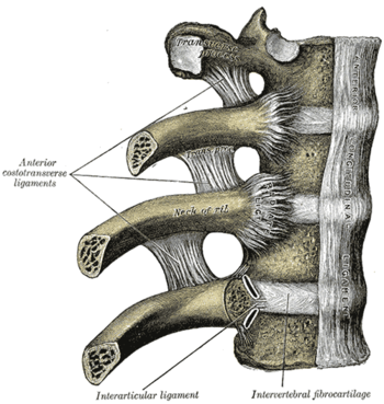
 
* '''Costotransverse ligament''' – Connects the neck of the rib and the transverse process.
 
* '''Lateral costotransverse ligament''' – Extends from the transverse process to the tubercle of the rib.
 
* '''Superior costotransverse ligament''' – Passes from the upper border of the neck of the rib to the transverse process of the vertebra superior to it.
 
===Physiology and Biomechanics===
The skeletal makeup of the thorax includes the thoracic vertebrae, intervertebral discs, ribs, sternum and costal cartilages . The contents of the hollow thorax consist of the lungs, heart and thymus gland, as well as the beginning of the great systemic artery (aorta) and the end of the great systemic veins (superior and inferior vena cava).<ref>O’Rahilly R. Philadelphia: Saunders; 1986. Basic Human Anatomy.</ref>
 
The costovertebral joint and costovertebral ligaments play a pivotal role in '''thoracic stabilization''', '''load bearing''', '''mobility''', '''protection and chest wall movement''', all while '''contributing heavily to respiratory effort'''.  
 
The thoracic architecture allows motion in all planes. In flexion-extension, the range of motion begins at four degrees at T1 and increases incrementally to 12 degrees at T12. Lateral twisting typically allows six to seven degrees per vertebral segment. Rotation occurs inversely to flexion. T1 rotates nine degrees and incrementally decreases to two degrees at T12. Rib-joint stiffness is greatest at T2 and weakest at T10 .
 
The thoracic cage plays an important role in load-bearing, providing between 30-40% of thoracic spine stiffness. ''Additionally'', the costovertebral complex serves as scaffolding for the musculature of the thoracic spine and shoulder. All of the above motions and loads cause stress to the costovertebral attachments at both the vertebral body and the transverse process. Fortunately, the strong costovertebral and costotransverse ligaments stabilize these joints and also add a degree of flexibility.<ref>Ganong WF. San Francisco: Lange; 1977. Review of Medical Physiology. </ref>
 
====The costovertebral complex is mandatory for respiratory movement of the chest wall.====
'''There are two basic mechanisms for lung expansion and contraction;'''
* one is the caudal and cephalic movement of the diaphragm, which lengthens or shortens the chest cavity. The contraction of the diaphragm during inspiration generates a negative intrathoracic pressure, allowing for expansion of the lungs. During quiet breathing, the intrapleural pressure is about -2.5 mm Hg (relative to atmospheric pressure) at the start of inspiration and decreases to approximately -6 mm Hg. Thus, generating a slightly negative pressure gradient allows air to flow in. When the diaphragm relaxes, the elastic recoil of the lungs, abdominal structures and chest wall compresses the lungs, causing expiration. The airway pressure becomes slightly positive at that point, allowing air to flow out of the lungs. During quiet expiration, contraction of the inspiratory muscles in the early stages exerts a braking action on the recoil forces and delays expiration . Quiet breathing occurs primarily by diaphragmatic movement. Even bilateral paralysis of the diaphragm does not lead to hypoventilation as long as the thorax remains mobile and the thoracic muscles and costovertebral joints are functioning well. However, the diaphragm is instrumental in the event the intercostal muscles are paralyzed or the thorax becomes immobile .<ref>Guyton AC. Philadelphia: Saunders; 1986. Textbook of Medical Physiology.</ref>
 
* The second method for lung expansion is the elevation of the ribs, which increases the anteroposterior and transverse diameter of the chest cavity. In the normal resting position, ribs are slanted downward allowing the sternum to be situated posteriorly. When the ribs are elevated, they are projected laterally and anteriorly leading to an increased transverse and anteroposterior diameter. With an increased diameter, maximum inspiration can increase the intrathoracic volume by as much as 20% .
 
* The muscles of deep inspiration involved in raising the rib cage are the external intercostals, anterior serrati, scalene, sternocleidomastoid, levatores costarum and the serratus posterior superior . As these muscles contract, the ribs and sternum are elevated and projected outward. In a forced inspiration, the muscles of deep inspiration are utilized concurrently with the levator scapulae, trapezius and rhomboids to raise and fix the scapula. Additionally, the pectoral muscles and serratus anterior muscles also aid in elevating the ribs. Strong inspiration can reduce intrapleural pressure as low as -30 mm Hg .<ref>Ganong WF. San Francisco: Lange; 1977. Review of Medical Physiology.</ref>
 
* The muscles of forced expiration that pull the rib cage downward are the internal intercostals and the abdominal recti primarily, with minor contributions from the quadratus lumborum, subcostals, transverse thoracic and serratus posterior inferior The posterior costovertebral joints and the anterior costochondral joints assist in making the downward movement possible. The range of motion at any one of the thoracic joints is small, but the frequency of movement of these joints is extremely great. The second through the sixth ribs each move around two axes. Movement at the costovertebral joint in a side-to-side (gliding) axis results in the raising and lowering of the sternal end of a rib. This is referred to as the “pump-handle” movement. Because the ribs are sloped downward, any elevation (e.g., during deep inspiration) will result in an upward (cephalic) and forward (anterior) movement of the sternum, thus increasing the anteroposterior diameter of the thorax. The lower ribs move laterally when they are elevated, which consequently increases the transverse diameter of the thorax. This motion primarily occurs at the seventh through 10th costotransverse joints. Since the articular tubercle of the joints is flat, the ribs move up and down and allow what is referred to as the “bucket-handle” movement.<ref>Thoracic spine: anatomical and biomechanical considerations for manual therapy. Edmonston SJ, Singer KP. Man Ther. 1997;2:132–143. </ref>
'''The axis of rotation at the first rib is unique compared to the other ribs.''' There is minimal movement at the first rib during quiet breathing. A slight rotation about its neck causes a small amount of rising and lowering of the sternum, which can result in a minute change of the anteroposterior diameter of the thoracic inlet.
 
The second through sixth ribs move in a similar axis to the seventh through 10th ribs. The costal cartilage deviates upwards on the second through sixth ribs, which causes a posterior movement of the sternum (the “pump handle” effect) during elevation . The length of the transverse process also has an effect on the costovertebral complex. As the length of the transverse process varies (with its concomitant costotransverse articular surface), the leverage that is exerted on the costovertebral joints varies as well . Movement a few millimeters anteriorly, cephalically or laterally of the thoracic wall can increase its volume by approximately one-half liter .
 
'''The costovertebral complex is an essential component of the biomechanics of chest wall movement.''' The costovertebral ligaments make the actions of the costovertebral joints and intervertebral movement possible. These ligaments function to affix, stabilize and allow some motion of the ribs on the thoracic vertebra at the costovertebral joint. Their presence helps with the load-bearing, protection, posture and scaffolding roles that the thoracic cage provides with their stabilization properties . More so, they allow and limit movement of the ribs at the transverse joint to allow for maximum expansion of the thoracic cavity as needed for respiratory demand. Lastly, their actions on both the costovertebral and intervertebral complexes allow lateral bending and axial rotation.
 
===Innervation of the costovertebral ligaments<ref name=":0">Muacevic. A & Adler.J ,2016,Ligaments of the Costovertebral Joints including Biomechanics, Innervations, and Clinical Applications: A Comprehensive Review with Application to Approaches to the Thoracic Spine,PMC5154401, V.8(11)
</ref> ===
Innervation of the costovertebral ligaments is supplied by the '''lateral branch of the thoracic dorsal rami of C8 and T1 to T11''' . The costovertebral joints receive this innervation in a segmental fashion with each joint receiving fibers from the level above and directly below it. Pain originating from the joints is well-localized and level specific . This seems to follow Hilton’s law, which states “that the innervation of a joint is the same innervation as the muscles which move the joint and the skin overlying the joint” .
 
Mechanoreceptors have been identified in the region of the middle costotransverse ligament . This innervation is characteristic of neurons providing nociception and mechanoreceptor activity. Activation of these mechanoreceptors, either by mechanic loading or inflammation, may cause pain and reflex muscular hypertonus. Tachykinins (substance P and calcitonin gene-related peptide), neuropeptides activated in response to nociceptive stimulation, have been found aside costovertebral ligaments. Thus, it appears that known neuropeptides are present along the costovertebral ligaments that can activate pain and propagate an inflammatory cascade .
 
Studies in rabbits have identified slowly adapting mechanoreceptors in the costovertebral complex that are capable of signaling the absolute rib position, direction and velocity of movement . These mechanoreceptors are also sensitive to tension and can alter the strain of the surrounding ligaments. There is a group of these receptors that is sensitive to caudal rib movement, known as “expiratory receptors,” and a group that is sensitive to rostral rib movement, or “inspiratory receptors” . There have been no similar studies in humans. Nonetheless, these findings provide a probable pathway to consider. In approximately 60% of individuals, there is a linkage of the brachial plexus to the first and/or second intercostal nerve, known as Kuntz’s nerve. Therefore, disorders affecting the first or second costovertebral joints can result in arm pain referred via this pathway .


==References==
==References==

Latest revision as of 13:30, 9 April 2024

Original Editor - Rewan Aloush


Top Contributors - Rewan Aloush, Lucinda hampton, Kim Jackson and George Prudden  

Introduction[edit | edit source]

The costovertebral joints describe two groups of synovial plane joints which connect the proximal end of the ribs with their corresponding thoracic vertebrae, enclosing the thoracic cage from the posterior side.

Costovertebral articulations. Anterior view.

Joining of ribs to the vertebrae occurs at two places

  1. Head - Two convex facets from the head attach to two adjacent vertebrae. This forms a synovial planar (gliding) joint, which is strengthened by the ligament of the head and the intercapital ligament.
  2. Tubercle of the rib - Articulation of the tubercle is to the transverse process of the adjacent vertebrae. This articulation is reinforced by the dorsal costotransverse ligament[1]

Anatomy[edit | edit source]

Costovertebral Joint[edit | edit source]

Costovertebral joint consists of the head of the rib (the head of a typical rib has two facets - each facet with a separate synovial joint separated by a ridge. The head of each rib articulates with:

  • The lower rib facet articulates with the upper costal facet of its own vertebra
  • The upper facet articulates with the lower facet of the vertebral body above. 
  • The first rib articulates with the T1 vertebra only and the lowest three ribs articulate only with their own vertebral body.

There are two types of ligaments:

  1. intra-articular ligament - attaches the intervertebral disc to the ridge in between the two facets of the head of the rib.
  2. radiate ligament - formed by three bands which connect the rib head to the vertebral bodies. A superior band runs to the vertebral body above and an inferior band runs to the vertebral body below. A central band runs deep to the anterior longitudinal ligament and blends into the intervertebral disc to join the ligament on the opposite side. In the first rib and the last three ribs, only two bands exist as they only articulate with their own vertebra[2].
Afbeelding 3.png

Costotransverse Joint[edit | edit source]

There are two facets of a tubercle of a rib, the medial and lateral.

  1. The hyaline cartilage-lined medial facet forms a plane synovial joint with the tip of the transverse process which is reinforced by a capsule.
  2. The lateral facet is attached to the transverse process through three ligaments:
  • Lateral costotransverse ligament - attaches the lateral facet to the tip of the transverse process of the vertebral body.
  • Costotransverse ligament - attaches the back of the neck of the rib to the front of the transverse process
  • Superior costotransverse ligaments - attaches the neck of the rib to the underside of the transverse process of the vertebra above

The lower two ribs are only attached by ligaments and do not form synovial joints with the transverse process[2].

Movements[edit | edit source]

The movements on these joints are called ‘pump-handle’ or ‘bucket-handle’ movements, and are limited to a small degree of gliding and rotation of the rib head.

  • The function of these movements is to enable lifting of the ribs upwards and outwards during breathing.
  • The end result is the increase of the lateral diameter of the thorax and subsequent expansion of the lung parenchyma as the air is being inhaled[3].

The costovertebral complex is an essential component of the biomechanics of chest wall movement. The costovertebral ligaments make the actions of the costovertebral joints and intervertebral movement possible. The ligaments function to:

  • Affix, stabilize and allow some motion of the ribs on the thoracic vertebra at the costovertebral joint. Their presence helps with the load-bearing, protection, posture and scaffolding roles that the thoracic cage provides with their stabilization properties .
  • Allow and limit movement of the ribs at the transverse joint to allow for maximum expansion of the thoracic cavity as needed for respiratory demand. Their actions on both the costovertebral and intervertebral complexes allow lateral bending and axial rotation.

Muscles That Act on the Costovertebral Joints[edit | edit source]

Image017.jpg

The prime movers of the costovertebral joints are the muscles of respiration.ie

However, all the muscles that attach to the ribs and which are sorted as accessory respiratory muscles can cause the movements on these joints;

sternocleidomastoid, scalene, serratus anterior, pectoralis major, pectoralis minor, latissimus dorsi and serratus posterior superior.

Innervation of the Costovertebral Ligaments[edit | edit source]

Both types of costovertebral joints are innervated by the lateral branches of the posterior rami of spinal nerves C8-T11.

  • The innervation has a segmental pattern
  • Each joint receives the fibers from a spinal nerve of its numerically equivalent level and from the level above.[3]

Clinical Significance[edit | edit source]

Costotransverse disorders are disorders affecting or involving the costotransverse and costovertebral joints and ligaments. Important considerations:

  • Often overlooked yet it can be responsible for pain in the thoracic region or functional impairments of the thoracic spine.
  • May occur due to trauma, degenerative changes, tumours, deformities or muscular spasm.
  • A diagnosis is usually made through a clinical examination with treatment consisting of mobilisation, exercise therapy, injections and/or fixation of unstable joints[4].

References[edit | edit source]

  1. Gray.H,Gray's anatomy 20th edition,page 299
  2. 2.0 2.1 Radiopedia Costrovert. Joint Available from:https://radiopaedia.org/articles/costovertebral-joint (last accessed 10.5.2020)
  3. 3.0 3.1 Kenhub CV joints Available from:https://www.kenhub.com/en/library/anatomy/costovertebral-joints (last accessed 10.5.2020)
  4. B A Young, H E Gill, R S Wainner, T W Flynn. Thoracic costotransverse joint pain patterns: a study in normal volunteers. BMC Musculoskelet disord. 2008; 9: 140