Costotransverse Disorders: Difference between revisions
No edit summary |
No edit summary |
||
| (46 intermediate revisions by 12 users not shown) | |||
| Line 1: | Line 1: | ||
<div class="editorbox"> | |||
'''Original Editors ''' - [[User:Yves Hubar|Yves Hubar]] | '''Original Editors ''' - [[User:Yves Hubar|Yves Hubar]] | ||
'''Top Contributors''' - {{Special:Contributors/{{FULLPAGENAME}}}} | '''Top Contributors''' - {{Special:Contributors/{{FULLPAGENAME}}}} | ||
</div> | </div> | ||
== Definition/Description == | == Definition/Description == | ||
Costotransverse disorders are disorders affecting or involving the costotransverse and [[Costovertebral Joints|costovertebral joints]] and ligaments <ref name=":0">B A Young, H E Gill, R S Wainner, T W Flynn. [https://scholar.google.com/scholar_url?url=https://bmcmusculoskeletdisord.biomedcentral.com/articles/10.1186/1471-2474-9-140&hl=en&sa=T&oi=gsb-ggp&ct=res&cd=0&d=13620971517754149219&ei=a3D_Y-_lOoWYywTYn4T4Ag&scisig=AAGBfm3wEEM12DzhEgbyGNROGK_mVIkv-g Thoracic costotransverse joint pain patterns]: a study in normal volunteers. BMC Musculoskelet disord. 2008; 9: 140 </ref> which are often overlooked during examination for pain source localisation in this area due to possible visceral pain referral and the complexities of the thoracic neural network.<ref name=":5" /> It is suggested that dysfunctions in these joints could account for pain in the thorax or functional impairments. <ref name=":0" /> <ref name=":2">D Aspegren, T Hyde, M Miller. [https://scholar.google.com/scholar_url?url=https://www.sciencedirect.com/science/article/pii/S0161475407000759&hl=en&sa=T&oi=gsb&ct=res&cd=0&d=6379619836327534304&ei=kHD_Y6zXEryR6rQP8fyloAI&scisig=AAGBfm2E50sx_Lw1bvuTm5BqA1wPSEXzlg Conservative treatment of a female collegiate volleybal player with costochondritis.] Journal of manipulative and physiological therapeutics (2007) 30(4): 321-325 </ref><ref name=":1">Edge-Hughes, L. [https://scholar.google.com/scholar_url?url=https://www.sciencedirect.com/science/article/pii/S1938973614000087&hl=en&sa=T&oi=gsb&ct=res&cd=0&d=4835174966929252867&ei=33D_Y-b6G8K1ywTR2b-ACw&scisig=AAGBfm0_NiE12NAJ6fxWp2LhTVd1yZ0_wA Canine Thoracic Costovertebral and Costotransverse Joints:] Three Case Reports of Dysfunction and Manual Therapy Guidelines for Assessment and Treatment of These Structures. Topics in Compan An Med; 29, 1: 1–5. </ref> | |||
== Clinically Relevant Anatomy == | == Clinically Relevant Anatomy == | ||
* The '''costotransverse joint''' is an articulation between the articular costal tubercle of the rib and the costal facet of the transverse process of a thoracic vertebra.<ref>DA. Kinesiology of the Musculoskeletal System: Foundations for Physical Rehabilitation. St. Louis: Mosby 2002. ISBN: 978-0-8151-6349-7</ref> The '''costovertebral joint''' is the articulation between the costal facte or demi-facets (formed by the caudal side of the superior vertebra and the cranial side of the inferior vertebra) and the head of the [[Ribs|rib]] <ref name=":1" /> These facets form a solid angle whose base consists of the annulus fibrosis of the intervertebral disc. A synovial joint ensures the connection between the rib and the thoracic vertebra. <ref name=":3">Irwin J. et al, [https://scholar.google.com/scholar_url?url=https://search.proquest.com/openview/595da2c2b3fe6c63ffa1b49f7313fbc6/1%3Fpq-origsite%3Dgscholar%26cbl%3D2026366%26diss%3Dy&hl=en&sa=T&oi=gsb&ct=res&cd=0&d=18107430632382169547&ei=FHH_Y56_FbyR6rQP8fyloAI&scisig=AAGBfm1aAB3BB1ubagalrg9BgYYc0F9CLA The effect of costovertebral adjustment versus ischaemic compression of rhomboid muscles for interscapular pain], university of johannesburg, 2015 </ref> Together with the thoracic cage, the costovertebral and costotransverse joints provide stability.<ref name=":1" /> | |||
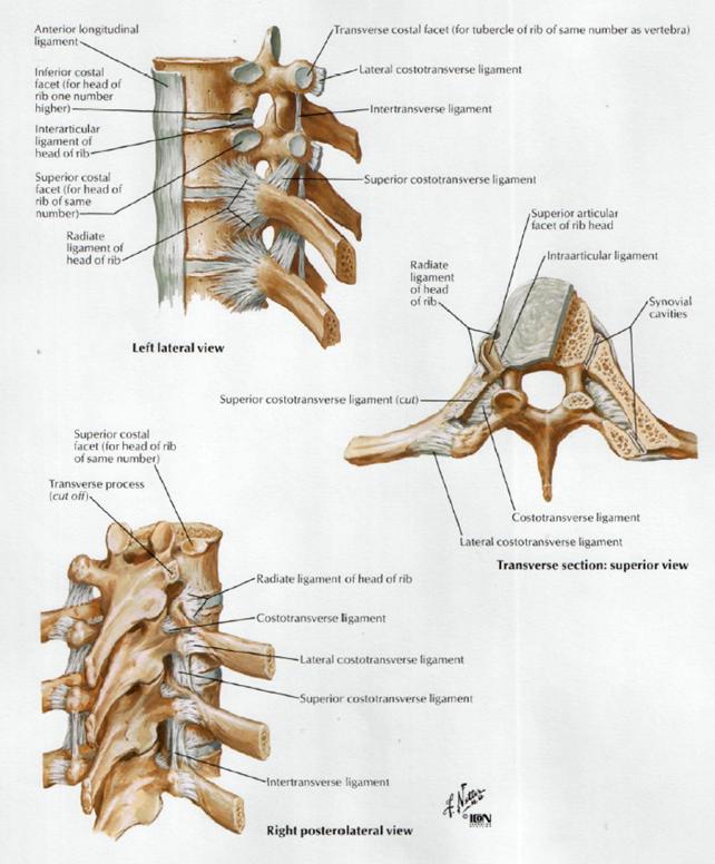
* Ligaments: The following costotransverse and costovertebral ligaments connect the two joints-<ref>A F Ibrahim, H H Darwish. [https://scholar.google.com/scholar_url?url=https://onlinelibrary.wiley.com/doi/abs/10.1002/ca.20102&hl=en&sa=T&oi=gsb&ct=res&cd=0&d=5831329189147282428&ei=MHH_Y-3qL5n4yATzjJS4CA&scisig=AAGBfm1-RTY94SMO_Mt59yFkifqAdPZqUw The costotransverse ligaments in human:] a detailed anatomical study. Clinical Anatomy 18:340-345 (2008) </ref> <ref>Agur A.M.R., Lee M.J. [https://scholar.google.com/scholar_url?url=https://books.google.com/books%3Fhl%3Den%26lr%3D%26id%3DH20V4pCpACYC%26oi%3Dfnd%26pg%3DPR3%26dq%3DAgur%2BA.M.R.,%2BLee%2BM.J.%2BGrant%2527s%2BAtlas%2Bof%2BAnatomy.%2BLippincott%2BWilliams%2B%2526%2BWilkins%2B1999%2B(10th%2Bed.)%2BISBN%2B%2B978%2B0683302646%26ots%3DUBvflIzs23%26sig%3DSG1aiHdERGI1IyOPawJAUXtEEYY&hl=en&sa=T&oi=gsb&ct=res&cd=0&d=8139934413421084040&ei=R3H_Y4TqEYmwywSdlrLQBQ&scisig=AAGBfm2vIF9V3QoX2mn2Xzb8noifzEbbSw Grant's Atlas of Anatomy.] Lippincott Williams & Wilkins 1999 (10th ed.) ISBN: 978-0683302646</ref> Ligamentum costotransversarium superius, Ligamentum costotransversarium., Ligamentum costotransversarium laterale, Ligamentum capitis costae radiatum, Inferior costotransverse ligaments, Posterior costotransverse ligaments, identified on the fifth to tenth ribs. These ligaments limit movement in the costotransverse joint to a minimal gliding movement. The ribs articulate posteriorly twice with the corresponding vertebra. The radiate ligament and the intra-articular ligament stabilise the head of ribs 2 to 9 and sometimes the head of the 10th rib in the costovertebral joint. | |||
* The head of ribs 2 to 9 and sometimes the 10th articulate with the vertebral body of two thoracic vertebrae at the costovertebral joint. The head of ribs 1, 11 and 12 articulate with the corresponding vertebrae. There is no intra-articular ligament in these joints. | |||
* The neck of ribs 1 to 10 articulate, through the tubercle, with the transverse process of their corresponding vertebrae and are stabilised with ligaments and the articular capsule. Ribs 11 and 12 do not articulate with the transverse processes. <ref>Andor W.J.M. Glaudemans et al. [https://scholar.google.com/scholar_url?url=https://books.google.com/books%3Fhl%3Den%26lr%3D%26id%3Dr17gCQAAQBAJ%26oi%3Dfnd%26pg%3DPR5%26dq%3DAndor%2BW.J.M.%2BGlaudemans%2Bet%2Bal.%2BNuclear%2BMedicine%2Band%2BRadiologic%2BImaging%2Bin%2BSports%2BInjuries.%2BSpringer,%2B2015,%2B259%2B260%26ots%3D7Uqv5uIjZB%26sig%3D5yHFWUqd-j-grq9Dx0JB-3ivU6w&hl=en&sa=T&oi=gsb&ct=res&cd=0&d=1276274689965722077&ei=Y3H_Y7XnMYjeygTN9LHQDg&scisig=AAGBfm1x3fZGpaQbQHhRrhj3kJc8mQJDLA Nuclear Medicine and Radiologic Imaging in Sports Injuries]. Springer, 2015, 259-260</ref> | |||
* Lateral flexion as well as rotation is limited by the ribcage therefore only small movements are possible in the costotransverse joints. There is no sagittal plane flexion and extension in these joints, however gliding movements within the joint have been seen. These gliding movements are mostly medially and laterally orientated. The medial and lateral glides are functional motions. <ref>Sharon Weiselfish-Giammatteo, [https://scholar.google.com/scholar_url?url=https://books.google.com/books%3Fhl%3Den%26lr%3D%26id%3DTck88Fs2XzAC%26oi%3Dfnd%26pg%3DIA14%26dq%3DSharon%2BWeiselfish%2BGiammatteo,%2BIntegrative%2BManual%2BTherapy%2Bfor%2BBiomechanics%2B%2BApplication%2Bof%2Bmuscle%2Benergy%2Band%2B%25E2%2580%259Cbeyond%25E2%2580%259D%2Btechnique.%2BNoth%2BAtlantic%2BBooks%2BBerkeley,%2BCalifornia,%2B2003,%2Bp%2B261%26ots%3DcK5_Ik74ya%26sig%3DmCo8nNFp8ACV3GdEtPHUzx6HzLk&hl=en&sa=T&oi=gsb&ct=res&cd=0&d=4338269428903992896&ei=fHH_Y40chZjLBNifhPgC&scisig=AAGBfm1MxEQqAWUEUyffm0crK58JKB4dlg Integrative Manual Therapy for Biomechanics:] Application of muscle energy and “beyond” technique. Noth Atlantic Books Berkeley, California, 2003, p 261</ref> | |||
< | <br> | ||
[[Image:Image017.jpg|center]]Figure 1: Ligaments connecting the ribs and vertebra | |||
Movement of the ribs at the costovertebral joints <ref name=":3" />. The axes of the ribs allow three basic types of motion: | |||
* | * Bucket-handle motion: One end of the rib is fixed at the vertebral end, the majority of the rib elevation occurs through upward excursion of the lateral position. This motion increases the transverse diameter of the rib cage. | ||
* Pump-handle motion: One end is fixed, and the free end describes an arc. When the ribs move around the axis, the anterioposterior diameter of the rib cage increases. | |||
* Caliper motion: The 11th and 12th ribs have only costovertebral articulations. The motion produces slight changes in both the transverse and the anteroposterior dimension. | |||
All twelve ribs have pump handle and bucket handle movements, but the upper ribs have a greater pump handle motion and the lower ones have more bucket handle type movements. | |||
[ | This [https://hal.bim.msu.edu/CMEonLine/RibCage/Biomechanics/start.html link] explains pump-handle and bucket-handle motion mechanics of the rib cage during respiration<ref>Rib Cage Biomechanics - Visualization Technology<nowiki/>https://hal.bim.msu.edu/CMEonLine/RibCage/Biomechanics/start.html</ref>. | ||
== Epidemiology /Etiology == | == Epidemiology /Etiology == | ||
* Local joint compression may occur as a result of a trauma or muscle spasm <ref>Thomas E. Hyde, Conservative Management of Sports Injuries. Jones and Barlett Publishers, 2007, p440. </ref> | |||
* It is more common in women <ref name=":2" /> <ref name=":17">Hudes, K. [https://scholar.google.com/scholar_url?url=https://www.ncbi.nlm.nih.gov/pmc/articles/PMC2597886/&hl=en&sa=T&oi=gsb-ggp&ct=res&cd=0&d=8311641053692702252&ei=p3H_Y9nWAoWYywTYn4T4Ag&scisig=AAGBfm2qRFvgUaWErGMtyEqWz5Mm_DUmDA Low-tech rehabilitation and management of a 64 year old male patient with acute idiopathic onset of costochondritis.] Family Chiropractic & Rehabilitation, (2008) 52 (4), 224-228. </ref> and can occur at any age | |||
* When subjected to severe trauma, these joints can subluxate or dislocate. Due to being at the top of the rib cage, the first costotransverse joint is the most vulnerable. <ref name=":8">Christensen EE, Dietz GW. I[https://scholar.google.com/scholar_url?url=https://pubs.rsna.org/doi/abs/10.1148/radiology.134.1.7350632&hl=en&sa=T&oi=gsb&ct=res&cd=0&d=8804118112602783570&ei=wHH_Y8DLAbyR6rQP8fyloAI&scisig=AAGBfm3354686HS9J7Yp-6_3_LKh3oITSA njuries of the costovertebral articulation]. Radiology 1980. Jan; 134(1): 41-3 </ref> | |||
* Though distinctly unusual at the costotransverse and costovertebral joint, rheumatoid arthritis can occur in these joints.<ref>MJ Cohen, J Ezekiel, RH Persellin. [https://scholar.google.com/scholar_url?url=https://ard.bmj.com/content/37/5/473.short&hl=en&sa=T&oi=gsb&ct=res&cd=0&d=2127597150573199627&ei=33H_Y7WXHsK1ywTR2b-ACw&scisig=AAGBfm3N_SFIipEol3pflkX_m-pXiD7-Yw Costovertebral and costotransverse joint involvement in rheumatoid artritis.] Ann Rheum Dis 1978 October; 37(5): 473-475 </ref> | |||
* The involvement of dysfunction at these joints are a source of referred pain at the thoracic spine, with suggested involvement of the costotransverse joint concerning [[T4 Syndrome|T4 syndrome]] <ref name=":0" /> | |||
* The costotransverse joint is known to be involved in patients with [[Ankylosing Spondylitis (Axial Spondyloarthritis)|ankylosing spondylitis]], which, combined with involvement of the costovertebral, sternoclavicular and sternomanubrial joints, would result in increased rigidity of the thorax and increased dorsal kyphosis. This does not result in decreased pulmonary function, which is possibly due to an increase in diaphragmatic breathing <ref>Z Unlu, M can, C Goktan, P Celik. [https://scholar.google.com/scholar_url?url=https://link.springer.com/article/10.1007/s100670200073&hl=en&sa=T&oi=gsb&ct=res&cd=0&d=10505433490924133379&ei=B3L_Y6bZNY7gygSPuIegDA&scisig=AAGBfm0sXAsXQYR1V0mPGDHShvYfaS1QWQ Lumbar stuffness but not thoracic radiographic changes relate to alteration of lung function tests in ankylosing spondylitis]. Clin Rheumatol (2002) 21(4):275-279</ref> | |||
* Due to the positive response to manual therapy directed towards posterior spinal structures, conditions diagnosed as costochondritis might actually be caused by neurogenic inflammation.<ref>M I Rabey. [https://scholar.google.com/scholar_url?url=https://www.sciencedirect.com/science/article/pii/S1356689X07000276/pdf%3Fmd5%3D66f8e1f8782d0029fdd9bd7ea0cd5378%26pid%3D1-s2.0-S1356689X07000276-main.pdf%26_valck%3D1&hl=en&sa=T&oi=gsb&ct=res&cd=0&d=12692838329674799845&ei=JXL_Y-GaDZn0yAT_xK_ADg&scisig=AAGBfm0llRAplShp4lWTKId9mmw8EIZ0uQ Costochondritis:] are the symptoms and signs due to neurogenic inflammation. Two cases that responded to manual therapy directed towards posterior spinal structures. Manual therapy (2008) 13(1): 82-86 </ref> <ref name=":2" /> | |||
== Characteristics/Clinical Presentation == | |||
== Characteristics/Clinical Presentation | |||
& | Possible symptoms <ref name=":5">Smith H S, Current Therapy in Pain. Saunders Elsevier, 2009: p199</ref><ref>Edge-Hughes, L. [https://scholar.google.com/scholar_url?url=https://www.sciencedirect.com/science/article/pii/S1938973614000087&hl=en&sa=T&oi=gsb&ct=res&cd=0&d=4835174966929252867&ei=VXL_Y_vUCM6bywTVg7Fo&scisig=AAGBfm0_NiE12NAJ6fxWp2LhTVd1yZ0_wA Canine Thoracic Costovertebral and Costotransverse Joints:] Three Case Reports of Dysfunction and Manual Therapy Guidelines for Assessment and Treatment of These Structures. Topics in Compan An Med; 29, 1: 1–5</ref>are: | ||
* Pain localised to the posterior thorax <ref name=":4">Fruth S J. [https://scholar.google.com/scholar_url?url=https://academic.oup.com/ptj/article-abstract/86/2/254/6160469&hl=en&sa=T&oi=gsb&ct=res&cd=0&d=13179047466588118112&ei=fXL_Y77QKYjeygTN9LHQDg&scisig=AAGBfm19B2k7KNtxJGiL0DjEFINBb6nTAw Differential diagnosis and treatment in a patient with posterior upper thoracic pain.] Physical therapy (2006) 86(2): 254-268 </ref> | |||
* Pain may radiate to the anterior chest wall, along the rib, sometimes into the shoulder and sometimes towards the upper limb <ref name=":5" /> | |||
* Unilateral symptoms <ref name=":4" /> | |||
* Pain with deep inspiration, coughing/sneezing/laughing | |||
* Increased pain with passive or active thoracolumbar flexion, rotation and ipsilateral side bending, lifting or twisting movements | |||
* Hypomobility of the costotransverse and costovertebral joint | |||
* Palpable tenderness and pain at costotransverse joint and rib angle | |||
* Movement in adjacent thoracic vertebral and rib segments is usually restricted and may stimulate or exacerbate protective muscle spasm | |||
* Increased muscle tension in paraspinal muscles, rhomboid muscles, trapezius muscles and levator scapula muscles | |||
* Neck pain, head ache or both | |||
* The sensation of having a useless or heavy limb | |||
* Referred pain originating from under the scapula and worsens with coughing, sneezing or deep breathing | |||
* Acute, atypical chest pain | |||
== Differential Diagnosis == | |||
& | Differential diagnostic possibilities have to be taken into consideration in cases of posterior upper thoracic and/or scapular pain and possible involvement of costovertebral and costotransverse joints. <ref name=":4" /> Possible musculoskeletal sources of thoracic pain are: <ref name=":6">McConaghy JR, Oza RS. [https://scholar.google.com/scholar_url?url=https://www.aafp.org/afp/2013/0201/p177.html&hl=en&sa=T&oi=gsb-ggp&ct=res&cd=0&d=5735340565193854856&ei=wHL_Y92bOYjeygTN9LHQDg&scisig=AAGBfm0sippv9g0U3SD-lP5sRxzhcaTR9w Outpatient diagnosis of acute chest pain in adults.] Am Fam Physician (2013) Feb 1;87(3):177-82</ref> <ref name=":4" /> | ||
* Muscle strain (erector spinae, lower and middle [[trapezius]], [[rhomboids]], latissimus dorsi, levator scapulae and intercostal muscles) | |||
* | * Vertebral or rib fracture | ||
* Zygapophyseal joint arthropathy | |||
& | * Active trigger points | ||
* [[Spinal Stenosis|Spinal stenosis]] | |||
* Intervertebral disc protrusion or herniation | |||
* Diffuse idiopathic skeletal hyperostosis (DISH) | |||
* Intercostal neuralgia: often follows injury or thoracic surgery, focal tenderness over affected intercostal region , burning pain and paraesthesia in thorax or abdomen that usually follow the nerve path <ref>Williams EH et al. Neurectomy for treatment of intercostal neuralgia. Ann Thorac Surg (2008) May;85(5):1766-70. </ref> | |||
* T4 syndrome | |||
* Ankylosing spondylitis | |||
* Costovertebral and costotransverse joint dysfunction | |||
* Thoracic radiculopathy <ref>Walter R, Essentials of Physical Medicine and Rehabilitation. Elsevier Saunders, Philadelphia, 2008, p 576. </ref> | |||
* Fractures | |||
Possible visceral sources of thoracic pain<ref name=":6" /> <ref name=":4" /> | |||
* Cancer | |||
* Cardiac conditions | |||
** substernal pain; | |||
** shortness of breath | |||
** increased pain with exertion; | |||
** frequent left shoulder, medial arm and jaw pain <ref name=":10">Haasenritter J et al. [https://scholar.google.com/scholar_url?url=https://hrcak.srce.hr/file/139793&hl=en&sa=T&oi=gsb-ggp&ct=res&cd=0&d=1985860266087180879&ei=NnP_Y_LHKY7gygSPuIegDA&scisig=AAGBfm0-7UdnNlUW3TGewyCjwr4jV5dGzg Does the patient with chest pain have a coronary heart disease?] Diagnostic value of single symptoms and signs – a meta-analysis. Croat Med J (2012) Oct;53(5):432-41.</ref> | |||
* Renal conditions | |||
** referred pain to the ipsilateral subcostal and costovertebral regions at T10-T12; | |||
** typically dull and aching pain; | |||
** possible changes in urinary frequency or output <ref name=":6" /> | |||
* Pulmonary conditions | |||
* Gastroesophageal conditions | |||
** band like pain around mid-thorax at the level of the lesion; | |||
** referred pain to the mid thoracic area; | |||
** nausea/vomiting; | |||
** weight loss, heartburn or substernal pain; | |||
** stabbing or burning chest pain <ref>Spiegel BM et al. [https://scholar.google.com/scholar_url?url=https://www.ncbi.nlm.nih.gov/pmc/articles/PMC4285435/&hl=en&sa=T&oi=gsb-ggp&ct=res&cd=0&d=16957654354022326688&ei=jnP_Y52zFLyR6rQP8fyloAI&scisig=AAGBfm1lo4sADt1gvHUd_xiNmL-L-8b6Lw Development of the NIH Patient-Reported Outcomes Measurement Information System (PROMIS) gastrointestinal symptom scales]. Am J Gastroenterol (2014) Nov;109(11):1804-14</ref> | |||
* Gall bladder conditions | |||
** pain in the right mid epigastric region (T8-T9 level); | |||
** referred pain at the mid back between the scapulae, right upper trapezius muscle and right subscapular area; | |||
** jaundice | |||
** fever | |||
** chills | |||
** indigestion | |||
** nausea/vomiting | |||
** intolerance of fatty foods <ref name=":7">Duncan CB, Riall TS. [https://scholar.google.com/scholar_url?url=https://link.springer.com/article/10.1007/s11605-012-2024-1&hl=en&sa=T&oi=gsb-ggp&ct=res&cd=0&d=5065531308935299425&ei=yXP_Y8qcA5n4yATzjJS4CA&scisig=AAGBfm2vktw0Xn34w1lSPfx1nT73LyQ25Q Evidence-based current surgical practice:] calculous gallbladder disease. J Gastrointest Surg (2012) Nov;16(11):2011-25</ref> | |||
* Hepatobiliary conditions | |||
** pain in the right upper abdomen quadrant | |||
** referred pain at right interscapular and subscapular areas | |||
** right shoulder pain | |||
** anorexia | |||
** nausea | |||
** vomiting | |||
** jaundice | |||
** ascites | |||
** significant fatigue <ref name=":7" /> | |||
== Diagnostic Procedures == | == Diagnostic Procedures == | ||
Clinical findings and patient presentation are used to diagnose costovertebral and costotransverse joint dysfunction | Clinical findings and patient presentation are used to diagnose costovertebral and costotransverse joint dysfunction. This is done through a thorough anamnesis and clinical examination <ref name="Scaringe">Scaringe JG, Ketner C. Manual methods for the treatment of rib dysfunctions and associated functional lesions. Topics in Clinical Chiropractic. (1999);6:20-38. </ref> <ref name="Triano">Triano JJ et al. Costoverebral and costotransverse joint pain: a commonly overlooked pain generator. Topics in Clinical Chiropractic. (1999);6:79-92. </ref> | ||
== Outcome Measures == | == Outcome Measures == | ||
Pain is measured with a numeric pain scale ([[Visual Analogue Scale| | Pain is measured with a numeric pain scale ([[Visual Analogue Scale|VAS]]), whilst function is measured with the Dallas Pain Questionnaire and Functional Rating Index.<ref name=":17" /> | ||
== Examination == | == Examination == | ||
The physical examination consists of the following components: | The physical examination consists of the following components:<ref name=":4" /> | ||
*Inspection of the posterior, lateral and anterior sides of the trunk, to check for deviations from an ideal posture (standing) | *Inspection of the posterior, lateral and anterior sides of the trunk, to check for deviations from an ideal posture (standing) <ref name=":4" /> | ||
{{#ev:youtube| | {{#ev:youtube|y0BhqpjW9OE|300}}<ref>Youtube (2012). Macleod’s examination of the thoracic and lumbar spine. Consulted on May 28, 2014 on https://www.youtube.com/watch?feature=player_detailpage&v=3UhKFcR1Wbs</ref> | ||
*Active range of motion of the cervical (seated), trunk (standing) and shoulder regions (seated) to determine pain | *Active range of motion of the cervical (seated), trunk (standing) and shoulder regions (seated) to determine pain provoking movements <ref name="Huygen">Huygen F et al. Painful shoulder complaints. Pain Pract. (2010) Jul-Aug;10(4):318-26. (LOE: 1A)</ref> | ||
{| width="100%" cellspacing="1" cellpadding="1" | {| width="100%" cellspacing="1" cellpadding="1" | ||
|- | |- | ||
| {{#ev:youtube|PrbmBMehYx0|300}} ( | | {{#ev:youtube|PrbmBMehYx0|300}} <ref>Youtube (2009). Range of Motion Test – Cervical ROM. Consulted on May 28, 2014 on https://www.youtube.com/watch?v=PrbmBMehYx0</ref> | ||
| {{#ev:youtube|_u0fPi2XBls|300}} ( | | {{#ev:youtube|_u0fPi2XBls|300}} <ref>Youtube (2012). Range of Motion Test – Thoracic ROM. Consulted on May 28, 2014 on https://www.youtube.com/watch?v=_u0fPi2XBls</ref> | ||
|} | |} | ||
{{#ev:youtube|d7HfaAlgaro|300}}( | {{#ev:youtube|d7HfaAlgaro|300}}<ref>Youtube (2009). Shoulder Exam (3 of 9): Range of motion. Consulted on May 28, 2014 on https://www.youtube.com/watch?v=d7HfaAlgaro</ref> | ||
*Manual muscle testing to assess upper extremities strength and intensity of possible elicited pain (seated) ( | *Manual muscle testing to assess upper extremities strength and intensity of possible elicited pain (seated) <ref name="Hislop">Hislop HJ, Avers D, Brown M. [https://scholar.google.com/scholar_url?url=https://books.google.com/books%3Fhl%3Den%26lr%3D%26id%3DpeNOAQAAQBAJ%26oi%3Dfnd%26pg%3DPP1%26dq%3DHislop%2BHJ,%2BAvers%2BD,%2BBrown%2BM.%2BDaniels%2Band%2BWorthingham%25E2%2580%2599s%2BMuscle%2BTesting%2B%2BTechniques%2Bof%2BManual%2BExamination%2Band%2BPerformance%2BTesting.%2B9th%2BEdition,%2BPa%2B%2BWB%2BSaunders%2BCo%253B%2B(2014)%2B(LOE%2B%2B3A)%26ots%3DmyHUAgY91w%26sig%3DoIsJ6ZSc3EiSw4X8m7tUrI5s8j4&hl=en&sa=T&oi=gsb&ct=res&cd=0&d=13640344623196258713&ei=HHT_Y7XSGMK1ywTR2b-ACw&scisig=AAGBfm3E1IHOGlYeOfdRFwM-J7t5ya9KTA Daniels and Worthingham’s Muscle Testing]: Techniques of Manual Examination and Performance Testing. 9th Edition, Pa: WB Saunders Co; (2014) (LOE: 3A)</ref> | ||
*Sensory examination of the upper extremities and thoracic area , in order to determine whether nerve root or peripheral nerve lesions are present | *Sensory examination of the upper extremities and thoracic area , in order to determine whether nerve root or peripheral nerve lesions are present <ref name=":4" /> | ||
{{#ev:youtube| | {{#ev:youtube|COToUselGpQ|300}} <ref>Youtube (2012). Macleod’s examination of the upper limbs sensation. Consulted on May 28, 2014 on https://www.youtube.com/watch?feature=player_detailpage&v=wSSCZeOKXBE</ref> | ||
* | *Accessory motion or joint movement of the thoracic spine (in prone position) to check for pain and mobility <ref name="Hicks">Hicks GE et al. [https://scholar.google.com/scholar_url?url=https://www.sciencedirect.com/science/article/pii/S0003999303003654&hl=en&sa=T&oi=gsb&ct=res&cd=0&d=11911785022224884698&ei=OnT_Y9XHBYWYywTYn4T4Ag&scisig=AAGBfm16XP1GXgdHm1GIpKf1W-3ZXha4bw Interrater reliability of clinical examination measures for identification of lumbar segmental instability.] Arch Phys Med Rehabil. (2003);84:1858-1864 (LOE: 2B)</ref> <ref name="Seffinger">Seffinger MA et al. [https://scholar.google.com/scholar_url?url=https://journals.lww.com/spinejournal/fulltext/2004/10010/reliability_of_spinal_palpation_for_diagnosis_of.22.aspx&hl=en&sa=T&oi=gsb&ct=res&cd=0&d=6972645237141383658&ei=UnT_Y42-CryR6rQP8fyloAI&scisig=AAGBfm3sV4VUNFqMdtJW7Z5WZnZ9oVu50w Reliability of spinal palpation for diagnosis of back and neck pain]: a systematic review of the literature. Spine. (2004);29:E413-425 (LOE: 2A)</ref> using the posterioranterior (PA) pressure test. <ref>Heiderscheit B., [https://scholar.google.com/scholar_url?url=https://www.tandfonline.com/doi/abs/10.1179/106698108790818369&hl=en&sa=T&oi=gsb&ct=res&cd=0&d=6860333891407900390&ei=aHT_Y_rlKZn0yAT_xK_ADg&scisig=AAGBfm0oX_nFmpK6Vte7Uf-CpXo46zFbQg Reliability of Joint Mobility and Pain Assessment of the Thoracic Spine and Rib Cage in Asymptomatic Individuals], J Man Manip Ther. 2008; 16(4): 210–216</ref> | ||
*Posterior (prone) and anterior (supine) costosternal joint | *Posterior (prone) and anterior (supine) costosternal joint movement to assess for pain and mobility <ref name="Hicks" /> <ref name="Seffinger" /> | ||
*Active and passive scapular mobility in case of reduced active shoulder range of motion and/or pain of the scapula region | *Active and passive scapular mobility in case of reduced active shoulder range of motion and/or pain of the scapula region <ref name="Huygen" /> | ||
{{#ev:youtube|pEY93k5XXL0|300}} ( | {{#ev:youtube|pEY93k5XXL0|300}} <ref>Youtube (2009). Shoulder Exam (4 of 9): Scapular control (Is there scapular dyskinesia?). Consulted on May 28, 2014 on https://www.youtube.com/watch?v=pEY93k5XXL0</ref> | ||
*Palpation of the cervical, upper trunk and shoulder regions to check for soreness or pain | *Palpation of the cervical, upper trunk and shoulder regions to check for soreness or pain<ref name=":8" /> <ref name=":10" /> | ||
{| width="100%" cellspacing="1" cellpadding="1" | {| width="100%" cellspacing="1" cellpadding="1" | ||
|- | |- | ||
| {{#ev:youtube| | | {{#ev:youtube|fAZYno9kO0A|300}} <ref>Youtube (2011). Macleod’s Examination of cervical spine (non traumatic). Consulted on May 28, 2014 on https://www.youtube.com/watch?v=qdi3VKA-wJI</ref> | ||
| {{#ev:youtube|Xf52jbNA7wg|300}} ( | | {{#ev:youtube|Xf52jbNA7wg|300}} <ref>Youtube (2009). Shoulder Exam (2 of 9): Inspection and Palpation. Consulted on on May 28 on https://www.youtube.com/watch?v=Xf52jbNA7wg</ref> | ||
|} | |} | ||
== Medical Management | == Medical Management == | ||
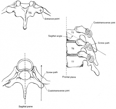
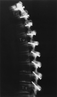
In an unstable thoracic spine (caused by trauma, degenerative conditions, tumors and deformities) posterior instrumentation and fusion are commonly used as treatment. Numerous techniques have been reported to restore spinal stability and correct spinal deformities. Posterior fixation of the thoracic spine through the costotransverse joint may be an alternative (figure 2). <br>Thanapipatsiri and Chan | In an unstable thoracic spine (caused by trauma, degenerative conditions, tumors and deformities) posterior instrumentation and fusion are commonly used as treatment. Numerous techniques have been reported to restore spinal stability and correct spinal deformities. Posterior fixation of the thoracic spine through the costotransverse joint may be an alternative (figure 2). <br>''Thanapipatsiri and Chan'' <ref name="Thanapipatsiri">Thanapipatsiri S, Chan DPK. [https://scholar.google.com/scholar_url?url=https://journals.lww.com/jspinaldisorders/Abstract/1996/08000/Safety_of_Thoracic_Transverse_Process_Fixation__An.4.aspx&hl=en&sa=T&oi=gsb&ct=res&cd=0&d=12197953438685398025&ei=k3T_Y7iXNc6bywTVg7Fo&scisig=AAGBfm2546AJUj1J21wrvy1sw_JrzAhFoQ Safety of thoracic transverse process fixation]: an anatomic study. J Spinal Disorders 1996;9:294–8. (Level of evidence 4)</ref> showed that posterior thoracic fixation through the transverse processes is safe as implants placed beneath the transverse processes are away from the parietal pleura and the intercostal vessels and nerves. ''Heller et al.'' demonstrated that transverse process screws were weaker biomechanically than pedicle screws in the upper thoracic spine, but safer technically <ref name="Heller">Heller JG, Shuster JK, Hutton A. [https://scholar.google.com/scholar_url?url=https://journals.lww.com/spinejournal/fulltext/1999/04010/pedicle_and_transverse_process_screws_of_the_upper.8.aspx&hl=en&sa=T&oi=gsb&ct=res&cd=0&d=2642528614440106169&ei=tHT_Y7PdCM6bywTVg7Fo&scisig=AAGBfm2CPNm0gFg9Ct2TKQxyO15X8yEKyg Pedicle and transverse process screws of the upper thoracic spine]. Spine 1999;24:654–8. (Level of evidence: 4)</ref> Fixation around the transverse processes in the thoracic spine appears to be simple and safe, but fixation through the costotransverse joint may be more rigid than through the transverse process. | ||
Risks of costotransverse screw fixation: | Risks of costotransverse screw fixation: | ||
| Line 172: | Line 166: | ||
*Fixation at multiple levels may result in limitation of movement of the thoracic cage and may affect the long function. | *Fixation at multiple levels may result in limitation of movement of the thoracic cage and may affect the long function. | ||
( | <ref name="Rongming">Rongming, X., Nabil, A.E., Yiangjia, O., Martin, S., Richard, A.Y. (2000). [https://scholar.google.com/scholar_url?url=https://www.sciencedirect.com/science/article/pii/S0090301900002032&hl=en&sa=T&oi=gsb&ct=res&cd=0&d=16834674028008284874&ei=3HT_Y4vKMsK1ywTR2b-ACw&scisig=AAGBfm3GoUugnHP3nyhBgidg8l-nWwMMdw Anatomic Considerations of Costotransverse Screw Placement in the Thoracic Spine]. Surg Neurol 2000;53:349 –55. (Level of evidence: 4)</ref> | ||
[[Image:Wervel operatie.jpg|400px]] [[Image:Rx spine.png|200px]] | [[Image:Wervel operatie.jpg|400px]] [[Image:Rx spine.png|200px]] | ||
Figure 2: | Figure 2: posterior costotransverse screw insertion technique. <ref name="Rongming" /> <br> | ||
== | Patients with upper back pain, pain between the ribs and thorax pain may be candidates for a costotransverse or costovertebral joint injection. The injections have two purposes: to confirm the diagnosis that these joints are the source of the back pain and secondly, to offer temporary pain relief.<ref>Baker R.; Costotransverse and costovertebral joint injections.; Spine-health.; Sept 2004 </ref><ref>Brian A Young et al.; [https://scholar.google.com/scholar_url?url=https://bmcmusculoskeletdisord.biomedcentral.com/articles/10.1186/1471-2474-9-140&hl=en&sa=T&oi=gsb-ggp&ct=res&cd=0&d=13620971517754149219&ei=_nT_Y5qzGMK1ywTR2b-ACw&scisig=AAGBfm3wEEM12DzhEgbyGNROGK_mVIkv-g Thoracic costotransvers joint pain patterns:]a study in normal volunteers. BMC Musculoskeletal Disorders2008 9:140 </ref> | ||
== Physical Therapy Management == | |||
*Mobilisation of the rib is | Physical therapy interventions have been shown to be effective at improving the prognosis of back pain in the thoracic region. <ref name=":14">. Johnson K. et al, Thoracic region self-mobilization: a clinical suggestion, The international journal of sports physical therapy volume 7 number 2, april 2012, P252-256 </ref> | ||
=== Mobilisation === | |||
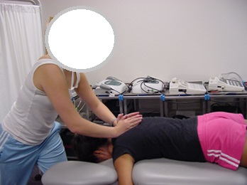
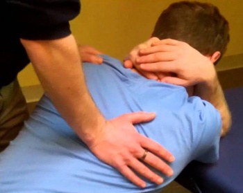
*Mobilisation of the rib is in an anteroposterior direction, using oscillations with contact on the dorsal surface of the rib, adjacent to the thoracic spinal articulation<ref name=":17" /> <ref name=":16">Hislop HJ, Avers D, Brown M. [https://scholar.google.com/scholar_url?url=https://books.google.com/books%3Fhl%3Den%26lr%3D%26id%3DpeNOAQAAQBAJ%26oi%3Dfnd%26pg%3DPP1%26dq%3DHislop%2BHJ,%2BAvers%2BD,%2BBrown%2BM.%2BDaniels%2Band%2BWorthingham%25E2%2580%2599s%2BMuscle%2BTesting%2B%2BTechniques%2Bof%2BManual%2BExamination%2Band%2BPerformance%2BTesting.%2B9th%2BEdition,%2BPa%2B%2BWB%2BSaunders%2BCo%253B%2B(2014)%26ots%3DmyHUAgYc1t%26sig%3DrZrZwnT9fjzjY0qvBEfLQ-rMO_w&hl=en&sa=T&oi=gsb&ct=res&cd=0&d=13640344623196258713&ei=RXX_Y7SYEcK1ywTR2b-ACw&scisig=AAGBfm0eFCAmDcdrIFz4H-klmJ1y1OfhgQ Daniels and Worthingham’s Muscle Testing]: Techniques of Manual Examination and Performance Testing. 9th Edition, Pa: WB Saunders Co; (2014)</ref> (figure 3). Rib distraction mobilisation can be an effective initial treatment. This technique is best done with the patient in a sitting or prone position. | |||
[[Image:Exer1.jpg]] | [[Image:Exer1.jpg]] | ||
<span style="font-size: 13.2799997329712px; line-height: 19.9200000762939px;">Figure 3 | <span style="font-size: 13.2799997329712px; line-height: 19.9200000762939px;">Figure 3 </span><ref name=":17" /> | ||
*A rib rotational glide is an effective technique and sometimes preferable to the rib distraction technique if direct pressure on the rib is too painful. This technique can be administered while the patient in standing, sitting, or in reclining position (figure 4) | *A rib rotational glide is an effective technique and sometimes preferable to the rib distraction technique if direct pressure on the rib is too painful. This technique can be administered while the patient in standing, sitting, or in reclining position (figure 4) | ||
| Line 192: | Line 189: | ||
[[Image:Exer2.jpg|350px]] | [[Image:Exer2.jpg|350px]] | ||
Figure 4 ( | Figure 4 <ref name="Maitland">Maitland, G., Hengeveld, E., Banks, K., English, K. (2005). Maitland' Vertebral Manipulation. 7th ed. Heinemann, Toronto: Elsevier Butterworth. (Level of evidence: 1B)</ref><br> | ||
Maitland suggests that mobilisations are effective for: | |||
*Treatment of stiffness <ref name="Maitland" /> | |||
*Treatment of stiffness | |||
*Treatment of pain, rather than stiffness. | *Treatment of pain, rather than stiffness. | ||
3 repetitions | 3 repetitions are usually performed, followed by retesting for pain on palpation. Mobilisations are repeated to any rib that was still painful until there is no longer palpable pain. <ref name="Maitland" /> | ||
Also General spinal mobilisations <ref name=":11">David L Aiken et al.; [https://scholar.google.com/scholar_url?url=https://www.tandfonline.com/doi/abs/10.1179/2042618612Y.0000000024&hl=en&sa=T&oi=gsb&ct=res&cd=0&d=8102289018380462918&ei=4HX_Y4ugHcK1ywTR2b-ACw&scisig=AAGBfm1OGacX_9uY4M_0MkhIFVVrvaSRsg The use of functional and traditional mobilization interventions in a patient with chronic thoracic pain;] J Man Manip Ther.; 2013 Aug; 21(3): 134–141</ref> and [[Elbow Mobilizations|Elbow mobilisations]] <ref name=":11" /> <br><br>'''Other manual techniques include:''' | |||
< | *Dorsal glides of the thoracic spinal facet joints (via the ribcage) <ref name=":12">Walter R, Essentials of Physical Medicine and Rehabilitation. Elsevier Saunders, Philadelphia, 2008, p 576</ref> . | ||
*Soft tissue techniques: massage ([[Deep Friction Massage|deep friction]], [[Friction Massage|friction and]] [[Massage|normal]]), stretching and trigger point release) <ref name=":12" /> | |||
*Massage of the scalene muscles <ref name=":13">Heiderscheit B., [https://scholar.google.com/scholar_url?url=https://www.tandfonline.com/doi/abs/10.1179/106698108790818369&hl=en&sa=T&oi=gsb&ct=res&cd=0&d=6860333891407900390&ei=CHb_Y8KJNY7gygSPuIegDA&scisig=AAGBfm0oX_nFmpK6Vte7Uf-CpXo46zFbQg Reliability of Joint Mobility and Pain Assessment of the Thoracic Spine and Rib Cage in Asymptomatic Individuals], J Man Manip Ther. 2008; 16(4): 210–216</ref> | |||
*[[Low-level laser therapy|Laser therapy]] of the ribs <ref name=":13" /> | |||
*[[Acupuncture|Acupuncture]] ([[Acupuncture contraindications|contra-indications]]) <ref name=":12" /> | |||
*High velocity, low amplitude chiropractic diversified adjustments in a anteroposterior direction with the patient in a supine position <ref name=":1" /> | |||
*High-velocity, low amplitude manipulations directed at the costovertebral, costotransverse and intervertebral zygapophyseal thoracic joints<ref name=":2" /> | |||
*Mobilisations of the costovertebral and costosternal joints: large amplitude P/A oscillations of the ribs in prone position. | |||
=== Exercise Therapy === | |||
*Chest lifts as a home exercise | |||
* | |||
Chest lifts may be a useful generic treatment technique for a rib articular dysfunction or thoracic facet joint dysfunction. With the patient in hand and knee position, reach underneath the chest with both hands, lace fingers together, and slowly and gently lift the torso. | |||
Repeat this manoeuvre a few times, slowly and rhythmically, lifting from different portions of the chest or sternum to create a movement at different segments of the thoracic spine. These techniques should be pain free. | |||
*Underwater treadmill therapy | |||
*Push-ups to activate the pectoral musculature (after 6th visit). 5 to 10 repetitions, 3 sets <ref name=":17" /> | |||
*Wall push ups (after 7th visit), progressively build up to full push ups. 5 to 10 repetitions, 3 sets <ref name=":17" /> | |||
*Rotation exercises while seated: sitting tall, back and neck straight and your arms across your chest. Keep the legs still and rotate to one side as far as able without pain, hold for 3 seconds and repeat 10-15 times to each side, alternating sides <ref name=":1" /> | |||
*Shoulder retraction: This exercise should first be performed while seated. In a later phase,a standing position can be adopted. Retract the shoulders as far as comfortably possible, without pain and hold for 3 seconds, repeating 10-15 times<ref name=":1" /> | |||
*Scapular stabilisation and postural re-education <ref name=":4" /><br> | |||
=== Self mobilisation techniques === | |||
Tape two tennis balls together to create an ellipse shaped ball.<ref name=":14" /> | |||
* | <br>Thoracic spine mini-crunches: | ||
* | * Place the balls on the segmental level below the on to be mobilised. | ||
* | * Lying on the balls and arms are crossed over the chest, which protracts the scapula, allowing the balls to make contact with the spine | ||
* Slowly lift the shoulders off the floor. Hold this position for 3 seconds. | |||
* Do 2-3 sets of 15-20 repetitions | |||
<br>Supine arm circles: | |||
* Place the balls at the point of discomfort | |||
* Lie down on the balls. Bring the shoulders to 90 degrees abduction. Draw circles with the wrist in a clockwise and then counterclockwise motion. | |||
* 2-3 sets of 30 to 60 seconds in both directions | |||
== Clinical Bottom Line == | == Clinical Bottom Line == | ||
This condition is often overlooked yet it can be responsible for pain in the thoracic region or functional impairments of the thoracic spine. It may occur due to trauma, degenerative changes, tumours, deformities or muscular spasm. Differential diagnosis are numerous and pain can be referred into the neck and/or head. A diagnosis is usually made through a clinical examination with treatment consisting of mobilisation, exercise therpay, injections and/or fixation of unstable joints. | |||
== References == | == References == | ||
[[Category:Primary Contact]] | |||
<references /> | |||
<references /> | |||
Latest revision as of 17:45, 1 March 2023
Original Editors - Yves Hubar
Top Contributors - Yves Hubar, Lisa De Donder, Scott Cornish, Simisola Ajeyalemi, Admin, Kim Jackson, Rachael Lowe, WikiSysop, Vidya Acharya, Chelsea Mclene, Maëlle Cormond, Claire Knott, 127.0.0.1, Laura Ritchie and Evan Thomas
Definition/Description[edit | edit source]
Costotransverse disorders are disorders affecting or involving the costotransverse and costovertebral joints and ligaments [1] which are often overlooked during examination for pain source localisation in this area due to possible visceral pain referral and the complexities of the thoracic neural network.[2] It is suggested that dysfunctions in these joints could account for pain in the thorax or functional impairments. [1] [3][4]
Clinically Relevant Anatomy[edit | edit source]
- The costotransverse joint is an articulation between the articular costal tubercle of the rib and the costal facet of the transverse process of a thoracic vertebra.[5] The costovertebral joint is the articulation between the costal facte or demi-facets (formed by the caudal side of the superior vertebra and the cranial side of the inferior vertebra) and the head of the rib [4] These facets form a solid angle whose base consists of the annulus fibrosis of the intervertebral disc. A synovial joint ensures the connection between the rib and the thoracic vertebra. [6] Together with the thoracic cage, the costovertebral and costotransverse joints provide stability.[4]
- Ligaments: The following costotransverse and costovertebral ligaments connect the two joints-[7] [8] Ligamentum costotransversarium superius, Ligamentum costotransversarium., Ligamentum costotransversarium laterale, Ligamentum capitis costae radiatum, Inferior costotransverse ligaments, Posterior costotransverse ligaments, identified on the fifth to tenth ribs. These ligaments limit movement in the costotransverse joint to a minimal gliding movement. The ribs articulate posteriorly twice with the corresponding vertebra. The radiate ligament and the intra-articular ligament stabilise the head of ribs 2 to 9 and sometimes the head of the 10th rib in the costovertebral joint.
- The head of ribs 2 to 9 and sometimes the 10th articulate with the vertebral body of two thoracic vertebrae at the costovertebral joint. The head of ribs 1, 11 and 12 articulate with the corresponding vertebrae. There is no intra-articular ligament in these joints.
- The neck of ribs 1 to 10 articulate, through the tubercle, with the transverse process of their corresponding vertebrae and are stabilised with ligaments and the articular capsule. Ribs 11 and 12 do not articulate with the transverse processes. [9]
- Lateral flexion as well as rotation is limited by the ribcage therefore only small movements are possible in the costotransverse joints. There is no sagittal plane flexion and extension in these joints, however gliding movements within the joint have been seen. These gliding movements are mostly medially and laterally orientated. The medial and lateral glides are functional motions. [10]
Figure 1: Ligaments connecting the ribs and vertebra
Movement of the ribs at the costovertebral joints [6]. The axes of the ribs allow three basic types of motion:
- Bucket-handle motion: One end of the rib is fixed at the vertebral end, the majority of the rib elevation occurs through upward excursion of the lateral position. This motion increases the transverse diameter of the rib cage.
- Pump-handle motion: One end is fixed, and the free end describes an arc. When the ribs move around the axis, the anterioposterior diameter of the rib cage increases.
- Caliper motion: The 11th and 12th ribs have only costovertebral articulations. The motion produces slight changes in both the transverse and the anteroposterior dimension.
All twelve ribs have pump handle and bucket handle movements, but the upper ribs have a greater pump handle motion and the lower ones have more bucket handle type movements.
This link explains pump-handle and bucket-handle motion mechanics of the rib cage during respiration[11].
Epidemiology /Etiology[edit | edit source]
- Local joint compression may occur as a result of a trauma or muscle spasm [12]
- It is more common in women [3] [13] and can occur at any age
- When subjected to severe trauma, these joints can subluxate or dislocate. Due to being at the top of the rib cage, the first costotransverse joint is the most vulnerable. [14]
- Though distinctly unusual at the costotransverse and costovertebral joint, rheumatoid arthritis can occur in these joints.[15]
- The involvement of dysfunction at these joints are a source of referred pain at the thoracic spine, with suggested involvement of the costotransverse joint concerning T4 syndrome [1]
- The costotransverse joint is known to be involved in patients with ankylosing spondylitis, which, combined with involvement of the costovertebral, sternoclavicular and sternomanubrial joints, would result in increased rigidity of the thorax and increased dorsal kyphosis. This does not result in decreased pulmonary function, which is possibly due to an increase in diaphragmatic breathing [16]
- Due to the positive response to manual therapy directed towards posterior spinal structures, conditions diagnosed as costochondritis might actually be caused by neurogenic inflammation.[17] [3]
Characteristics/Clinical Presentation[edit | edit source]
- Pain localised to the posterior thorax [19]
- Pain may radiate to the anterior chest wall, along the rib, sometimes into the shoulder and sometimes towards the upper limb [2]
- Unilateral symptoms [19]
- Pain with deep inspiration, coughing/sneezing/laughing
- Increased pain with passive or active thoracolumbar flexion, rotation and ipsilateral side bending, lifting or twisting movements
- Hypomobility of the costotransverse and costovertebral joint
- Palpable tenderness and pain at costotransverse joint and rib angle
- Movement in adjacent thoracic vertebral and rib segments is usually restricted and may stimulate or exacerbate protective muscle spasm
- Increased muscle tension in paraspinal muscles, rhomboid muscles, trapezius muscles and levator scapula muscles
- Neck pain, head ache or both
- The sensation of having a useless or heavy limb
- Referred pain originating from under the scapula and worsens with coughing, sneezing or deep breathing
- Acute, atypical chest pain
Differential Diagnosis[edit | edit source]
Differential diagnostic possibilities have to be taken into consideration in cases of posterior upper thoracic and/or scapular pain and possible involvement of costovertebral and costotransverse joints. [19] Possible musculoskeletal sources of thoracic pain are: [20] [19]
- Muscle strain (erector spinae, lower and middle trapezius, rhomboids, latissimus dorsi, levator scapulae and intercostal muscles)
- Vertebral or rib fracture
- Zygapophyseal joint arthropathy
- Active trigger points
- Spinal stenosis
- Intervertebral disc protrusion or herniation
- Diffuse idiopathic skeletal hyperostosis (DISH)
- Intercostal neuralgia: often follows injury or thoracic surgery, focal tenderness over affected intercostal region , burning pain and paraesthesia in thorax or abdomen that usually follow the nerve path [21]
- T4 syndrome
- Ankylosing spondylitis
- Costovertebral and costotransverse joint dysfunction
- Thoracic radiculopathy [22]
- Fractures
Possible visceral sources of thoracic pain[20] [19]
- Cancer
- Cardiac conditions
- substernal pain;
- shortness of breath
- increased pain with exertion;
- frequent left shoulder, medial arm and jaw pain [23]
- Renal conditions
- referred pain to the ipsilateral subcostal and costovertebral regions at T10-T12;
- typically dull and aching pain;
- possible changes in urinary frequency or output [20]
- Pulmonary conditions
- Gastroesophageal conditions
- band like pain around mid-thorax at the level of the lesion;
- referred pain to the mid thoracic area;
- nausea/vomiting;
- weight loss, heartburn or substernal pain;
- stabbing or burning chest pain [24]
- Gall bladder conditions
- pain in the right mid epigastric region (T8-T9 level);
- referred pain at the mid back between the scapulae, right upper trapezius muscle and right subscapular area;
- jaundice
- fever
- chills
- indigestion
- nausea/vomiting
- intolerance of fatty foods [25]
- Hepatobiliary conditions
- pain in the right upper abdomen quadrant
- referred pain at right interscapular and subscapular areas
- right shoulder pain
- anorexia
- nausea
- vomiting
- jaundice
- ascites
- significant fatigue [25]
Diagnostic Procedures[edit | edit source]
Clinical findings and patient presentation are used to diagnose costovertebral and costotransverse joint dysfunction. This is done through a thorough anamnesis and clinical examination [26] [27]
Outcome Measures[edit | edit source]
Pain is measured with a numeric pain scale (VAS), whilst function is measured with the Dallas Pain Questionnaire and Functional Rating Index.[13]
Examination[edit | edit source]
The physical examination consists of the following components:[19]
- Inspection of the posterior, lateral and anterior sides of the trunk, to check for deviations from an ideal posture (standing) [19]
- Active range of motion of the cervical (seated), trunk (standing) and shoulder regions (seated) to determine pain provoking movements [29]
| [30] | [31] |
- Manual muscle testing to assess upper extremities strength and intensity of possible elicited pain (seated) [33]
- Sensory examination of the upper extremities and thoracic area , in order to determine whether nerve root or peripheral nerve lesions are present [19]
- Accessory motion or joint movement of the thoracic spine (in prone position) to check for pain and mobility [35] [36] using the posterioranterior (PA) pressure test. [37]
- Posterior (prone) and anterior (supine) costosternal joint movement to assess for pain and mobility [35] [36]
- Active and passive scapular mobility in case of reduced active shoulder range of motion and/or pain of the scapula region [29]
| [39] | [40] |
Medical Management[edit | edit source]
In an unstable thoracic spine (caused by trauma, degenerative conditions, tumors and deformities) posterior instrumentation and fusion are commonly used as treatment. Numerous techniques have been reported to restore spinal stability and correct spinal deformities. Posterior fixation of the thoracic spine through the costotransverse joint may be an alternative (figure 2).
Thanapipatsiri and Chan [41] showed that posterior thoracic fixation through the transverse processes is safe as implants placed beneath the transverse processes are away from the parietal pleura and the intercostal vessels and nerves. Heller et al. demonstrated that transverse process screws were weaker biomechanically than pedicle screws in the upper thoracic spine, but safer technically [42] Fixation around the transverse processes in the thoracic spine appears to be simple and safe, but fixation through the costotransverse joint may be more rigid than through the transverse process.
Risks of costotransverse screw fixation:
- Injury of the intercostal neurovascular structures.
- Injury of the parietal pleura.
- Fixation at multiple levels may result in limitation of movement of the thoracic cage and may affect the long function.
Figure 2: posterior costotransverse screw insertion technique. [43]
Patients with upper back pain, pain between the ribs and thorax pain may be candidates for a costotransverse or costovertebral joint injection. The injections have two purposes: to confirm the diagnosis that these joints are the source of the back pain and secondly, to offer temporary pain relief.[44][45]
Physical Therapy Management[edit | edit source]
Physical therapy interventions have been shown to be effective at improving the prognosis of back pain in the thoracic region. [46]
Mobilisation[edit | edit source]
- Mobilisation of the rib is in an anteroposterior direction, using oscillations with contact on the dorsal surface of the rib, adjacent to the thoracic spinal articulation[13] [47] (figure 3). Rib distraction mobilisation can be an effective initial treatment. This technique is best done with the patient in a sitting or prone position.
Figure 3 [13]
- A rib rotational glide is an effective technique and sometimes preferable to the rib distraction technique if direct pressure on the rib is too painful. This technique can be administered while the patient in standing, sitting, or in reclining position (figure 4)
Figure 4 [48]
Maitland suggests that mobilisations are effective for:
- Treatment of stiffness [48]
- Treatment of pain, rather than stiffness.
3 repetitions are usually performed, followed by retesting for pain on palpation. Mobilisations are repeated to any rib that was still painful until there is no longer palpable pain. [48]
Also General spinal mobilisations [49] and Elbow mobilisations [49]
Other manual techniques include:
- Dorsal glides of the thoracic spinal facet joints (via the ribcage) [50] .
- Soft tissue techniques: massage (deep friction, friction and normal), stretching and trigger point release) [50]
- Massage of the scalene muscles [51]
- Laser therapy of the ribs [51]
- Acupuncture (contra-indications) [50]
- High velocity, low amplitude chiropractic diversified adjustments in a anteroposterior direction with the patient in a supine position [4]
- High-velocity, low amplitude manipulations directed at the costovertebral, costotransverse and intervertebral zygapophyseal thoracic joints[3]
- Mobilisations of the costovertebral and costosternal joints: large amplitude P/A oscillations of the ribs in prone position.
Exercise Therapy[edit | edit source]
- Chest lifts as a home exercise
Chest lifts may be a useful generic treatment technique for a rib articular dysfunction or thoracic facet joint dysfunction. With the patient in hand and knee position, reach underneath the chest with both hands, lace fingers together, and slowly and gently lift the torso.
Repeat this manoeuvre a few times, slowly and rhythmically, lifting from different portions of the chest or sternum to create a movement at different segments of the thoracic spine. These techniques should be pain free.
- Underwater treadmill therapy
- Push-ups to activate the pectoral musculature (after 6th visit). 5 to 10 repetitions, 3 sets [13]
- Wall push ups (after 7th visit), progressively build up to full push ups. 5 to 10 repetitions, 3 sets [13]
- Rotation exercises while seated: sitting tall, back and neck straight and your arms across your chest. Keep the legs still and rotate to one side as far as able without pain, hold for 3 seconds and repeat 10-15 times to each side, alternating sides [4]
- Shoulder retraction: This exercise should first be performed while seated. In a later phase,a standing position can be adopted. Retract the shoulders as far as comfortably possible, without pain and hold for 3 seconds, repeating 10-15 times[4]
- Scapular stabilisation and postural re-education [19]
Self mobilisation techniques[edit | edit source]
Tape two tennis balls together to create an ellipse shaped ball.[46]
Thoracic spine mini-crunches:
- Place the balls on the segmental level below the on to be mobilised.
- Lying on the balls and arms are crossed over the chest, which protracts the scapula, allowing the balls to make contact with the spine
- Slowly lift the shoulders off the floor. Hold this position for 3 seconds.
- Do 2-3 sets of 15-20 repetitions
Supine arm circles:
- Place the balls at the point of discomfort
- Lie down on the balls. Bring the shoulders to 90 degrees abduction. Draw circles with the wrist in a clockwise and then counterclockwise motion.
- 2-3 sets of 30 to 60 seconds in both directions
Clinical Bottom Line[edit | edit source]
This condition is often overlooked yet it can be responsible for pain in the thoracic region or functional impairments of the thoracic spine. It may occur due to trauma, degenerative changes, tumours, deformities or muscular spasm. Differential diagnosis are numerous and pain can be referred into the neck and/or head. A diagnosis is usually made through a clinical examination with treatment consisting of mobilisation, exercise therpay, injections and/or fixation of unstable joints.
References[edit | edit source]
- ↑ 1.0 1.1 1.2 B A Young, H E Gill, R S Wainner, T W Flynn. Thoracic costotransverse joint pain patterns: a study in normal volunteers. BMC Musculoskelet disord. 2008; 9: 140
- ↑ 2.0 2.1 2.2 Smith H S, Current Therapy in Pain. Saunders Elsevier, 2009: p199
- ↑ 3.0 3.1 3.2 3.3 D Aspegren, T Hyde, M Miller. Conservative treatment of a female collegiate volleybal player with costochondritis. Journal of manipulative and physiological therapeutics (2007) 30(4): 321-325
- ↑ 4.0 4.1 4.2 4.3 4.4 4.5 Edge-Hughes, L. Canine Thoracic Costovertebral and Costotransverse Joints: Three Case Reports of Dysfunction and Manual Therapy Guidelines for Assessment and Treatment of These Structures. Topics in Compan An Med; 29, 1: 1–5.
- ↑ DA. Kinesiology of the Musculoskeletal System: Foundations for Physical Rehabilitation. St. Louis: Mosby 2002. ISBN: 978-0-8151-6349-7
- ↑ 6.0 6.1 Irwin J. et al, The effect of costovertebral adjustment versus ischaemic compression of rhomboid muscles for interscapular pain, university of johannesburg, 2015
- ↑ A F Ibrahim, H H Darwish. The costotransverse ligaments in human: a detailed anatomical study. Clinical Anatomy 18:340-345 (2008)
- ↑ Agur A.M.R., Lee M.J. Grant's Atlas of Anatomy. Lippincott Williams & Wilkins 1999 (10th ed.) ISBN: 978-0683302646
- ↑ Andor W.J.M. Glaudemans et al. Nuclear Medicine and Radiologic Imaging in Sports Injuries. Springer, 2015, 259-260
- ↑ Sharon Weiselfish-Giammatteo, Integrative Manual Therapy for Biomechanics: Application of muscle energy and “beyond” technique. Noth Atlantic Books Berkeley, California, 2003, p 261
- ↑ Rib Cage Biomechanics - Visualization Technologyhttps://hal.bim.msu.edu/CMEonLine/RibCage/Biomechanics/start.html
- ↑ Thomas E. Hyde, Conservative Management of Sports Injuries. Jones and Barlett Publishers, 2007, p440.
- ↑ 13.0 13.1 13.2 13.3 13.4 13.5 Hudes, K. Low-tech rehabilitation and management of a 64 year old male patient with acute idiopathic onset of costochondritis. Family Chiropractic & Rehabilitation, (2008) 52 (4), 224-228.
- ↑ 14.0 14.1 Christensen EE, Dietz GW. Injuries of the costovertebral articulation. Radiology 1980. Jan; 134(1): 41-3
- ↑ MJ Cohen, J Ezekiel, RH Persellin. Costovertebral and costotransverse joint involvement in rheumatoid artritis. Ann Rheum Dis 1978 October; 37(5): 473-475
- ↑ Z Unlu, M can, C Goktan, P Celik. Lumbar stuffness but not thoracic radiographic changes relate to alteration of lung function tests in ankylosing spondylitis. Clin Rheumatol (2002) 21(4):275-279
- ↑ M I Rabey. Costochondritis: are the symptoms and signs due to neurogenic inflammation. Two cases that responded to manual therapy directed towards posterior spinal structures. Manual therapy (2008) 13(1): 82-86
- ↑ Edge-Hughes, L. Canine Thoracic Costovertebral and Costotransverse Joints: Three Case Reports of Dysfunction and Manual Therapy Guidelines for Assessment and Treatment of These Structures. Topics in Compan An Med; 29, 1: 1–5
- ↑ 19.0 19.1 19.2 19.3 19.4 19.5 19.6 19.7 19.8 Fruth S J. Differential diagnosis and treatment in a patient with posterior upper thoracic pain. Physical therapy (2006) 86(2): 254-268
- ↑ 20.0 20.1 20.2 McConaghy JR, Oza RS. Outpatient diagnosis of acute chest pain in adults. Am Fam Physician (2013) Feb 1;87(3):177-82
- ↑ Williams EH et al. Neurectomy for treatment of intercostal neuralgia. Ann Thorac Surg (2008) May;85(5):1766-70.
- ↑ Walter R, Essentials of Physical Medicine and Rehabilitation. Elsevier Saunders, Philadelphia, 2008, p 576.
- ↑ 23.0 23.1 Haasenritter J et al. Does the patient with chest pain have a coronary heart disease? Diagnostic value of single symptoms and signs – a meta-analysis. Croat Med J (2012) Oct;53(5):432-41.
- ↑ Spiegel BM et al. Development of the NIH Patient-Reported Outcomes Measurement Information System (PROMIS) gastrointestinal symptom scales. Am J Gastroenterol (2014) Nov;109(11):1804-14
- ↑ 25.0 25.1 Duncan CB, Riall TS. Evidence-based current surgical practice: calculous gallbladder disease. J Gastrointest Surg (2012) Nov;16(11):2011-25
- ↑ Scaringe JG, Ketner C. Manual methods for the treatment of rib dysfunctions and associated functional lesions. Topics in Clinical Chiropractic. (1999);6:20-38.
- ↑ Triano JJ et al. Costoverebral and costotransverse joint pain: a commonly overlooked pain generator. Topics in Clinical Chiropractic. (1999);6:79-92.
- ↑ Youtube (2012). Macleod’s examination of the thoracic and lumbar spine. Consulted on May 28, 2014 on https://www.youtube.com/watch?feature=player_detailpage&v=3UhKFcR1Wbs
- ↑ 29.0 29.1 Huygen F et al. Painful shoulder complaints. Pain Pract. (2010) Jul-Aug;10(4):318-26. (LOE: 1A)
- ↑ Youtube (2009). Range of Motion Test – Cervical ROM. Consulted on May 28, 2014 on https://www.youtube.com/watch?v=PrbmBMehYx0
- ↑ Youtube (2012). Range of Motion Test – Thoracic ROM. Consulted on May 28, 2014 on https://www.youtube.com/watch?v=_u0fPi2XBls
- ↑ Youtube (2009). Shoulder Exam (3 of 9): Range of motion. Consulted on May 28, 2014 on https://www.youtube.com/watch?v=d7HfaAlgaro
- ↑ Hislop HJ, Avers D, Brown M. Daniels and Worthingham’s Muscle Testing: Techniques of Manual Examination and Performance Testing. 9th Edition, Pa: WB Saunders Co; (2014) (LOE: 3A)
- ↑ Youtube (2012). Macleod’s examination of the upper limbs sensation. Consulted on May 28, 2014 on https://www.youtube.com/watch?feature=player_detailpage&v=wSSCZeOKXBE
- ↑ 35.0 35.1 Hicks GE et al. Interrater reliability of clinical examination measures for identification of lumbar segmental instability. Arch Phys Med Rehabil. (2003);84:1858-1864 (LOE: 2B)
- ↑ 36.0 36.1 Seffinger MA et al. Reliability of spinal palpation for diagnosis of back and neck pain: a systematic review of the literature. Spine. (2004);29:E413-425 (LOE: 2A)
- ↑ Heiderscheit B., Reliability of Joint Mobility and Pain Assessment of the Thoracic Spine and Rib Cage in Asymptomatic Individuals, J Man Manip Ther. 2008; 16(4): 210–216
- ↑ Youtube (2009). Shoulder Exam (4 of 9): Scapular control (Is there scapular dyskinesia?). Consulted on May 28, 2014 on https://www.youtube.com/watch?v=pEY93k5XXL0
- ↑ Youtube (2011). Macleod’s Examination of cervical spine (non traumatic). Consulted on May 28, 2014 on https://www.youtube.com/watch?v=qdi3VKA-wJI
- ↑ Youtube (2009). Shoulder Exam (2 of 9): Inspection and Palpation. Consulted on on May 28 on https://www.youtube.com/watch?v=Xf52jbNA7wg
- ↑ Thanapipatsiri S, Chan DPK. Safety of thoracic transverse process fixation: an anatomic study. J Spinal Disorders 1996;9:294–8. (Level of evidence 4)
- ↑ Heller JG, Shuster JK, Hutton A. Pedicle and transverse process screws of the upper thoracic spine. Spine 1999;24:654–8. (Level of evidence: 4)
- ↑ 43.0 43.1 Rongming, X., Nabil, A.E., Yiangjia, O., Martin, S., Richard, A.Y. (2000). Anatomic Considerations of Costotransverse Screw Placement in the Thoracic Spine. Surg Neurol 2000;53:349 –55. (Level of evidence: 4)
- ↑ Baker R.; Costotransverse and costovertebral joint injections.; Spine-health.; Sept 2004
- ↑ Brian A Young et al.; Thoracic costotransvers joint pain patterns:a study in normal volunteers. BMC Musculoskeletal Disorders2008 9:140
- ↑ 46.0 46.1 . Johnson K. et al, Thoracic region self-mobilization: a clinical suggestion, The international journal of sports physical therapy volume 7 number 2, april 2012, P252-256
- ↑ Hislop HJ, Avers D, Brown M. Daniels and Worthingham’s Muscle Testing: Techniques of Manual Examination and Performance Testing. 9th Edition, Pa: WB Saunders Co; (2014)
- ↑ 48.0 48.1 48.2 Maitland, G., Hengeveld, E., Banks, K., English, K. (2005). Maitland' Vertebral Manipulation. 7th ed. Heinemann, Toronto: Elsevier Butterworth. (Level of evidence: 1B)
- ↑ 49.0 49.1 David L Aiken et al.; The use of functional and traditional mobilization interventions in a patient with chronic thoracic pain; J Man Manip Ther.; 2013 Aug; 21(3): 134–141
- ↑ 50.0 50.1 50.2 Walter R, Essentials of Physical Medicine and Rehabilitation. Elsevier Saunders, Philadelphia, 2008, p 576
- ↑ 51.0 51.1 Heiderscheit B., Reliability of Joint Mobility and Pain Assessment of the Thoracic Spine and Rib Cage in Asymptomatic Individuals, J Man Manip Ther. 2008; 16(4): 210–216
