Costochondritis: Difference between revisions
Kim Jackson (talk | contribs) No edit summary |
Kim Jackson (talk | contribs) No edit summary |
||
| (20 intermediate revisions by 3 users not shown) | |||
| Line 1: | Line 1: | ||
<div class="editorbox"> '''Original Editor '''- [[User:User Name|Nick Demunter]] '''Top Contributors''' - {{Special:Contributors/{{FULLPAGENAME}}}}</div> | <div class="editorbox"> '''Original Editor '''- [[User:User Name|Nick Demunter]] '''Top Contributors''' - {{Special:Contributors/{{FULLPAGENAME}}}}</div> | ||
Palpation of the affected chondrosternal joints of the chest wall elicits tenderness <ref name=":0" /> and pain is reproduced by palpation of the affected cartilage segments which may radiate out into the chest wall. | == Definition/Description == | ||
<br>Costochondritis is a self-limiting condition defined as painful chronic inflammation of the costochondral junctions of [[ribs]] or chondrosternal joints of the anterior chest wall.<ref name=":0">PROULX A and TERESA W.; Costochondritis: Diagnosis and Treatment; ''Am Fam Physician.'' 2009 Sep 15;80(6):617-620</ref> | |||
* It is a clinical diagnosis and does not require specific diagnostic testing in the absence of concomitant cardiopulmonary symptoms or risk factors. | |||
* Costochondritis is often confused with [[Tietzes|Tietze syndrome]]. | |||
* Palpation of the affected chondrosternal joints of the chest wall elicits tenderness <ref name=":0" /> and pain is reproduced by palpation of the affected cartilage segments which may radiate out into the chest wall. | |||
== Clinically Relevant Anatomy == | |||
The thoracic wall consists of the | |||
* Sternum anteriorly, | |||
* 12 thoracic vertebrae posteriorly, | |||
* 12 paired ribs and associated costal cartilages.<ref name=":3">Clemens WM. et al. ; Introduction to Chest Wall Reconstruction : Anatomy and Physiology of the Chest and Indications for Chest Wall Reconstruction ; Semin Plast Surg. ; 2011 ; 25(1) : 5-15</ref> | |||
Ribs consist of [[bone]] and [[cartilage]], with cartilage serving as an elastic bridge between the bony portion of the rib and the sternum. | |||
[[ | According to their attachment to the sternum, the ribs are classified into 3 groups: true, false, and floating ribs. | ||
[ | # True ribs are the ribs that directly articulate with the sternum with their costal [[cartilage]]<nowiki/>s - ribs 1-7. They articulate with the sternum by the sternocostal joints. The first rib is an exception to that rule; it is a [[Joint Classification|synarthrosis]] and the first rib could uniquely articulate with the clavicle by the costoclavicular joint | ||
# The false ribs (8,9,10) are the ribs that indirectly articulate with the sternum, as their costal cartilages connect with the seventh costal cartilage by the costochondral joint. | |||
# The floating ribs (11,12) do not articulate with the sternum at all (distal two ribs)<ref>Safarini OA, Bordoni B. [https://www.ncbi.nlm.nih.gov/books/NBK538328/ Anatomy, Thorax, Ribs]. InStatPearls [Internet] 2019 Feb 19. StatPearls Publishing.Available from:https://www.ncbi.nlm.nih.gov/books/NBK538328/ (last accessed 14.4.2020)</ref>. | |||
<br><span>The ribs move with respiration and with truncal motion or movement of the upper extremities.</span><ref name=":3" /> | |||
== | == Aetiology == | ||
Costochondritis is inflammatory. It is caused by inflammation of the costal cartilages and their sternal articulations, also known as the costochondral junctions<ref name=":1">Schumann JA, Parente JJ. [https://www.ncbi.nlm.nih.gov/books/NBK532931/ Costochondritis].Available from:https://www.ncbi.nlm.nih.gov/books/NBK532931/ (last accessed 29.4.2020)</ref>. | |||
=== Epidemiology === | |||
The epidemiology of costochondritis is not well established. | |||
* In a small study published in 1994, there was a higher frequency of costochondritis seen in females and Hispanics. | |||
* In a group of 122 patients presenting to the emergency department with chest pain not due to malignancy, fever, or trauma, costochondritis was the diagnosis in 36 of the patients (30%)<ref name=":1" /> | |||
* Can affect children as well as adults. A study of chest pain in an outpatient adolescent clinic found that 31 percent of adolescents had musculoskeletal causes, with costochondritis accounting for 14 percent of adolescent patients with chest pain<ref name=":0" />. | |||
== Characteristics/Clinical Presentation == | == Characteristics/Clinical Presentation == | ||
[[File:Ashkan-forouzani-oxaIBWkrGXE-unsplash.jpg|right|frameless]] | |||
Consider pursuing other causes of chest pain prior to establishing a costochondritis diagnosis as Costochondritis is a diagnoses of exclusion. Cardiac and respiratory causes will need to be ruled out. If the patient complains of radiating pain, shortness of breath, dizziness, exertional chest pain, fever, or productive cough these are symptoms that may indicate different and more serious causes of chest pain. If there has been trauma an occult rib fracture should also be considered. If cardiopulmonary causes and trauma have been excluded the below findings should be present to varying degrees<ref name=":1" />. | |||
Possible findings include | |||
* Patient will give a history of the pain worsening with movement and certain positions. The pain will also typically be worse when the patient takes a deep breath. | |||
* Pain quality is variable, but it may be described as a sharp or dull pain. | |||
* Patients report a gradual or rapid onset of pain and swelling of the upper costal cartilage of the costochondral junction. | |||
* Pain is usually reproducible by mild-to-moderate palpation. Often, there is point tenderness where one or two ribs meet the sternum (a pitfall of the typical physical exam findings is that pain due to acute coronary syndrome can also be described as reproducible)<ref name=":1" />. | |||
* Symptoms may occur gradually and can disappear spontaneously after a few days, but equally it may take years to disappear. <ref name=":4">Fam A.G., Smythe H.A.,Musculoskeletal chest wall pain, Can Med Assoc J. Sept 19851; 133(5):379-389</ref> <ref>Gregory P.L., BISWAS A.C., Batt M.E.,Musculoskeletal problems of the chest wall in athletes, Sports Med., 2002;32(4):235-50.</ref> Even after the symptoms have resolved, they may return at the same location or at another rib level. <ref name=":2">Hurst J.W., Morris D.C., Williams B.R. “Chest Pain” in patients with costochondritis or Tietze's syndrome, Wiley-Blackwell, 2001, p23-29</ref> | |||
* There may be hypomobility of the upper thoracic spine, costovertebral joints, and the lateral ribs.<ref name=":5">Han J N et al.; Respiratory function of the rib cage muscles; European Respiratory Journal ISSN 0903 1993. </ref> | |||
== Evaluation == | |||
Costochondritis is usually self-limited and benign - should be distinguished from other, more serious causes of chest pain. | |||
</ref> | * [[Coronary Artery|Coronary artery disease]] is present in 3 to 6 percent of adult patients with chest pain and chest wall tenderness to palpation. | ||
* History and physical examination of the chest that document reproducible pain by palpation over the costal cartilages are usually all that is needed to make the diagnosis in children, adolescents, and young adults. | |||
* Patients older than 35 years, those with a history or risk of coronary artery disease, and any patient with cardiopulmonary symptoms should have an electrocardiograph and possibly a chest radiograph<ref name=":6" /> | |||
* Consider further testing to rule out cardiac causes if clinically indicated by age or cardiac risk status<ref name=":6">Proulx AM, Zryd TW. [https://www.ncbi.nlm.nih.gov/pubmed/19817327 Costochondritis: diagnosis and treatment.] American family physician. 2009 Sep 15;80(6):617-20.Available from:https://www.ncbi.nlm.nih.gov/pubmed/19817327 (last accessed 29.4.2020)</ref> | |||
== Differential Diagnosis == | == Differential Diagnosis == | ||
The differential diagnosis for costochondritis is rather long. Some of the diagnoses included are associated with major morbidity and mortality. eg | |||
* [[Acute Coronary Syndrome]] (ACS) | |||
* | * [[Pneumothorax]] | ||
* | * [[Pneumonia]] | ||
* [[Pulmonary Embolism]] | |||
* [[Tietzes|Tietze’s]] syndrome, much less common than costochondritis, and it tends to cause chest swelling in addition to the other symptoms, | |||
* Xiphoidalgia: painful swelling and discomfort of the xiphoid process of the sternum <ref>Brian E Udermann et al.; Slipping Rib Syndrome in a Collegiate Swimmer: A Case Report; J Athl Train. 2005 Apr-Jun; 40(2): 120–122</ref> | * Xiphoidalgia: painful swelling and discomfort of the xiphoid process of the sternum <ref>Brian E Udermann et al.; Slipping Rib Syndrome in a Collegiate Swimmer: A Case Report; J Athl Train. 2005 Apr-Jun; 40(2): 120–122</ref> | ||
* Slipping | * [[Slipping Rib Syndrome]]: hypermobility of the anterior ends of the false rib costal cartilages <ref name=":8">Brenda M. Birmann et al.; Prediagnosis biomarkers of insulin-like growth factor-1, insulin, and interleukin-6 dysregulation and multiple myeloma risk in the Multiple Myeloma Cohort Consortium. Blood. 2012 Dec 13; 120(25): 4929–4937.</ref><sup></sup><sup></sup> | ||
<sup></sup | |||
<sup></sup> | |||
== Outcome Measures == | == Outcome Measures == | ||
[http://www.physio-pedia.com/Patient_Specific_Functional_Scale Patient-specific functional scale ( PSFS)]: specific questionnaires for costochondritis have not yet been produced, but the PSFS is a valid, reproducable, and responsive outcome measure for patients with neck pain, back pain, and upper quarter complaints 19 | [http://www.physio-pedia.com/Patient_Specific_Functional_Scale Patient-specific functional scale ( PSFS)]: specific questionnaires for costochondritis have not yet been produced, but the PSFS is a valid, reproducable, and responsive outcome measure for patients with neck pain, back pain, and upper quarter complaints 19 | ||
[http://www.tandfonline.com/doi/pdf/10.1179/jmt.2009.17.3.163?redirect=1&#.V2bdQo9OLmK The Global rating of change (GROC)]: to measure the patient’s subjective rate of improvement .<ref name=":0" /> | [http://www.tandfonline.com/doi/pdf/10.1179/jmt.2009.17.3.163?redirect=1&#.V2bdQo9OLmK The Global rating of change (GROC)]: to measure the patient’s subjective rate of improvement.improvement .<ref name=":0" /> | ||
Measurement of thoracic and cervical mobility:<ref>FREESTON J; Can Early Diagnosis and Management of Costochondritis Reduce Acute Chest Pain Admissions?; The Journal of Rheumatology November 2004, 31 (11) 2269-2271</ref> | |||
* Rotation of the thoracolumbar spine (TR): TR has high validity and sensitivity ranks and improvement of the measurement technology would probably result in a superior test for the follow-up. | * Rotation of the thoracolumbar spine (TR): TR has high validity and sensitivity ranks and improvement of the measurement technology would probably result in a superior test for the follow-up. | ||
* Finger to floor distance (FFD): high reliability and sensitivity, but poor correlation with spinal changes | * [[Fingertips to Floor Distance - Special Test|Finger to floor distance]] (FFD): high reliability and sensitivity, but poor correlation with spinal changes | ||
* [http://www.physio-pedia.com/Schober_test The Schober test] | * [http://www.physio-pedia.com/Schober_test The Schober test] | ||
* Thoracolumbar flexion | * Thoracolumbar flexion | ||
* Occiput to wall distance <ref>Viitanena J, H. Kautiainena, J. Sunia, M. L. Kokkoa & K. Lehtinena; The Relative Value of Spinal and Thoracic Mobility Measurements in Ankylosing Spondylitis; Scandinavian Journal of Rheumatology; Volume 24, 1995 - Issue 2</ref> | * [[Occiput to Wall Distance OWD|Occiput to wall]] distance <ref>Viitanena J, H. Kautiainena, J. Sunia, M. L. Kokkoa & K. Lehtinena; The Relative Value of Spinal and Thoracic Mobility Measurements in Ankylosing Spondylitis; Scandinavian Journal of Rheumatology; Volume 24, 1995 - Issue 2</ref> | ||
== Examination == | == Examination == | ||
Patients with Costochondritis will present with | [[File:Katherine-hanlon-QgcdtM9rA5s-unsplash.jpg|alt=|right|frameless]] | ||
Patients with Costochondritis will present with: | |||
Palpation should be performed with 1 digit, on the anterior, posterior and lateral side of the chest, the clavicle, the cervical and thoracic spine. When on the affected area it reveals a | * Chest pain reproducible by palpation of the affected area, with ribs 2 to 5 mostly affected. | ||
* Aggravating factors can be slouching or exercise. | |||
Motion palpation is a manual process of moving a joint into its maximal end range of motion, after which it is challenged with a light springing movement. This end point of joint movement forms the basis for determining the normal or abnormal joint movement. When motion palpation is reduced, the joint is considered fixated or hypokinetic.<ref name=":9" /> | * Often occurs after a recent illness with coughing or after intense exercise and it mostly of unilateral origin.<ref name=":0" /> | ||
* May be an associated restriction of the corresponding costovertebral and costotransverse on examination. | |||
Cardiac causes should be ruled out in patients who present with a high risk. | * Loss of normal spinal movement associated with the chest pain.<ref name=":9">Aspegren D; Conservative Treatment of a Female Collegiate Volleyball Player with Costochondritis ; Journal of Manipulative and Physiological Therapeutics ; May 2007 Volume 30, Issue 4, Pages 321–325 </ref> | ||
* Palpation should be performed with 1 digit, on the anterior, posterior, and lateral side of the chest, the clavicle, the cervical and thoracic spine. When on the affected area it reveals a reproducible pain which might suggest Costochondritis, but it cannot entirely concluded.<ref name=":0" /> | |||
* Motion palpation is a manual process of moving a joint into its maximal end range of motion, after which it is challenged with a light springing movement. This end point of joint movement forms the basis for determining the normal or abnormal joint movement. When motion palpation is reduced, the joint is considered fixated or hypokinetic.<ref name=":9" /> | |||
* Cardiac causes should be ruled out in patients who present with a high risk. | |||
== Medical Management == | == Medical Management == | ||
Treatment consists of conservative management and is usually symptomatic, <ref name=":12">Grindstaff L.T. et al. ; Treatment of a female collegiate rower with costochondritis : a case report ; J Man Manip Ther. ;2010 ;18(2) : 64-68 </ref> | Treatment consists of conservative management and is usually symptomatic, <ref name=":12">Grindstaff L.T. et al. ; Treatment of a female collegiate rower with costochondritis : a case report ; J Man Manip Ther. ;2010 ;18(2) : 64-68 </ref> | ||
Local injections with steroid into the joint, tendon sheath or around the nerve, inhibits inflammation, reduces swelling and pain to improve movement. <ref>Kamel M. et al. ; Ultrasonographic assessement of local steroid injection in Tietze’s syndrome ; Br J Rheumatol ; 1997 ;36(5) : 547-50 </ref> Alternative | Management includes | ||
* Reassurance | |||
* Topical or oral analgesics.<ref name=":12" /> | |||
* Local injections with steroid into the joint, tendon sheath or around the nerve, inhibits inflammation, reduces swelling and pain to improve movement. <ref>Kamel M. et al. ; Ultrasonographic assessement of local steroid injection in Tietze’s syndrome ; Br J Rheumatol ; 1997 ;36(5) : 547-50 </ref> | |||
* If patients have severe or refractory costochondritis, refer for outpatient follow-up. Physical therapy is a treatment option for refractory costochondritis<ref name=":1" /> | |||
* Alternative treatments may also include: ice, acupuncture, manual therapy, exercise, and other medications such as sulfasalazine which may have an additional long-term benefit in the management of costochondritis <ref>Freeston J. et al. ; Can early diagnosis and management of costochondritis reduce acute chest pain admissions ?; J Rheumatol ; 2004 ; 31(11)-2269-71 </ref> | |||
== Physical Therapy Management == | == Physical Therapy Management == | ||
May Include: | |||
[[File:Toa-heftiba-a9pFSC8dTlo-unsplash.jpg|right|frameless]] | |||
* Education - reassure the patient by explaining the condition <ref name=":10">Massin MM, Bourguignont A, Coremans C, Comté L, Lepage P, Gérard P. Chest pain in pediatric patients presenting to an emergency department or to a cardiac clinic. Clin Pediatr 2004;43(3):231-238</ref> | |||
[[File: | |||
* Minimising activities that provoke the symptoms (e.g. reducing the frequency or intensity of exercise or work activities) | |||
* A course of [[Trigger Points|trigger point]] therapy to reduce pain - eg.cross fibre friction [[massage]] | |||
* Heat or cold therapies can be used as conservative management strategies to reduce pain levels <ref name=":13">Hudes K, Low-tech rehabilitation and management of a 64 year old male patient with acute idiopathic onset of costochondritis. J Can Chiropr Assoc. 2008 December; 52(4): 224–228</ref> | |||
* Postural exercises - Re-train proper [[posture]] in functional positions (Neuro-muscular control). Functional training is all about using the right muscles at the right time, to sustain the correct posture, in daily activities. Simple activities like eg. correct standing posture, sit to stand and walking up stairs all need be addressed to ensure correct technique and muscle recruitment. | |||
* [[Thoracic Manual Techniques and Exercises|Thoracic manual therapies]] directed at the lateral and posterior rib structures to improve rib and thoracic spine mobility<ref name=":5" /> | |||
* Exercises in the range of motion should be induced as soon as possible. The patient may not have pain when he is doing the exercises eg.rotation exercises for thoracic spine. Do not invoke pain. | |||
* Progressive stretches. They can begin with simple mobility exercises as tolerated <ref name=":13" />eg <ref>Rovetta G, Sessarego P, Monteforte P. Stretching exercises for costochondritis pain. G Ital Med Lav Ergon. Apr-Jun 2009;31(2):169-71 </ref> Stretching of the M. pectoralis major can be helpful (stretch the M. pectoralis major, stand in a corner for 10 sec with both of your hands against the wall (like when you do a push-up)repeat it a few times a day for 1 or 2 minutes). | |||
* Mobilisation of the spine and ribs to improve thorax mobility and to reduce symptoms. <ref name=":11">Buntinx F, Knockaert D, Bruyninckx R, et al. Chest pain in general practice or in the hospital emergency department: is it the same? Fam Pract. 2001;18(6):586-589. | |||
</ref> | |||
* On the painful area they can use transcutaneous electrical stimulation and electroacupuncture. The acupuncture needle (also known as a solid filiform needle) is placed within the involved spinal segment. Than low-frequency electrical currents are applied on the inserted needle.<ref>Imamura ST., et al., syndrome de tietze, Cossermeli W., Terapêutica em reumatologia, Sao Paulo, lemos editorial, p773-777, 2000.</ref> | |||
* Dry needling can help to reduce pain levels, however this treatment should only be performed by a qualified and experienced provider<ref>Richard B, Westrick P., Evaluation and treatment of musculoskeletal chest wall pain in military athlete. The International Journal of Sports Physical Therapy, 2012, Volume 7(3) Available from:https://www.ncbi.nlm.nih.gov/pmc/articles/PMC3362990/ (last accessed 30.4.2020)</ref><br> | |||
== Clinical Bottom Line <u></u> == | == Clinical Bottom Line <u></u> == | ||
Costochondritis | Costochondritis should be a diagnosis of exclusion. Rule out other causes of chest pain that are associated with increased morbidity and mortality. | ||
* Patients typically present with chest pain worse with breathing, and it is often positional. | |||
* Costochondritis is a self-limited disease. | |||
* It should be reproducible on a physical exam, and the patient's vital signs should be within normal limits. If ordered, labs, ECG, and chest x-ray should also be normal. | |||
* Diagnosis is confirmed by a scan or bone scintigraphy and by a physical assessment of the affected costal cartilage. | |||
* The treatment of costochondritis consists of conservative management and is usually symptomatic <ref name=":13" />. | |||
* Physiotherapy is often ordered if the condition does not respond to treatment (see physiotherapy section for details). | |||
<u></u> | |||
== References == | == References == | ||
<references /> | <references /> | ||
[[Category:Conditions]] | [[Category:Conditions]] | ||
[[Category:Thoracic Spine - Conditions]] | |||
[[Category:Thoracic Spine]] | [[Category:Thoracic Spine]] | ||
[[Category:Thoracic Spine - Conditions]] | |||
Latest revision as of 12:51, 2 May 2024
Definition/Description[edit | edit source]
Costochondritis is a self-limiting condition defined as painful chronic inflammation of the costochondral junctions of ribs or chondrosternal joints of the anterior chest wall.[1]
- It is a clinical diagnosis and does not require specific diagnostic testing in the absence of concomitant cardiopulmonary symptoms or risk factors.
- Costochondritis is often confused with Tietze syndrome.
- Palpation of the affected chondrosternal joints of the chest wall elicits tenderness [1] and pain is reproduced by palpation of the affected cartilage segments which may radiate out into the chest wall.
Clinically Relevant Anatomy[edit | edit source]
The thoracic wall consists of the
- Sternum anteriorly,
- 12 thoracic vertebrae posteriorly,
- 12 paired ribs and associated costal cartilages.[2]
Ribs consist of bone and cartilage, with cartilage serving as an elastic bridge between the bony portion of the rib and the sternum.
According to their attachment to the sternum, the ribs are classified into 3 groups: true, false, and floating ribs.
- True ribs are the ribs that directly articulate with the sternum with their costal cartilages - ribs 1-7. They articulate with the sternum by the sternocostal joints. The first rib is an exception to that rule; it is a synarthrosis and the first rib could uniquely articulate with the clavicle by the costoclavicular joint
- The false ribs (8,9,10) are the ribs that indirectly articulate with the sternum, as their costal cartilages connect with the seventh costal cartilage by the costochondral joint.
- The floating ribs (11,12) do not articulate with the sternum at all (distal two ribs)[3].
The ribs move with respiration and with truncal motion or movement of the upper extremities.[2]
Aetiology[edit | edit source]
Costochondritis is inflammatory. It is caused by inflammation of the costal cartilages and their sternal articulations, also known as the costochondral junctions[4].
Epidemiology[edit | edit source]
The epidemiology of costochondritis is not well established.
- In a small study published in 1994, there was a higher frequency of costochondritis seen in females and Hispanics.
- In a group of 122 patients presenting to the emergency department with chest pain not due to malignancy, fever, or trauma, costochondritis was the diagnosis in 36 of the patients (30%)[4]
- Can affect children as well as adults. A study of chest pain in an outpatient adolescent clinic found that 31 percent of adolescents had musculoskeletal causes, with costochondritis accounting for 14 percent of adolescent patients with chest pain[1].
Characteristics/Clinical Presentation[edit | edit source]
Consider pursuing other causes of chest pain prior to establishing a costochondritis diagnosis as Costochondritis is a diagnoses of exclusion. Cardiac and respiratory causes will need to be ruled out. If the patient complains of radiating pain, shortness of breath, dizziness, exertional chest pain, fever, or productive cough these are symptoms that may indicate different and more serious causes of chest pain. If there has been trauma an occult rib fracture should also be considered. If cardiopulmonary causes and trauma have been excluded the below findings should be present to varying degrees[4].
Possible findings include
- Patient will give a history of the pain worsening with movement and certain positions. The pain will also typically be worse when the patient takes a deep breath.
- Pain quality is variable, but it may be described as a sharp or dull pain.
- Patients report a gradual or rapid onset of pain and swelling of the upper costal cartilage of the costochondral junction.
- Pain is usually reproducible by mild-to-moderate palpation. Often, there is point tenderness where one or two ribs meet the sternum (a pitfall of the typical physical exam findings is that pain due to acute coronary syndrome can also be described as reproducible)[4].
- Symptoms may occur gradually and can disappear spontaneously after a few days, but equally it may take years to disappear. [5] [6] Even after the symptoms have resolved, they may return at the same location or at another rib level. [7]
- There may be hypomobility of the upper thoracic spine, costovertebral joints, and the lateral ribs.[8]
Evaluation[edit | edit source]
Costochondritis is usually self-limited and benign - should be distinguished from other, more serious causes of chest pain.
- Coronary artery disease is present in 3 to 6 percent of adult patients with chest pain and chest wall tenderness to palpation.
- History and physical examination of the chest that document reproducible pain by palpation over the costal cartilages are usually all that is needed to make the diagnosis in children, adolescents, and young adults.
- Patients older than 35 years, those with a history or risk of coronary artery disease, and any patient with cardiopulmonary symptoms should have an electrocardiograph and possibly a chest radiograph[9]
- Consider further testing to rule out cardiac causes if clinically indicated by age or cardiac risk status[9]
Differential Diagnosis[edit | edit source]
The differential diagnosis for costochondritis is rather long. Some of the diagnoses included are associated with major morbidity and mortality. eg
- Acute Coronary Syndrome (ACS)
- Pneumothorax
- Pneumonia
- Pulmonary Embolism
- Tietze’s syndrome, much less common than costochondritis, and it tends to cause chest swelling in addition to the other symptoms,
- Xiphoidalgia: painful swelling and discomfort of the xiphoid process of the sternum [10]
- Slipping Rib Syndrome: hypermobility of the anterior ends of the false rib costal cartilages [11]
Outcome Measures[edit | edit source]
Patient-specific functional scale ( PSFS): specific questionnaires for costochondritis have not yet been produced, but the PSFS is a valid, reproducable, and responsive outcome measure for patients with neck pain, back pain, and upper quarter complaints 19
The Global rating of change (GROC): to measure the patient’s subjective rate of improvement.improvement .[1]
Measurement of thoracic and cervical mobility:[12]
- Rotation of the thoracolumbar spine (TR): TR has high validity and sensitivity ranks and improvement of the measurement technology would probably result in a superior test for the follow-up.
- Finger to floor distance (FFD): high reliability and sensitivity, but poor correlation with spinal changes
- The Schober test
- Thoracolumbar flexion
- Occiput to wall distance [13]
Examination[edit | edit source]
Patients with Costochondritis will present with:
- Chest pain reproducible by palpation of the affected area, with ribs 2 to 5 mostly affected.
- Aggravating factors can be slouching or exercise.
- Often occurs after a recent illness with coughing or after intense exercise and it mostly of unilateral origin.[1]
- May be an associated restriction of the corresponding costovertebral and costotransverse on examination.
- Loss of normal spinal movement associated with the chest pain.[14]
- Palpation should be performed with 1 digit, on the anterior, posterior, and lateral side of the chest, the clavicle, the cervical and thoracic spine. When on the affected area it reveals a reproducible pain which might suggest Costochondritis, but it cannot entirely concluded.[1]
- Motion palpation is a manual process of moving a joint into its maximal end range of motion, after which it is challenged with a light springing movement. This end point of joint movement forms the basis for determining the normal or abnormal joint movement. When motion palpation is reduced, the joint is considered fixated or hypokinetic.[14]
- Cardiac causes should be ruled out in patients who present with a high risk.
Medical Management[edit | edit source]
Treatment consists of conservative management and is usually symptomatic, [15]
Management includes
- Reassurance
- Topical or oral analgesics.[15]
- Local injections with steroid into the joint, tendon sheath or around the nerve, inhibits inflammation, reduces swelling and pain to improve movement. [16]
- If patients have severe or refractory costochondritis, refer for outpatient follow-up. Physical therapy is a treatment option for refractory costochondritis[4]
- Alternative treatments may also include: ice, acupuncture, manual therapy, exercise, and other medications such as sulfasalazine which may have an additional long-term benefit in the management of costochondritis [17]
Physical Therapy Management[edit | edit source]
May Include:
- Education - reassure the patient by explaining the condition [18]
- Minimising activities that provoke the symptoms (e.g. reducing the frequency or intensity of exercise or work activities)
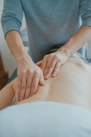
- A course of trigger point therapy to reduce pain - eg.cross fibre friction massage
- Heat or cold therapies can be used as conservative management strategies to reduce pain levels [19]
- Postural exercises - Re-train proper posture in functional positions (Neuro-muscular control). Functional training is all about using the right muscles at the right time, to sustain the correct posture, in daily activities. Simple activities like eg. correct standing posture, sit to stand and walking up stairs all need be addressed to ensure correct technique and muscle recruitment.
- Thoracic manual therapies directed at the lateral and posterior rib structures to improve rib and thoracic spine mobility[8]
- Exercises in the range of motion should be induced as soon as possible. The patient may not have pain when he is doing the exercises eg.rotation exercises for thoracic spine. Do not invoke pain.
- Progressive stretches. They can begin with simple mobility exercises as tolerated [19]eg [20] Stretching of the M. pectoralis major can be helpful (stretch the M. pectoralis major, stand in a corner for 10 sec with both of your hands against the wall (like when you do a push-up)repeat it a few times a day for 1 or 2 minutes).
- Mobilisation of the spine and ribs to improve thorax mobility and to reduce symptoms. [21]
- On the painful area they can use transcutaneous electrical stimulation and electroacupuncture. The acupuncture needle (also known as a solid filiform needle) is placed within the involved spinal segment. Than low-frequency electrical currents are applied on the inserted needle.[22]
- Dry needling can help to reduce pain levels, however this treatment should only be performed by a qualified and experienced provider[23]
Clinical Bottom Line [edit | edit source]
Costochondritis should be a diagnosis of exclusion. Rule out other causes of chest pain that are associated with increased morbidity and mortality.
- Patients typically present with chest pain worse with breathing, and it is often positional.
- Costochondritis is a self-limited disease.
- It should be reproducible on a physical exam, and the patient's vital signs should be within normal limits. If ordered, labs, ECG, and chest x-ray should also be normal.
- Diagnosis is confirmed by a scan or bone scintigraphy and by a physical assessment of the affected costal cartilage.
- The treatment of costochondritis consists of conservative management and is usually symptomatic [19].
- Physiotherapy is often ordered if the condition does not respond to treatment (see physiotherapy section for details).
References[edit | edit source]
- ↑ 1.0 1.1 1.2 1.3 1.4 1.5 PROULX A and TERESA W.; Costochondritis: Diagnosis and Treatment; Am Fam Physician. 2009 Sep 15;80(6):617-620
- ↑ 2.0 2.1 Clemens WM. et al. ; Introduction to Chest Wall Reconstruction : Anatomy and Physiology of the Chest and Indications for Chest Wall Reconstruction ; Semin Plast Surg. ; 2011 ; 25(1) : 5-15
- ↑ Safarini OA, Bordoni B. Anatomy, Thorax, Ribs. InStatPearls [Internet] 2019 Feb 19. StatPearls Publishing.Available from:https://www.ncbi.nlm.nih.gov/books/NBK538328/ (last accessed 14.4.2020)
- ↑ 4.0 4.1 4.2 4.3 4.4 Schumann JA, Parente JJ. Costochondritis.Available from:https://www.ncbi.nlm.nih.gov/books/NBK532931/ (last accessed 29.4.2020)
- ↑ Fam A.G., Smythe H.A.,Musculoskeletal chest wall pain, Can Med Assoc J. Sept 19851; 133(5):379-389
- ↑ Gregory P.L., BISWAS A.C., Batt M.E.,Musculoskeletal problems of the chest wall in athletes, Sports Med., 2002;32(4):235-50.
- ↑ Hurst J.W., Morris D.C., Williams B.R. “Chest Pain” in patients with costochondritis or Tietze's syndrome, Wiley-Blackwell, 2001, p23-29
- ↑ 8.0 8.1 Han J N et al.; Respiratory function of the rib cage muscles; European Respiratory Journal ISSN 0903 1993.
- ↑ 9.0 9.1 Proulx AM, Zryd TW. Costochondritis: diagnosis and treatment. American family physician. 2009 Sep 15;80(6):617-20.Available from:https://www.ncbi.nlm.nih.gov/pubmed/19817327 (last accessed 29.4.2020)
- ↑ Brian E Udermann et al.; Slipping Rib Syndrome in a Collegiate Swimmer: A Case Report; J Athl Train. 2005 Apr-Jun; 40(2): 120–122
- ↑ Brenda M. Birmann et al.; Prediagnosis biomarkers of insulin-like growth factor-1, insulin, and interleukin-6 dysregulation and multiple myeloma risk in the Multiple Myeloma Cohort Consortium. Blood. 2012 Dec 13; 120(25): 4929–4937.
- ↑ FREESTON J; Can Early Diagnosis and Management of Costochondritis Reduce Acute Chest Pain Admissions?; The Journal of Rheumatology November 2004, 31 (11) 2269-2271
- ↑ Viitanena J, H. Kautiainena, J. Sunia, M. L. Kokkoa & K. Lehtinena; The Relative Value of Spinal and Thoracic Mobility Measurements in Ankylosing Spondylitis; Scandinavian Journal of Rheumatology; Volume 24, 1995 - Issue 2
- ↑ 14.0 14.1 Aspegren D; Conservative Treatment of a Female Collegiate Volleyball Player with Costochondritis ; Journal of Manipulative and Physiological Therapeutics ; May 2007 Volume 30, Issue 4, Pages 321–325
- ↑ 15.0 15.1 Grindstaff L.T. et al. ; Treatment of a female collegiate rower with costochondritis : a case report ; J Man Manip Ther. ;2010 ;18(2) : 64-68
- ↑ Kamel M. et al. ; Ultrasonographic assessement of local steroid injection in Tietze’s syndrome ; Br J Rheumatol ; 1997 ;36(5) : 547-50
- ↑ Freeston J. et al. ; Can early diagnosis and management of costochondritis reduce acute chest pain admissions ?; J Rheumatol ; 2004 ; 31(11)-2269-71
- ↑ Massin MM, Bourguignont A, Coremans C, Comté L, Lepage P, Gérard P. Chest pain in pediatric patients presenting to an emergency department or to a cardiac clinic. Clin Pediatr 2004;43(3):231-238
- ↑ 19.0 19.1 19.2 Hudes K, Low-tech rehabilitation and management of a 64 year old male patient with acute idiopathic onset of costochondritis. J Can Chiropr Assoc. 2008 December; 52(4): 224–228
- ↑ Rovetta G, Sessarego P, Monteforte P. Stretching exercises for costochondritis pain. G Ital Med Lav Ergon. Apr-Jun 2009;31(2):169-71
- ↑ Buntinx F, Knockaert D, Bruyninckx R, et al. Chest pain in general practice or in the hospital emergency department: is it the same? Fam Pract. 2001;18(6):586-589.
- ↑ Imamura ST., et al., syndrome de tietze, Cossermeli W., Terapêutica em reumatologia, Sao Paulo, lemos editorial, p773-777, 2000.
- ↑ Richard B, Westrick P., Evaluation and treatment of musculoskeletal chest wall pain in military athlete. The International Journal of Sports Physical Therapy, 2012, Volume 7(3) Available from:https://www.ncbi.nlm.nih.gov/pmc/articles/PMC3362990/ (last accessed 30.4.2020)


