Corticospinal Tract: Difference between revisions
Wendy Walker (talk | contribs) No edit summary |
Evan Thomas (talk | contribs) mNo edit summary |
||
| Line 6: | Line 6: | ||
== Description == | == Description == | ||
The corticospinal tract is a descending tract of the spinal cord which contains bundles of axons which originate in the cerebral cortex and descend to synapse within the brainstem or spinal cord. The neurons are called "upper motor neurons". <ref name="Bear 2001">Bear MF, Connors BW, Paradiso. Neuroscience: Exploring the Brain Neuroscience: Exploring the Brain, Michael A. Paradiso. Edition 2, illustrated. Lippincott Williams &amp;amp;amp;amp;amp;amp;amp;amp;amp;amp;amp;amp;amp; Wilkins, 2001</ref> | The corticospinal tract is a descending tract of the spinal cord which contains bundles of axons which originate in the cerebral cortex and descend to synapse within the brainstem or spinal cord. The neurons are called "upper motor neurons". <ref name="Bear 2001">Bear MF, Connors BW, Paradiso. Neuroscience: Exploring the Brain Neuroscience: Exploring the Brain, Michael A. Paradiso. Edition 2, illustrated. Lippincott Williams &amp;amp;amp;amp;amp;amp;amp;amp;amp;amp;amp;amp;amp;amp;amp; Wilkins, 2001</ref> | ||
== Anatomy == | == Anatomy == | ||
| Line 12: | Line 12: | ||
=== [[Image:Corticospinal.jpg|frame|right]]Origin === | === [[Image:Corticospinal.jpg|frame|right]]Origin === | ||
*60% of fibres originate from the primary motor area, the premotor area, and the supplementary motor area of the frontal lobe | |||
*Other fibres originate from the primary <br>sensory area, the parietal cortex and the parietal operculum <ref name="Bear 2001" /><br> | |||
=== Course / Path === | |||
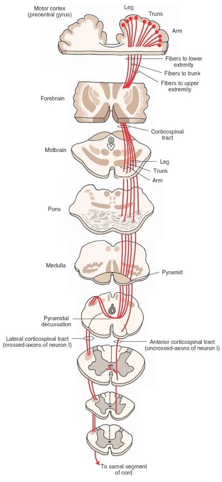
The descending corticospinal tract descends from the origin: | |||
*Through the corona radiata | |||
*Posterior half of lateral ventrical (lower limb represented by posterior fibres, face most anterior) | |||
*Posterior limb of internal capsule (lower limb represented by posterior fibres, face most anterior) | |||
*Enters midbrain through cerebral peduncle (face represented by medial fibres, foot lateral and hand in the middle | |||
*Enter medulla where they form medullary pyramids on either side of midline: | |||
#'''Lateral fibres (lateral corticospinal tract)''' are contralateral fibres. These make up between 75-90% fibres. They descend in the posterior part of the lateral funiculus. This tract is detected through to the lumbosacral spine and fibres synapse either directly on anterior horn cells of the contralateral side to their origin (ipsilateral to their side of descent in the spinal cord), or on interneurones of layers within this same side. | |||
#'''Anterior fibres (Anterior corticospinal tract)''' makes up between 10-25% of fibres. They descend ipsilaterally, however decussate near to their termination. Therefore these fibres continue to innteravate the contralateral side of the spinal cord. <ref name="Crossman" /><ref name="Bear 2001" /> | |||
Of all corticospinal fibres approximately 20% terminate at thoracic levels, 25% at lumbosacral levels and 55% at cervical levels. Many of the fibres that originate from the motor cortex then terminate in the ventral horn of the spinal cord. <ref name="Crossman">Crossman AR, Neary D. Neuroanatomy: An Illustrated Colour Text. Third Edition. London: Elsevier, 2004</ref> | Of all corticospinal fibres approximately 20% terminate at thoracic levels, 25% at lumbosacral levels and 55% at cervical levels. Many of the fibres that originate from the motor cortex then terminate in the ventral horn of the spinal cord. <ref name="Crossman">Crossman AR, Neary D. Neuroanatomy: An Illustrated Colour Text. Third Edition. London: Elsevier, 2004</ref> | ||
| Line 24: | Line 32: | ||
== Function == | == Function == | ||
'''Anterior Corticospinal Tract''' | '''Anterior Corticospinal Tract''' | ||
*responsible for the control of the proximal musculature.<br> | |||
'''Lateral Corticospinal Tract''' | |||
*responsible for the control of the distal musculature <ref name="Masri">Masri OA. An Essay on the Human Corticospinal Tract: History, Development, Anatomy, and Connections. Neuroanatomy 2011; 10:1-4</ref><br> | |||
'''Lateral Corticospinal Tract ''' | *fine control of movements of the hand | ||
== Pathology == | == Pathology == | ||
| Line 58: | Line 63: | ||
== References == | == References == | ||
<references /><br> | |||
<references /> | |||
[[Category:Neurology]][[Category: | [[Category:Anatomy]] [[Category:Neurology]] [[Category:Brain]] [[Category:Nerves]] | ||
Revision as of 22:21, 2 April 2017
Original Editor - Kate Sampson
Top Contributors - Kate Sampson, Matt Ross, Lucinda hampton, Kim Jackson, Anas Mohamed, Joao Costa, WikiSysop, Vidya Acharya, Carina Therese Magtibay, 127.0.0.1, Wendy Walker and Evan Thomas
Description[edit | edit source]
The corticospinal tract is a descending tract of the spinal cord which contains bundles of axons which originate in the cerebral cortex and descend to synapse within the brainstem or spinal cord. The neurons are called "upper motor neurons". [1]
Anatomy[edit | edit source]
 Origin[edit | edit source]

- 60% of fibres originate from the primary motor area, the premotor area, and the supplementary motor area of the frontal lobe
- Other fibres originate from the primary
sensory area, the parietal cortex and the parietal operculum [1]
Course / Path[edit | edit source]
The descending corticospinal tract descends from the origin:
- Through the corona radiata
- Posterior half of lateral ventrical (lower limb represented by posterior fibres, face most anterior)
- Posterior limb of internal capsule (lower limb represented by posterior fibres, face most anterior)
- Enters midbrain through cerebral peduncle (face represented by medial fibres, foot lateral and hand in the middle
- Enter medulla where they form medullary pyramids on either side of midline:
- Lateral fibres (lateral corticospinal tract) are contralateral fibres. These make up between 75-90% fibres. They descend in the posterior part of the lateral funiculus. This tract is detected through to the lumbosacral spine and fibres synapse either directly on anterior horn cells of the contralateral side to their origin (ipsilateral to their side of descent in the spinal cord), or on interneurones of layers within this same side.
- Anterior fibres (Anterior corticospinal tract) makes up between 10-25% of fibres. They descend ipsilaterally, however decussate near to their termination. Therefore these fibres continue to innteravate the contralateral side of the spinal cord. [2][1]
Of all corticospinal fibres approximately 20% terminate at thoracic levels, 25% at lumbosacral levels and 55% at cervical levels. Many of the fibres that originate from the motor cortex then terminate in the ventral horn of the spinal cord. [2]
Function[edit | edit source]
Anterior Corticospinal Tract
- responsible for the control of the proximal musculature.
Lateral Corticospinal Tract
- responsible for the control of the distal musculature [3]
- fine control of movements of the hand
Pathology[edit | edit source]
Damage can occur to the upper motor neurones of the corticospinal tract resulting in the upper motor neurone syndrome. Damage to the upper motor neurones can result can lead to presentations of "paralysis (or paresis), hypertonia, hyperreflexia, clonus, up-going plantar reflexes (Babinski’s sign) and spasticity". [4]
Prognosis[edit | edit source]
Stinear et al (2007) suggested that Corticospinal Tract integrity could be used to identify the likely extent of motor recovery and may enable appropriate selection of rehabilitation strategies for individuals recovering from stroke [5]. In a further study conducted by Stinear et al (2012) they trialled the use of the PREP(predicting motor recovery) algorithm to assess the likelihood of upper limb recovery. By utilising the SAFE score (sum of the shoulder abduction and finger extension) 72 hours after stroke, Transcranial magnetic stimulation, motor
evoked potentials in affected upper limb or the Asymmetry Index (measured with diffusion-weighted MRI) they were able to predict whether there could be a complete- no recovery. It was suggested from these finding that clinicians using the PREP algorithm may be able to predict the likely extent of upper limb recovery and may be able to therefore manage of patient expectations from an earlier period.[6]
Recent Related Research (from Pubmed)[edit | edit source]
Failed to load RSS feed from http://www.ncbi.nlm.nih.gov/entrez/eutils/erss.cgi?rss_guid=1Ds1JEbG0OWeBfrOW6IlPezs91fvq6o4kXlcDk6mk5F8IN-QBp|charset=UTF-8|short|max=10: Error parsing XML for RSS
Resources[edit | edit source]
References[edit | edit source]
- ↑ 1.0 1.1 1.2 Bear MF, Connors BW, Paradiso. Neuroscience: Exploring the Brain Neuroscience: Exploring the Brain, Michael A. Paradiso. Edition 2, illustrated. Lippincott Williams &amp;amp;amp;amp;amp;amp;amp;amp;amp;amp;amp;amp;amp;amp; Wilkins, 2001
- ↑ 2.0 2.1 Crossman AR, Neary D. Neuroanatomy: An Illustrated Colour Text. Third Edition. London: Elsevier, 2004
- ↑ Masri OA. An Essay on the Human Corticospinal Tract: History, Development, Anatomy, and Connections. Neuroanatomy 2011; 10:1-4
- ↑ Ivanhoe CB, Reistetter TA. Spasticity: the misunderstood part of the upper motor neuron syndrome.Am. J. Phys. Med. Rehabil. 2004; 83(10 Suppl): S3–9
- ↑ Stinear CM, Barber PA, Smale PR, Coxon JP, Fleming MK, Byblow WD. Functional potential in chronic stroke patients depends on corticospinal tract integrity. Brain. 2007 Jan 1;130(1):170-80.
- ↑ Stinear CM, Barber PA, Petoe M, Anwar S, Byblow WD. The PREP algorithm predicts potential for upper limb recovery after stroke. Brain. 2012 Aug 1;135(8):2527-35.
- ↑ 3D Neuroanatomy and Neurology. Neuroanatomy - The Corticospinal Tract in 3D. https://www.youtube.com/watch?v=9BaWBGRVxp8 (accessed 31/3/2016)
- ↑ Handwritten Tutorials. Spinal Pathways 4 - Corticospinal Tract. https://www.youtube.com/watch?v=dZ5H6PesskA (accessed on 31/3/2016)

