Compartment Syndrome of the Foot
Original Editors - Jessie Tourwe
Top Contributors - Jessie Tourwe, Admin, Scott Cornish, Shaimaa Eldib, Tomer Yona, Rachael Lowe, Kim Jackson, Samrah khan, Leana Louw, Kevin Vandebroucq, Karen Wilson, Wanda van Niekerk, 127.0.0.1 and Sigrid Bortels
Definition/Description[edit | edit source]
This syndrome is a condition that can appear in many parts of the body: foot, leg, thigh, forearm, hand, buttocks etc.[1] A compartment syndrome occurs when the muscles along with nerves and blood vessels are compressed in a compartment.
The developing of swelling and/or a hematoma causes the pressure to increase and because the fascia – made of inelastic connective tissue – can’t extend, the blood flow is disrupted. Tissue death can take place if the concentration of oxygen drops too low for too long.[2]
Clinically Relevant Anatomy[edit | edit source]
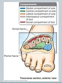
Anatomical studies of muscles and tendons show that the foot is divided into 4 large compartments (interosseous, medial, lateral, central) each including muscles, nerves and arteries. Early researches identified 9 compartments. However, it is very impractical to divide the foot into more than four compartments. That’s why most of the recent studies still refer to the foot as a whole of four compartments.[3]
| -Interosseous compartment: Dorsal interossei muscles Plantar interossei muscles Plantar lateral artery, vein and nerve
|
- Medial compartment: Abductor hallucis Flexor hallucis brevis Tendon of flexor hallucis longus Medial plantar arteries, veins and nerves
|
| - Lateral compartment: Abductor digiti minimi Flexor digiti minimi Opponens digiti minimi Branches of the lateral plantar artery vein and nerve
|
- Central compartment (3 levels): |
Epidemiology /Etiology [edit | edit source]
Chronic (exertional) compartment syndromes take place when athletes make too many efforts during a sport causing an overuse injury. The muscles get tired and irritated resulting in an inflammation and swelling. Sports like soccer, biking, running, tennis, gymnastics can be risk factors.[5]
It is possible that athletes don’t have the appropriate training program so that they overstrain their muscles. Use of inappropriate footwear.[6]
Other causes can be biomechanical faults in a person’s anatomy.[5] Limb length differences, muscle weakness, tightness in specific joints etc
Acute compartment syndromes can be produced by many different events. Crush injuries cover the majority of compartment syndromes of the foot[3], next to this fact one notices snake bites, burns, metatarsal fractures, talus or calcaneus fractures, dislocation of the Chopart and/or Lisfranc joints etc.[3] [2]
Steroids: Using steroids or creatine makes the muscles increase in volume.[7] (Level of evidence A1, B)
Bandages: If a tape, bandage or cast is too tight fitted, it may lead to a compartment syndrome.
Characteristics/Clinical Presentation[edit | edit source]
The most specific signs are:
- The skin appears pale and tensely swollen on the spot of tissue damage.
- Pain occurs when squeezing and/or touching the affected compartments.
- Pain when applying passive stretching to ankle, metatarsal joints and toes.
- Increased pain on dorsal flexion of the metatarsophalangeal joints.
- Muscle weakness of the intrinsic foot muscles when moving the foot in any way.
- Enlarged soreness radiating to the toes when moving them actively up and down.
Late findings are:
- It is possible the pulses are not palpable because the foot is very swollen.
- Neurological deficits: when a nerve is damaged the patient can report a decreased sensation.[2]
Considering the 5 P’s: Pain, Pallor, Paresthesia, Paralysis, Pulselessness[4]
Diagnostic Procedures[edit | edit source]
In order to diagnose a compartment syndrome there should be an awareness of the signs and symptoms specific to this syndrome as described above.
The only valuable test to diagnose this syndrome is an invasive measurement of the absolute compartment pressures. Otherwise known as Intracompartmental pressure monitoring (ICP).
Medical Management
[edit | edit source]
Emergency decompressive fasciotomy is being conducted in acute compartment syndrome.
- Indication: decompressive fasciotomy is indicated when the intracompartmental pressure measurement with absolute value of 30-45 mm Hg.
- Techniques:
- Dual dorsal incision is a gold standard technique, in which dorsal medial and lateral incision approach is being applied to release the compartments.
- Single medial incision is applied through medial approach to release all compartments but it is technically challenging.
- Complications: following fasciotomy care must be taken, otherwise chronic pain and hypersensitivity are the complications difficult to manage. Sometimes claw toes (fixed flexion deformity of digits) develops.[8]
Physical Therapy Management
[edit | edit source]
Overall nonoperative treatment has been generally unsuccessful.[9] After undergoing an operation the patient gets the advice to use ice packs and anti-inflammatory medication to reduce the swelling and to get enough rest. A physiotherapist can provide postoperative exercises to improve the muscle weakness and stimulate proprioceptive sensors. (Level of evidence B)
Soft tissue massage [5]
- Effleurages, petrissages
- Lymphatic drainage
Passive mobilization of the ankle joint, the metatarsals and phalanges [5]
- Tractions
Use of orthotics [5] [6]
To correct biomechanical defaults
For example: orthopedic soles for pronated feet, flat feet etc.
Stretch exercises to improve flexibility [10]
- Dorsal and plantar flexion - Let the patient move the feet up and down as far as possible. Repeating 10-20 times.
- 2.Inversion and eversion - Let the patient move the feet in and out as far as possible. Repeating 10-20 times.
- 3. Rotation - Let the patient move the feet in circles as large as possible. Repeating 10-20 times.
Strength exercises for intrinsic foot muscles
- Toe curl: Place a towel beneath the feet of the patient; he must pull the towel towards him by curling his toes into the towel. [11]
- Picking up marbles or other small objects: The patient has to claw his toes to be able to pick up the object from the floor.
- Walking: Early postoperative exercises involve walking with crutches. Once the patient can painless put weight on his foot and is comfortable in proper shoes, he/she may start to walk.
- Toe squeeze: Put some soft objects between the toes of the patient. Now he/she has to squeeze the toes and hold for 5 seconds, repeating 10 times.
- Toe raises, toe curls: to improve dorsal and plantar flexion of the toes, the patient can actively move the toes up and down. This exercise can be performed dynamic or static.
- Strength exercise for plantar flexion: rotations of the feet (feet must be kept together during the exercise) [11]
- Strength exercise for dorsal flexion: cycling in the air, feet must unroll properly.[11]
- Resistance band exercise: the patient can practice dorsal and plantar flexion, inversion and eversion. [10]
A low-key return to activities [9]
- Walking
How far can the patient rely on his feet?
In case of immobilization, the patient learns to walk with two crutches (no support on foot), with 1 crutch and eventually walk without crutches.
- When the patient can walk pain free, he/she can start constructive running.
- Once the patient can run pain free, he/she may participate other sports.
Important! If pain or swelling occurs during or after exercise, elevate the foot and use ice packs to reduce the swelling.
References[edit | edit source]
see adding references tutorial.
- ↑ Abraham T Rasul Jr. Compartment syndrome. eMedicine. 11 March 2009 http://emedicine.medscape.com/article/307668-overview (accessed on november/december 2010) Level of evidence: A1
- ↑ 2.0 2.1 2.2 Frink M, Hildebrand F, Krettek C, Brand J, Hankemeier S. Compartment syndrome of the lower leg and foot. The Association of bone and joint surgeons. 27 may 2009 http://emedicine.medscape.com/article/140002-overview (accessed november/december 2010) Level of evidence: B
- ↑ 3.0 3.1 3.2 Haddad S L, Managing risk: compartment syndromes of the foot. American Academy of Orthopaedics Surgeons, Jan/Feb 2007 http://www.aaos.org/news/bulletin/janfeb07/clinical1.asp (accessed on november/december 2010) Level of evidence: A1
- ↑ 4.0 4.1 Schünke M, Schulte E, Schumacher U, Voll M, Wesker K. Prometheus. Bohn Stafleu Van Loghum, Houten 2005. Pg 463
- ↑ 5.0 5.1 5.2 5.3 5.4 http://www.physioadvisor.com.au/10513350/compartment-syndrome-chronic-compartment-syndrom.htm
- ↑ 6.0 6.1 http://orthoinfo.aaos.org/topic.cfm?topic=a00204
- ↑ Tucker Alicia K. Chronic exertional compartment syndrome of the leg. Current Reviews in Musculoskeletal Medicine. 2 September 2010 http://ukpmc.ac.uk/articles/PMC2941579/ (accessed on november/december 2010) Level of evidence: A1
- ↑ Karadsheh M. Foot Compartment Syndrome. http://www.orthobullets.com (accessed 27 December 2016).
- ↑ 9.0 9.1 Matthew R. Bong, M.D., Daniel B. Polatsch, M.D., Laith M. Jazrawi, M.D. and Andrew S. Rokito, M.D. Chronic Exertional Compartment Syndrome. Diagnosis and Management. Bulletin, Hospital for joint diseases. Volume 62, N° 3, 4. 2005 Level of evidence: B
- ↑ 10.0 10.1 http://www.physioadvisor.com.au/8047989/ankle-flexibility-exercises-ankle-sprains-ankle.htm
- ↑ 11.0 11.1 11.2 Cite error: Invalid
<ref>tag; no text was provided for refs namedBRON 11


