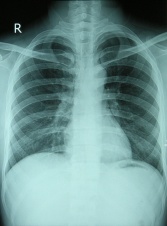
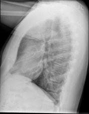
Chest X-Rays
Be the first to edit this page and have your name permanently included as the originating editor, see the editing pages tutorial for help.
Purpose
[edit | edit source]
Chest X-rays is a painless, non-invasive test and is the most commonly preferred diagnostic examination to produce images of heart, lungs, airways, blood vessels and the bones of the spine and chest.
Technique
[edit | edit source]
An X-ray uses electromagnetic waves and ionizing radiation to create pictures of the inside of your body.
The procedures involves positioning the body between the machine that produces the X-rays and a plate that that creates the image digitally or with X-ray film.
- For the front view: The patient is asked to stand against the plate, by holding her arms up or to the sides and roll your shoulders forward. The X-ray technician may then ask the patient to take few deep breaths and hold it for a couple of seconds. This tecniques of holding your breath generally helps to get a clear picture of your heart and lungs on the image.
- For the side-view: The patient is asked to turn and place one shoulder on the plate and raise her hands over her head. The technician may again ask the patient to take a deep breath and hold it.
The following video explains how to analyse a Chest- x-ray in nutshell
Evidence[edit | edit source]
A study conducted by Tape and Mushlin (1988) to study the effect of routine chest x-rays of pre-operative patients at risk for postoperative disease. Patient records from 341 admissions were reviewed to determine the relationship between chest x-ray results and postoperative chest complications. Patients who had major abnormalities had a 40% postoperative complication rate, compared with 9% for those with normal x-rays; but only 13% of the complications occurred in patients with major abnormalities. Nine patients had x-ray findings that led to clinical action: three with potentially beneficial management changes (congestive heart failure in 2, fibrosis in 1) and six with potentially detrimental clinical action (false diagnosis of tuberculosis in 2, false diagnosis of nodules in 2, falsely normal chest x-ray in 2). None of 50 surgical cancellations occurred as a result of an abnormal x-ray. All the beneficial effects attributable to preoperative chest x-rays accrued to patients who had clinical evidence of chest disease. The authors conclude that routine chest x-rays were not helpful in improving patient outcomes. They recommend ordering preoperative chest x-rays based on clinical indications so that the likelihood of false positives and false negatives and their associated detrimental effects can be minimized (Tape and Mushlin, 1988).
Resources
[edit | edit source]
How to read a Chest X-ray?
http://www.med.upenn.edu/normalcxr/
References
[edit | edit source]
[1]https://www.nhlbi.nih.gov/health/health-topics/topics/cxray


