Chest X-Rays: Difference between revisions
No edit summary |
No edit summary |
||
| Line 39: | Line 39: | ||
== Evidence == | == Evidence == | ||
Provide the evidence for this technique here | Provide the evidence for this technique here | ||
== Resources == | == Resources == | ||
Revision as of 00:32, 2 December 2016
Be the first to edit this page and have your name permanently included as the originating editor, see the editing pages tutorial for help.
Purpose
[edit | edit source]
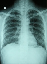
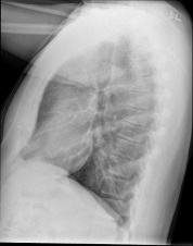
Chest X-rays is a painless, non-invasive test and is the most commonly preferred diagnostic examination to produce images of heart, lungs, airways, blood vessels and the bones of the spine and chest.
Technique
[edit | edit source]
An X-ray uses electromagnetic waves and ionizing radiation to create pictures of the inside of your body.
The procedures involves positioning the body between the machine that produces the X-rays and a plate that that creates the image digitally or with X-ray film.
- For the front view: The patient is asked to stand against the plate, by holding her arms up or to the sides and roll your shoulders forward. The X-ray technician may then ask the patient to take few deep breaths and hold it for a couple of seconds. This tecniques of holding your breath generally helps to get a clear picture of your heart and lungs on the image.
- For the side-view: The patient is asked to turn and place one shoulder on the plate and raise her hands over her head. The technician may again ask the patient to take a deep breath and hold it.
Evidence[edit | edit source]
Provide the evidence for this technique here
Resources[edit | edit source]
add any relevant resources here


