Cellulitis: Difference between revisions
Kim Jackson (talk | contribs) mNo edit summary |
(text, refs format, links) |
||
| Line 4: | Line 4: | ||
'''Top Contributors''' - {{Special:Contributors/{{FULLPAGENAME}}}} | '''Top Contributors''' - {{Special:Contributors/{{FULLPAGENAME}}}} | ||
</div> | </div> | ||
== | == Introduction == | ||
[[Image: | Cellulitis is a common bacterial skin infection, with over 14 million cases occurring in the United States annually.[[Image:Severe cellulitis.jpg|250x200px|thumb|A severe case of cellulitis that developed under a cast]]Cellulitis typically presents as a poorly demarcated, warm, erythematous area with associated edema and tenderness to palpation. | ||
* | * It is an acute bacterial infection causing inflammation of the deep dermis and surrounding subcutaneous tissue. | ||
* | * The infection is without an abscess or purulent discharge. | ||
* Beta-hemolytic streptococci typically cause cellulitis, generally group A streptococcus (i.e., ''Streptococcus pyogenes''), followed by methicillin-sensitive ''Staphylococcus aureus''. | |||
* Patients who are immunocompromised, colonized with methicillin-resistant Staphylococcus aureus, bitten by animals, or have comorbidities such as diabetes mellitus may become infected with other bacteria. | |||
* If the clinician correctly identifies and promptly treats cellulitis, it typically resolves with appropriate antibiotic treatment. | |||
* and | |||
== | === Etiology === | ||
The skin serves as a protective barrier preventing normal skin flora and other microbial pathogens from reaching the subcutaneous tissue and lymphatic system. When a break in the [[The Skin and Wound Healing|skin]] occurs, it allows for normal skin flora and other bacteria to enter into the dermis and subcutaneous tissue, these bacteria below the skin surface can lead to an acute superficial infection affecting the deep dermis and subcutaneous tissue, causing cellulitis. Cellulitis most commonly results from infection with group A beta-hemolytic streptococcus (i.e., ''Streptococcus pyogenes''). | |||
* | Risk factors for cellulitis include any culprit that could cause a breakdown in the skin barrier such as | ||
* | * skin injuries, | ||
* | * surgical incisions, | ||
* | * intravenous site punctures, | ||
* | * fissures between toes, | ||
* insect and animal bites | |||
* other skin infections. | |||
* Patients with comorbidities such as [[Diabetes Mellitus Type 2|diabetes mellitus]], venous insufficiency, peripheral arterial disease, and lymphedema are at higher risk of developing cellulitis[[Image:Cellulitis and lymphedema.JPG|right|400x400px]] | |||
Studies show lymphedema is a major risk factor for the development of cellulitis. There is known to be a link between the two, but it is not known which of the two comes first. Patients with lymphedema or chronic edema are more prone to infection due to damage to lymphatic vessels and immune deficiency in that area. Cellulitis on the other hand, can cause damage to the lymphatics and the development of lymphedema.<ref name="Riches">Riches K, Keeley V. Cellulitis in patients with chronic oedema. Nursing & Residential Care [serial on the Internet]. (2012, Mar), [cited March 31, 2017]; 14(3): 122-127. Available from: CINAHL with Full Text.</ref> | |||
== | == Epidemiology == | ||
* Cellulitis is relatively common, and most often occurs in middle-aged and older adults. | |||
* When comparing men and women, there is no statistically significant difference in the incidence of cellulitis. | |||
* There are approximately 50 cases per 1000 patient-years | |||
== Pathophysiology == | |||
Cellulitis is characterized by erythema, warmth, edema, and tenderness to palpation resulting from cytokine and neutrophil response from bacteria breaching the epidermis. | |||
The cytokines and neutrophils are recruited to the affected area after bacteria have penetrated the skin leading to an epidermal response. | |||
This response includes the production of antimicrobial peptides and keratinocyte proliferation producing the characteristic exam findings in cellulitis. | |||
Group A Streptococci, the most common bacteria to cause cellulitis, can also produce virulence factors such as pyrogenic exotoxins (A, B, C, and F) and streptococcal superantigen that can lead to a more pronounced and invasive disease<ref name=":0">Brown BD, Watson KL. [https://www.ncbi.nlm.nih.gov/books/NBK549770/ Cellulitis]. InStatPearls [Internet] 2019 Nov 6. StatPearls Publishing. Available from:https://www.ncbi.nlm.nih.gov/books/NBK549770/ (last accessed 12.2.2020)</ref> | |||
== Clinical Presentation == | |||
[[Image:Classic celulitis.PNG|thumb|A classic presentation of cellulitis: poorly demarcated erythema<ref name="Bailey">Bailey E, Kroshinsky D. Cellulitis: Diagnosis and Management. Dermatologic Therapy. 2011;24:229–39.http://onlinelibrary.wiley.com/doi/10.1111/j.1529-8019.2011.01398.x/full (accessed 15 Mar 2017).</ref> |258x258px]]Typical symptoms include acute poorly demarcated and spreading erythema along with pain, swelling, and warmth of the lower extremity but can occur on any area of skin or underlying subcutaneous tissue.<ref name="Tsai">Tsai C-YL, Calvin MK, Chung C, Susan Shin-Jung L, Yao-Shen C, Hung C. Development of a prediction model for bacteremia in hospitalized adults with cellulitis to aid in the efficient use of blood cultures: a retrospective cohort study. BMC Infectious Diseases. 2016;16(1):581.</ref><ref name="Raff">Raff AB, Kroshinsky D. Cellulitis: A Review. JAMA. 2016;316(3):325-37. http://jamanetwork.com/pdfaccess.ashx?url=/data/journals/jama/935437/ (accessed 27 Feb 2017).</ref> | |||
| | |||
| | |||
Symptoms may include fever, nausea, vomiting, and rigors.<ref name="Raff" /><ref name="Kilburn">Kilburn SA, Featherstone P, Higgins B, Brindle R. Interventions for cellulitis and erysipelas. (Cochrane review). Cochrane Database Syst Rev. 2010(6):CD004299.</ref> | |||
Other features include proximal dilated and edematous skin lymphatics and bulla formation. | |||
Cellulitis predominantly has a unilateral presentation, most commonly in the lower extremity.<ref name="Raff" /> | |||
== Examination == | |||
Patients with cellulitis will reveal an affected skin area typically with a poorly demarcated area of erythema. The erythematous area is often warm to the touch with associated swelling and tenderness to palpation. The patient may present with constitutional symptoms of generalized malaise, fatigue, and fevers. | |||
* Ask for a complete history of the presenting illness, focusing on the context in which the patient noticed the skin changes or how the cellulitis began to occur. | |||
* It is essential to ask patients if they: recently traveled, experienced any trauma or injuries, have a history of intravenous drug use, and/or have had insect or animal bites to the affected area. | |||
* A complete and thorough past medical history should additionally be conducted to evaluate for possible chronic medical conditions that predispose patients to cellulitis, such as [[Diabetes|diabetes mellitus]], venous stasis, [[Peripheral Arterial Disease|peripheral vascular disease]], chronic tinea pedis, and [[Lymphatic Obstruction (Lymphedema)|lymphedema]]. Diabetes Mellitus is one of most common comorbidities among those hospitalized for acute bacterial infections including cellulitis. Following a cellulitis infection, those with diabetes require a longer course of antibiotic therapy and are more likely to have an outpatient follow-up visit.<ref>Jenkins TC. Comparison of the Microbiology and Antibiotic Treatment among Diabetic and Non-Diabetic Patients Hospitalized for Cellulitis or Cutaneous Abscess. J Hosp Med. 9ADADDec12;:788–94.https://www.ncbi.nlm.nih.gov/pmc/articles/PMC4256165/ (accessed 28 Feb 2017).</ref>.<ref name="Riches" /> | |||
* Inspected area to look for any area of skin breakdown. The area should be demarcated with a marker to monitor for continuous spread. The area should be palpated to feel for fluctuance that could indicate the formation of a possible abscess. | |||
* Gently palpate the affected area, be sure to note any presence of warmth, tenderness, or purulent drainage. | |||
* Cellulitis can present on any area of the body, but most often affects the lower extremities. It is rarely bilateral. | |||
* In lower extremity cellulitis, careful examination between interspaces of the toes should take place. | |||
* Check for proper sensation and verify pulses are intact to monitor closely for compartment syndrome. | |||
* Note if there are developing vesicles, bullae, or the presence of peau d'orange and lymphadenopathy.<ref name=":0" /> | |||
== Diagnostic Tests/Lab Tests/Lab Values == | |||
Cultures are typically not beneficial in making a cellulitis diagnosis. It is most commonly diagnosed by history and physical examination alone. | |||
* Certain laboratory tests can indicate the presence of infection, but are not specific to cellulitis alone. | |||
* Imaging studies can identify more severe infections to differentiate from cellulitis, but are not a reliable diagnostic tool for cellulitis itself.<ref name="Raff" /> | |||
* Identification of the cause of the infection through blood, needle aspiration or punch biopsy are not recommended unless the patient has a complication or abnormal exposure history eg immunosuppressants, a diagnosis of chronic liver disease, aquatic soft tissue injury, animal and human bites, or being in contact with various bacterias.<ref name="Raff" /> | |||
* If a biopsy and culture are warranted, a histopathologic evaluation will be performed on the sample.<ref name="McNamara">McNamara DR, Tleyjeh IM, Berbari EF, Lahr BD, Martinez J, Mirzoyev SA, Baddour LM. A predictive model of recurrent lower extremity cellulitis in a population-based cohort. Arch Intern Med. 2007;167(7):709-715 http://jamanetwork.com/journals/jamainternalmedicine/fullarticle/412163 (accessed 15 March 2017)</ref> | |||
== Medical Management == | == Medical Management == | ||
Patients presenting with mild cellulitis and displaying no systemic signs of infection should be covered with antibiotics that target the treatment of streptococcal species. | |||
The duration of oral antibiotic therapy should be for a minimum duration of 5 days. | |||
* In nonpurulent cellulitis, patients should receive cephalexin 500 mg every 6 hours. If they have a severe allergic reaction to beta-lactamase inhibitors, treat with clindamycin 300 mg to 450 mg every 6 hours. | |||
* In patients with purulent cellulitis, methicillin-resistant staph aureus colonization, cellulitis associated with an abscess or extensive puncture wounds, or a history of intravenous drug use, patients should receive antibiotics that cover against methicillin-resistant staph aureus as well. | |||
* Cellulitis with MRSA risk factors should be treated with trimethoprim-sulfamethoxazole 800 mg/160 mg twice daily for 5 days in addition to cephalexin 500 mg every 6 hours. If a patient has an allergy to trimethoprim-sulfamethoxazole, treat with clindamycin 300 mg to 450 mg every 6 hours. A longer duration of antibiotic treatment may be a consideration in patients who show minimal improvement with antibiotic therapy within 48 hours. | |||
* Hospitalization with the induction of systemic antibiotics may be necessary for patients who: present with systemic signs of infection*, have failed outpatient treatment, are immunocompromised, exhibit rapidly progressing erythema, are unable to tolerate oral medications, or have cellulitis overlying or near an indwelling medical device. | |||
* If patients have significant edema with a known cause for the edema, the underlying condition should receive proper treatment to decrease the amount of edema and prevent future episodes of cellulitis. Patients should be instructed to keep the affected area elevated.<ref name=":0" /> | |||
NB It is beyond a physiotherapists scope to know the individual antibiotics and best choices. The pharmacist ideally will have a board specialty in infectious disease to assist and work with the clinician on the best antibiotic selection.<ref name=":0" /> | |||
== Physical Therapy Management == | == Physical Therapy Management == | ||
While there is lack of evidence that discusses specific physical therapy interventions for cellulitis, therapists should be aware of the signs and symptoms in order to refer the patient appropriately. Physical therapists should have awareness of risk factors and various causes of cellulitis, in addition to signs and symptoms.<br> | While there is lack of evidence that discusses specific physical therapy interventions for cellulitis, therapists should be aware of the signs and symptoms in order to refer the patient appropriately. Physical therapists should have awareness of risk factors and various causes of cellulitis, in addition to signs and symptoms.<br>Modalities that physical therapists can use for a patient with cellulitis include | ||
* Rest and elevation of the affected limb is important and can help alleviate pain. | |||
* The application of cool, wet, sterile bandages is also recommended for pain relief, and ice can be used as well. | |||
Preventative measures | |||
* Massage to promote lymphatic drainage, may help prevent cellulitis (not be used during an active cellulitis infection).<ref name="UMMC" /> | |||
* Compression stockings | |||
* Exercise promotion and specific exercises eg calf pumps whilst standing in lines etc.<ref name="UMMC">University of Maryland Medical Center. http://umm.edu/health/medical/altmed/condition/cellulitis (accessed 25 March 2017).</ref> | |||
* educate patients of the importance of maintaining good hand hygiene and adequately clean any future abrasions in their skin.<ref name=":0" /> | |||
== Differential Diagnosis == | |||
Cellulitis is a frequently encountered infection of the deep dermis and subcutaneous tissue, mainly affecting the lower extremities, but it can have many mimickers. | |||
Common differential diagnoses for Cellulitis include [[Deep Vein Thrombosis]], | Common differential diagnoses for Cellulitis include | ||
* Dermatitis | |||
* Erythema Migrans. | |||
* Erysipelas is sometimes considered a form of cellulitis. However, it is a more superficial infection affecting the upper dermis and superficial lymphatic system. | |||
* Chronic venous stasis dermatitis is a long-standing, bilateral, inflammatory dermatosis secondary to chronic venous insufficiency and typically involves the medial malleoli. It appears on the lower extremities and manifests as erythema with scaling, peripheral edema, and hyperpigmentation. | |||
* Necrotizing fasciitis is a rare infection of the fascia that leads to necrosis of the subcutaneous tissue. Its characteristic presentation includes fevers, erythema, edema, pain out of proportion to the exam, and crepitus. It qualifies as a surgical emergency and requires surgical debridement immediately. | |||
* Septic arthritis, or an infected joint, can involve any joint but typically involves the knee joint. Patients present with joint swelling, warmth, pain, and decreased mobility of the joint. Septic arthritis treatment is by joint aspiration and antibiotics directed at the most common pathogens. | |||
* [[Deep Vein Thrombosis]], is typically unilateral and presents with tenderness, erythema, warmth, and edema. It often affects the lower extremities. Patients commonly have the presence of risk factors for DVT, such as a history of immobility, active cancer, or a family history of venous thromboembolism. Deep vein thrombosis rarely manifests with fevers or leukocytosis, but they can be present. Ultrasound imaging is used to confirm the diagnosis.<ref name=":0" /> | |||
== Complications == | |||
Without prompt diagnosis and treatment, cellulitis could lead to several complications. | |||
Cellulitis that leads to bacteremia, endocarditis, or osteomyelitis will require a longer duration of antibiotics and possibly surgery.<ref name=":0" /> | |||
* Locally, cellulitis often results in significant tissue damage in the involved area.<ref name="Raff" /> | |||
* Cellulitis can spread systemically through the lymphatics and blood stream, which can lead to further complications.<ref name="Medscape">Medscape. Cellulitis. http://emedicine.medscape.com/article/214222-overview (accessed 27 Feb 2017).</ref> | |||
* If cellulitis does spread systemically through one of these systems, it can cause flu-like symptoms such as fever, rigors, nausea, and vomiting.<ref name="Kilburn" /><ref name="Riches" /> | |||
* Though rare, there is a risk for severe sepsis, gangrene, or necrotizing fasciitis if cellulitis spreads systemically and is left untreated.<ref name="Kilburn" /> | |||
== Prognosis == | |||
Overall, cellulitis has a good prognosis | |||
* If promptly diagnosed cellulitis with correct antibiotic treatment, patients can expect to notice an improvement in signs and symptoms within 48 hours. | |||
* Annual recurrence of cellulitis occurs in about 8 to 20% of patients, with overall reoccurrence rates reaching as high as 49% | |||
* Recurrence is preventable with prompt treatment of cuts or abrasions, proper hand hygiene, as well as effectively treating any underlying comorbidities. | |||
* There is approximately an 18% failure rate with initial antibiotic treatment. <ref name=":0" /> | |||
== Resources == | == Resources == | ||
Revision as of 08:25, 12 February 2020
Original Editors - Students from Bellarmine University's Pathophysiology of Complex Patient Problems project.
Top Contributors - Kacie McClendon, Elaine Lonnemann, Erica Shelley, Kim Jackson, Lucinda hampton, Fasuba Ayobami, 127.0.0.1, Evan Thomas, WikiSysop, Karen Wilson, Vidya Acharya and Claire Knott
Introduction[edit | edit source]
Cellulitis is a common bacterial skin infection, with over 14 million cases occurring in the United States annually.

Cellulitis typically presents as a poorly demarcated, warm, erythematous area with associated edema and tenderness to palpation.
- It is an acute bacterial infection causing inflammation of the deep dermis and surrounding subcutaneous tissue.
- The infection is without an abscess or purulent discharge.
- Beta-hemolytic streptococci typically cause cellulitis, generally group A streptococcus (i.e., Streptococcus pyogenes), followed by methicillin-sensitive Staphylococcus aureus.
- Patients who are immunocompromised, colonized with methicillin-resistant Staphylococcus aureus, bitten by animals, or have comorbidities such as diabetes mellitus may become infected with other bacteria.
- If the clinician correctly identifies and promptly treats cellulitis, it typically resolves with appropriate antibiotic treatment.
Etiology[edit | edit source]
The skin serves as a protective barrier preventing normal skin flora and other microbial pathogens from reaching the subcutaneous tissue and lymphatic system. When a break in the skin occurs, it allows for normal skin flora and other bacteria to enter into the dermis and subcutaneous tissue, these bacteria below the skin surface can lead to an acute superficial infection affecting the deep dermis and subcutaneous tissue, causing cellulitis. Cellulitis most commonly results from infection with group A beta-hemolytic streptococcus (i.e., Streptococcus pyogenes).
Risk factors for cellulitis include any culprit that could cause a breakdown in the skin barrier such as
- skin injuries,
- surgical incisions,
- intravenous site punctures,
- fissures between toes,
- insect and animal bites
- other skin infections.
- Patients with comorbidities such as diabetes mellitus, venous insufficiency, peripheral arterial disease, and lymphedema are at higher risk of developing cellulitis

Studies show lymphedema is a major risk factor for the development of cellulitis. There is known to be a link between the two, but it is not known which of the two comes first. Patients with lymphedema or chronic edema are more prone to infection due to damage to lymphatic vessels and immune deficiency in that area. Cellulitis on the other hand, can cause damage to the lymphatics and the development of lymphedema.[1]
Epidemiology[edit | edit source]
- Cellulitis is relatively common, and most often occurs in middle-aged and older adults.
- When comparing men and women, there is no statistically significant difference in the incidence of cellulitis.
- There are approximately 50 cases per 1000 patient-years
Pathophysiology[edit | edit source]
Cellulitis is characterized by erythema, warmth, edema, and tenderness to palpation resulting from cytokine and neutrophil response from bacteria breaching the epidermis.
The cytokines and neutrophils are recruited to the affected area after bacteria have penetrated the skin leading to an epidermal response.
This response includes the production of antimicrobial peptides and keratinocyte proliferation producing the characteristic exam findings in cellulitis.
Group A Streptococci, the most common bacteria to cause cellulitis, can also produce virulence factors such as pyrogenic exotoxins (A, B, C, and F) and streptococcal superantigen that can lead to a more pronounced and invasive disease[2]
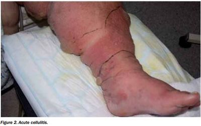
Clinical Presentation[edit | edit source]

Typical symptoms include acute poorly demarcated and spreading erythema along with pain, swelling, and warmth of the lower extremity but can occur on any area of skin or underlying subcutaneous tissue.[4][5]
Symptoms may include fever, nausea, vomiting, and rigors.[5][6]
Other features include proximal dilated and edematous skin lymphatics and bulla formation.
Cellulitis predominantly has a unilateral presentation, most commonly in the lower extremity.[5]
Examination[edit | edit source]
Patients with cellulitis will reveal an affected skin area typically with a poorly demarcated area of erythema. The erythematous area is often warm to the touch with associated swelling and tenderness to palpation. The patient may present with constitutional symptoms of generalized malaise, fatigue, and fevers.
- Ask for a complete history of the presenting illness, focusing on the context in which the patient noticed the skin changes or how the cellulitis began to occur.
- It is essential to ask patients if they: recently traveled, experienced any trauma or injuries, have a history of intravenous drug use, and/or have had insect or animal bites to the affected area.
- A complete and thorough past medical history should additionally be conducted to evaluate for possible chronic medical conditions that predispose patients to cellulitis, such as diabetes mellitus, venous stasis, peripheral vascular disease, chronic tinea pedis, and lymphedema. Diabetes Mellitus is one of most common comorbidities among those hospitalized for acute bacterial infections including cellulitis. Following a cellulitis infection, those with diabetes require a longer course of antibiotic therapy and are more likely to have an outpatient follow-up visit.[7].[1]
- Inspected area to look for any area of skin breakdown. The area should be demarcated with a marker to monitor for continuous spread. The area should be palpated to feel for fluctuance that could indicate the formation of a possible abscess.
- Gently palpate the affected area, be sure to note any presence of warmth, tenderness, or purulent drainage.
- Cellulitis can present on any area of the body, but most often affects the lower extremities. It is rarely bilateral.
- In lower extremity cellulitis, careful examination between interspaces of the toes should take place.
- Check for proper sensation and verify pulses are intact to monitor closely for compartment syndrome.
- Note if there are developing vesicles, bullae, or the presence of peau d'orange and lymphadenopathy.[2]
Diagnostic Tests/Lab Tests/Lab Values[edit | edit source]
Cultures are typically not beneficial in making a cellulitis diagnosis. It is most commonly diagnosed by history and physical examination alone.
- Certain laboratory tests can indicate the presence of infection, but are not specific to cellulitis alone.
- Imaging studies can identify more severe infections to differentiate from cellulitis, but are not a reliable diagnostic tool for cellulitis itself.[5]
- Identification of the cause of the infection through blood, needle aspiration or punch biopsy are not recommended unless the patient has a complication or abnormal exposure history eg immunosuppressants, a diagnosis of chronic liver disease, aquatic soft tissue injury, animal and human bites, or being in contact with various bacterias.[5]
- If a biopsy and culture are warranted, a histopathologic evaluation will be performed on the sample.[8]
Medical Management[edit | edit source]
Patients presenting with mild cellulitis and displaying no systemic signs of infection should be covered with antibiotics that target the treatment of streptococcal species.
The duration of oral antibiotic therapy should be for a minimum duration of 5 days.
- In nonpurulent cellulitis, patients should receive cephalexin 500 mg every 6 hours. If they have a severe allergic reaction to beta-lactamase inhibitors, treat with clindamycin 300 mg to 450 mg every 6 hours.
- In patients with purulent cellulitis, methicillin-resistant staph aureus colonization, cellulitis associated with an abscess or extensive puncture wounds, or a history of intravenous drug use, patients should receive antibiotics that cover against methicillin-resistant staph aureus as well.
- Cellulitis with MRSA risk factors should be treated with trimethoprim-sulfamethoxazole 800 mg/160 mg twice daily for 5 days in addition to cephalexin 500 mg every 6 hours. If a patient has an allergy to trimethoprim-sulfamethoxazole, treat with clindamycin 300 mg to 450 mg every 6 hours. A longer duration of antibiotic treatment may be a consideration in patients who show minimal improvement with antibiotic therapy within 48 hours.
- Hospitalization with the induction of systemic antibiotics may be necessary for patients who: present with systemic signs of infection*, have failed outpatient treatment, are immunocompromised, exhibit rapidly progressing erythema, are unable to tolerate oral medications, or have cellulitis overlying or near an indwelling medical device.
- If patients have significant edema with a known cause for the edema, the underlying condition should receive proper treatment to decrease the amount of edema and prevent future episodes of cellulitis. Patients should be instructed to keep the affected area elevated.[2]
NB It is beyond a physiotherapists scope to know the individual antibiotics and best choices. The pharmacist ideally will have a board specialty in infectious disease to assist and work with the clinician on the best antibiotic selection.[2]
Physical Therapy Management[edit | edit source]
While there is lack of evidence that discusses specific physical therapy interventions for cellulitis, therapists should be aware of the signs and symptoms in order to refer the patient appropriately. Physical therapists should have awareness of risk factors and various causes of cellulitis, in addition to signs and symptoms.
Modalities that physical therapists can use for a patient with cellulitis include
- Rest and elevation of the affected limb is important and can help alleviate pain.
- The application of cool, wet, sterile bandages is also recommended for pain relief, and ice can be used as well.
Preventative measures
- Massage to promote lymphatic drainage, may help prevent cellulitis (not be used during an active cellulitis infection).[9]
- Compression stockings
- Exercise promotion and specific exercises eg calf pumps whilst standing in lines etc.[9]
- educate patients of the importance of maintaining good hand hygiene and adequately clean any future abrasions in their skin.[2]
Differential Diagnosis[edit | edit source]
Cellulitis is a frequently encountered infection of the deep dermis and subcutaneous tissue, mainly affecting the lower extremities, but it can have many mimickers.
Common differential diagnoses for Cellulitis include
- Dermatitis
- Erythema Migrans.
- Erysipelas is sometimes considered a form of cellulitis. However, it is a more superficial infection affecting the upper dermis and superficial lymphatic system.
- Chronic venous stasis dermatitis is a long-standing, bilateral, inflammatory dermatosis secondary to chronic venous insufficiency and typically involves the medial malleoli. It appears on the lower extremities and manifests as erythema with scaling, peripheral edema, and hyperpigmentation.
- Necrotizing fasciitis is a rare infection of the fascia that leads to necrosis of the subcutaneous tissue. Its characteristic presentation includes fevers, erythema, edema, pain out of proportion to the exam, and crepitus. It qualifies as a surgical emergency and requires surgical debridement immediately.
- Septic arthritis, or an infected joint, can involve any joint but typically involves the knee joint. Patients present with joint swelling, warmth, pain, and decreased mobility of the joint. Septic arthritis treatment is by joint aspiration and antibiotics directed at the most common pathogens.
- Deep Vein Thrombosis, is typically unilateral and presents with tenderness, erythema, warmth, and edema. It often affects the lower extremities. Patients commonly have the presence of risk factors for DVT, such as a history of immobility, active cancer, or a family history of venous thromboembolism. Deep vein thrombosis rarely manifests with fevers or leukocytosis, but they can be present. Ultrasound imaging is used to confirm the diagnosis.[2]
Complications[edit | edit source]
Without prompt diagnosis and treatment, cellulitis could lead to several complications.
Cellulitis that leads to bacteremia, endocarditis, or osteomyelitis will require a longer duration of antibiotics and possibly surgery.[2]
- Locally, cellulitis often results in significant tissue damage in the involved area.[5]
- Cellulitis can spread systemically through the lymphatics and blood stream, which can lead to further complications.[10]
- If cellulitis does spread systemically through one of these systems, it can cause flu-like symptoms such as fever, rigors, nausea, and vomiting.[6][1]
- Though rare, there is a risk for severe sepsis, gangrene, or necrotizing fasciitis if cellulitis spreads systemically and is left untreated.[6]
Prognosis[edit | edit source]
Overall, cellulitis has a good prognosis
- If promptly diagnosed cellulitis with correct antibiotic treatment, patients can expect to notice an improvement in signs and symptoms within 48 hours.
- Annual recurrence of cellulitis occurs in about 8 to 20% of patients, with overall reoccurrence rates reaching as high as 49%
- Recurrence is preventable with prompt treatment of cuts or abrasions, proper hand hygiene, as well as effectively treating any underlying comorbidities.
- There is approximately an 18% failure rate with initial antibiotic treatment. [2]
Resources[edit | edit source]
A video link by dermatologist Dr. Noah Craft MD, PhD, DTMH discusses Cellulitis from the provider point of view and includes case studies, differential diagnosis, and treatment approaches.
References[edit | edit source]
- ↑ 1.0 1.1 1.2 Riches K, Keeley V. Cellulitis in patients with chronic oedema. Nursing & Residential Care [serial on the Internet]. (2012, Mar), [cited March 31, 2017]; 14(3): 122-127. Available from: CINAHL with Full Text.
- ↑ 2.0 2.1 2.2 2.3 2.4 2.5 2.6 2.7 Brown BD, Watson KL. Cellulitis. InStatPearls [Internet] 2019 Nov 6. StatPearls Publishing. Available from:https://www.ncbi.nlm.nih.gov/books/NBK549770/ (last accessed 12.2.2020)
- ↑ Bailey E, Kroshinsky D. Cellulitis: Diagnosis and Management. Dermatologic Therapy. 2011;24:229–39.http://onlinelibrary.wiley.com/doi/10.1111/j.1529-8019.2011.01398.x/full (accessed 15 Mar 2017).
- ↑ Tsai C-YL, Calvin MK, Chung C, Susan Shin-Jung L, Yao-Shen C, Hung C. Development of a prediction model for bacteremia in hospitalized adults with cellulitis to aid in the efficient use of blood cultures: a retrospective cohort study. BMC Infectious Diseases. 2016;16(1):581.
- ↑ 5.0 5.1 5.2 5.3 5.4 5.5 Raff AB, Kroshinsky D. Cellulitis: A Review. JAMA. 2016;316(3):325-37. http://jamanetwork.com/pdfaccess.ashx?url=/data/journals/jama/935437/ (accessed 27 Feb 2017).
- ↑ 6.0 6.1 6.2 Kilburn SA, Featherstone P, Higgins B, Brindle R. Interventions for cellulitis and erysipelas. (Cochrane review). Cochrane Database Syst Rev. 2010(6):CD004299.
- ↑ Jenkins TC. Comparison of the Microbiology and Antibiotic Treatment among Diabetic and Non-Diabetic Patients Hospitalized for Cellulitis or Cutaneous Abscess. J Hosp Med. 9ADADDec12;:788–94.https://www.ncbi.nlm.nih.gov/pmc/articles/PMC4256165/ (accessed 28 Feb 2017).
- ↑ McNamara DR, Tleyjeh IM, Berbari EF, Lahr BD, Martinez J, Mirzoyev SA, Baddour LM. A predictive model of recurrent lower extremity cellulitis in a population-based cohort. Arch Intern Med. 2007;167(7):709-715 http://jamanetwork.com/journals/jamainternalmedicine/fullarticle/412163 (accessed 15 March 2017)
- ↑ 9.0 9.1 University of Maryland Medical Center. http://umm.edu/health/medical/altmed/condition/cellulitis (accessed 25 March 2017).
- ↑ Medscape. Cellulitis. http://emedicine.medscape.com/article/214222-overview (accessed 27 Feb 2017).

