Methicillin-Resistant Staphylococcus Aureus: Difference between revisions

No edit summary
No edit summary
 
(75 intermediate revisions by 8 users not shown)
Line 1: Line 1:
&nbsp;<div class="noeditbox">Welcome to [[Pathophysiology of Complex Patient Problems|PT 635 Pathophysiology of Complex Patient Problems]] This is a wiki created by and for the students in the School of Physical Therapy at Bellarmine University in Louisville KY. Please do not edit unless you are involved in this project, but please come back in the near future to check out new information!!</div><div class="editorbox">
<div class="editorbox">
'''Original Editors '''- [[Pathophysiology of Complex Patient Problems|Students from Bellarmine University's&nbsp;Pathophysiology of Complex Patient Problems project.]]  
'''Original Editors '''- [[Pathophysiology of Complex Patient Problems|Students from Bellarmine University's Pathophysiology of Complex Patient Problems project.]]  


'''Top Contributors''' - {{Special:Contributors/{{FULLPAGENAME}}}} &nbsp; 
'''Top Contributors''' - {{Special:Contributors/{{FULLPAGENAME}}}}  
</div>
</div>  
== Definition/Description ==
== Introduction ==
[[File:Scanning electron micrograph of Methicillin-resistant Staphylococcus aureus (MRSA) and a dead Human neutrophil - NIAID.jpg|right|frameless]]
Methicillin-resistant Staphylococcus aureus (MRSA) is a [[Bacterial Infections|bacterium]] that causes infections in different parts of the body. It's tougher to treat than most strains of staphylococcus aureus -- or staph -- because it's resistant to some commonly used antibiotics. R image - scanning electronic microscope of MRSA and a dead neutrophil.


add text here<br>
MRSA infection is one of the leading causes of hospital-acquired infections and is commonly associated with significant morbidity, mortality, length of stay, and cost burden. MRSA infections can be further divided into hospital-associated (HA-MRSA) infections and community-associated (CA-MRSA) infections. They differ not only in respect to their clinical features and molecular biology but also to their antibiotic susceptibility and treatment<ref name=":0">Siddiqui AH, Koirala J. [https://www.ncbi.nlm.nih.gov/books/NBK482221/ Methicillin resistant Staphylococcus aureus (MRSA]). Available from:https://www.ncbi.nlm.nih.gov/books/NBK482221/ (last accessed 7.3.2020)</ref>


== Prevalence  ==
The bacteria from which MRSA arises, staphylococcus aureus, is found in the skin and in the nostrils of one third of all people whom do not show any symptoms of having been exposed to the bacteria. These carriers of the bacteria are then exposing the bacteria to all of the items that they touch as well as expelling it into the air where it will remain until the item is next cleaned.<ref name="K4">Robinson J, Edgley A, Morrell J. MRSA care in the community: why patient education matters. British Journal of Community Nursing. 2014;19(9):436-441. http://dx.doi.org.libproxy.bellarmine.edu/10.12968/bjcn.2014.19.9.436&amp;nbsp; (accessed 2 April 2016).</ref>


add text here <br>
=== Prevention ===
[[Image:Management of MRSA.jpg|right|frameless]]Prevention and control of MRSA infections include necessary infection-control steps like strict hand hygiene and adequate contact precautions. Hand hygiene means washing hands with soap and water or an alcohol-based cleanser before and after contact with patients who have MRSA infection. Contact precautions include the use of gowns, gloves, and possibly masks during clinical encounters with patients with MRSA infection. Infection control also may include keeping patients in isolated rooms or the same rooms of other patients who have an MRSA<ref name=":0" /> infection.<ref name="K4" />


== Characteristics/Clinical Presentation  ==
== Etiology ==
Methicillin resistance has occurred in ''S. aureus'' by mutation of a penicillin-binding protein, a chromosome-encoded protein. This type of resistance is transferred between ''S. aureus'' organisms by bacteriophages. This is one of the only medically relevant examples of chromosome-mediated drug resistance by phage transduction.<ref name=":0" />


add text here <br>
== Epidemiology ==
The history of MRSA infection goes back to 1961 when it was first described. Since then, the incidence and prevalence of MRSA infection have been increasing dramatically across the United States. Recently, some population studies have hinted at reducing HA-MRSA incidences in the United States but at the expense of a growing prevalence of CA-MRSA. The reported incidence of  MRSA infection ranges from 7% to 60%.


== Associated Co-morbidities  ==
'''Risk Factors'''
* The commonly associated risk factors for MRSA infection are prolonged hospitalization, intensive care admission, recent hospitalization, recent antibiotic use, MRSA colonization, invasive procedures, HIV infection, admission to nursing homes, open wounds, hemodialysis, and discharge with long-term central venous access or long-term indwelling urinary catheter.
* A higher incidence of MRSA infection is also seen among healthcare workers who come in direct contact with patients infected with this organism.
* Although advancing age by itself is not considered a risk factor for MRSA infection, age more than 65 years is a significant risk factor for hospitalization.
* Living in an area with a high prevalence of CA-MRSA or admission to a hospital with a high prevalence of HA-MRSA also is considered a significant risk factor for MRSA colonization.<ref name=":0" />


add text here <br>
== Pathophysiology ==
The key reason for MRSA resistance to beta-lactam antibiotics is due to the presence of the mecA gene sequence, which is known to generate transpeptidase PB2a that lowers the affinity of the organism to bind to beta-lactam antibiotics.<ref name=":0" />


== Medications  ==
== Clinical Presentation ==
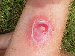
[[Image:Definition of MRSA.jpg|MRSA infection.|right|frameless|250x250px]]MRSA can cause a range of organ-specific infections, the most common being the skin and subcutaneous tissues, followed by invasive infections like osteomyelitis, meningitis, pneumonia, lung abscess, and empyema. Infective endocarditis caused by MRSA is associated with an increased morbidity and mortality compared to any other organism and is linked to intravenous drug abuse.


add text here <br>
'''Skin and soft tissue infections (SSTI):''' CA-MRSA is a predominant organism associated with SSTIs like cellulitis, necrotizing fasciitis, and diabetic foot ulcers. It also is increasingly associated with more invasive disease than those due to non-MRSA. More frequently these infections are multidrug-resistant leading to frequent recurrence, increased hospitalization, and mortality.


== Diagnostic Tests/Lab Tests/Lab Values  ==
'''Bone and joint infection:''' Staphylococci are the most common cause of bone and joint infections. MRSA can cause osteomyelitis of spine, long bones of upper and lower extremities by extension of local infection from a wound or as a part of hematogenous infection. Similarly, MRSA can cause septic arthritis of both native and prosthetic joints.
 
'''Pneumonia:''' Staphylococcal pneumonia - CA-MRSA can cause life-threatening necrotizing pneumonia in otherwise healthy individuals. It is characterized by severe respiratory symptoms, high fevers, hemoptysis, and hypotension. It rapidly progresses to sepsis and septic shock with leukopenia and elevated C-reactive protein. Multilobar cavitating alveolar infiltrates in a clinical setting like this is consistent with CA-MRSA infection.
 
MRSA is also a leading cause of hospital-acquired and ventilator-associated pneumonia.
 
'''Bacteremia:''' Bacteremia due to ''S. aureus'' has been reported to be associated with mortality rates of 15% to 60%. MRSA bacteremia is commonly seen in intensive care unit patients with central line insertions. Infective endocarditis is associated with MRSA bacteremia and should be ruled out in any patient with MRSA in the bloodstream. The outcomes related to MRSA bacteremia are worse than other MRSA infections because of the decreased response to vancomycin in these patients.
 
'''Endocarditis:''' MRSA is an important cause of bacterial endocarditis which can cause mortality in about a third of the infected patients (30-37%). Right-sided MRSA endocarditis is commonly associated with intravenous drug use and intravenous catheters. Patients with tricuspid valve vegetations may have septic pulmonary emboli causing nodular infiltrates and cavitating lesions in the lungs. Similarly, patients with involvement of mitral and aortic valves may have secondary infections in distant foci such as bones and joints, kidneys, brain, and other organs. It is important to take history and perform a thorough examination of these patients combined with necessary labs and radiological tests.<ref name=":0" />
 
== Characteristics  ==
 
• Chest pain<br>• Cough<br>• Shortness of breath<br>• Fatigue<br>• Fever and chills<br>• General ill feeling<br>• Headache<br>• Rash<br>• Wounds that do not heal 
 
Differences in Characteristics Between HA-MRSA and CA-MRSA<ref name="K2">Millar B, Loughrey A, Elborn J, Moore J. Proposed definitions of community-associated meticillin-resistant Staphylococcus aureus (CA-MRSA). Journal of Hospital Infection. 2007;67(2):109-113. http://eds.b.ebscohost.com.libproxy.bellarmine.edu/ehost/detail/detail?vid=14&amp;sid=71ee6b7e-199d-4adc-8e31-bc94eca59a09%40sessionmgr113&amp;hid=112&amp;bdata=JmxvZ2luLmFzcCZzaXRlPWVob3N0LWxpdmUmc2NvcGU9c2l0ZQ%3d%3d#AN=105828672&amp;db=cin20 (accessed 2 April 2016).</ref>


add text here <br>
{| width="75%" border="1" cellpadding="1" cellspacing="1"
|-
| '''Characteristic'''
| '''HA-MRSA'''
| '''CA-MRSA'''
|-
| Site of Infection
| Bacteraemia, wound infections, respiratory tract, urinary tract
| Mainly skin (abscesses, cellulitis, furunculosis
|-
| Risk Factors
| Indwelling devices, catheters, lines, hemodialysis, prolonged hospitalization, long-term antibiotic use
| Close physical contact, abrasion injuries, poor hygiene
|-
| Transmission
| Person-to-person (healthcare staff, visitors, patients), environment-to-person (hospital equipment
| Person-to-person (contact sports), environment-to-person (shared facilities, shared sports equipment)
|}   


== Etiology/Causes ==
== Medical Management ==
The selection of empiric antibiotic therapy for the treatment of MRSA infection depends on the type of disease, local ''S. aureus'' resistance patterns, availability of the drug, side effect profile, and individual patient profile.


add text here <br>
'''SSTIs:''' For most uncomplicated SSTIs suspected of MRSA infection, empirical treatment is with oral antibiotics like trimethoprim/sulfamethoxazole, tetracyclines, such as doxycycline or minocycline, and clindamycin.


== Systemic Involvement  ==
Parenteral antibiotics are indicated for invasive SSTIs or with signs of systemic involvement, inadequate response to oral therapy, or if an SSTI occurs adjacent to an indwelling device.


add text here
Intravenous vancomycin is the drug of choice for most MRSA infections seen in hospitalized patients. It can be used both as empiric and definitive therapy as most MRSA infections are susceptible to vancomycin. There are sporadic cases of vancomycin-resistant MRSA.


== Medical Management (current best evidence)  ==
Daptomycin is a suitable parenteral alternative when vancomycin is not available or not being tolerated. Other short-acting options include ceftaroline and telavancin. Long-acting treatment options include dalbavancin and oritavancin. Regardless of the initial empiric antibiotic choice, subsequent therapy should be tailored based on the careful review of culture and susceptibility data.


add text here
The duration of therapy for treatment of MRSA SSTIs may range from 5 to 14 days depending on the extent of infection and response to treatment.<ref name="K4" />


== Physical Therapy Management (current best evidence) ==
== Diagnostic Tests/Lab Tests/Lab Values ==


add text here
Clinical Laboratories for MRSA, according to the Clinical and Laboratory Standards Institute (CLSI)<ref>Centers for Disease Control and Prevention. Laboratory Testing for MRSA. http://www.cdc.gov/mrsa/lab/ (accessed 8 April 2016).</ref><br>


== Differential Diagnosis  ==
Broth Microdultion Testing with the addition of one of the following:


add text here
*A plate containing 6 μg/ml of oxacillin in Mueller-Hinton agar supplemented with 4% NaCl as alternative methods of testing for MRSA
*Latex Agglutination Test for PBP2 - tests for the mecA gene (Staphylococcal resistance to oxacillin/methicillin happens when an altered penicillin-binding protein, PBP2a, is produced.)
*Cefoxitin Disk Screen Test<br>
Typically the broth test and agar test are the main tests for detecting MRSA, and the cefoxitin disk screen test can be used as a back-up test. <br>• Detecting oxacillin/methicillin resistance can be difficult because of the presence of multiple populations (susceptible and resistant) may coexist within one culture of staphylococci.<br>• Oxacillin and cefoxitin are tested instead of methicillin because methicillin isn’t as readily available in the US as it once was. Oxacillin detects subpopulations well and cefoxitin activates mecA gene better, making it a more accurate test than other using oxacillin.<br>• Methicillin and oxacillin are in the same class of drugs. Methicillin has historically been the drug of choice for detecting resistance, hence the name MRSA. Even though the preferred drug of choice for testing has changed, the name had remained the same.<br>According to Centers for Disease Control and Prevention, there are additional tests to detect oxacillin/methicillin resistance but the most common and reliable tests have been listed above.
== Physical Therapy Management  ==


== Case Reports/ Case Studies  ==
MRSA is not primarily managed by physical therapy. Instead it is the physical therapist’s role to identify comorbidities that are inhibiting functional activity and treat the patient’s symptoms. When treating a patient with MRSA, it’s important for the physical therapist to know proper disinfecting techniques to prevent the spread of the disease. Precautions to be taken when treating a patient with MRSA include but are not limited to:<ref>Centers for Disease Control and Prevention. Cleaning &amp; Disinfecting Athlete Facilities. http://www.cdc.gov/mrsa/community/environment/athletic-facilities.html (accessed 8 April 2016).</ref><ref>Centers for Disease Control and Prevention. Laundry. http://www.cdc.gov/mrsa/community/environment/laundry.html (accessed 8 April 2016).</ref><ref name="Medline">MedlinePlus Medical Encyclopedia. MRSA. https://www.nlm.nih.gov/medlineplus/ency/article/007261.htm (accessed 7 April 2016).</ref>


add links to case studies here (case studies should be added on new pages using the [[Template:Case Study|case study template]])<br>
*Buy disinfectants that are registered by the Environmental Protection Agency (EPA)
*Note how to properly apply each product, how long it needs to be left on the surface, if the surface needs to be rinsed prior, if it’s only for specific surfaces, etc.
*Laundering specific clothes, towels and linens separately would be appropriate but not always necessary – wash and dry in the warmest temperate recommended on each piece of linen individually
*All surfaces ad equipment that came in contact with the patient should be cleaned, sometimes individual equipment for the patient with MRSA may be necessary if equipment cannot be properly cleaned
*Use barriers between the patient’s skin and surface when possible
*Physical therapist should wash body parts immediately after making contact with patient’s skin
*Patient should keep all open wounds clean and covered at all times until healed<br>
There has also been evidence to support the use of low-frequency ultrasound on bacteria, including MRSA. According to an article posted in 2010, low-frequency ultrasound delivered at 35 kHz reduced colony forming units of bacteria and alters colonial characteristics of MRSA.<ref>Conner-Kerr T, Alston G, Stovall A, Vernon T, Winter D, Meixner J et al. The Effects of Low-frequency Ultrasound (35kHz) on Methicillin-resistant Staphylococcus aureus (MRSA) in vitro. Ostomy Wound Manage. 2010;56(5):32–42. http://www.o-wm.com/content/effects-low-frequency-ultrasound-35-khz-methicillin-resistant-staphylococcus-aureus-mrsa-vit (accessed 9 April 2016).</ref>


== Resources <br> ==
=== Differential Diagnosis ===
The differential diagnosis for MRSA includes the following infections:<ref>Staphylococcus Aureus Infection Differential Diagnoses [Internet]. Emedicine.medscape.com. 2016.&amp;nbsp;http://emedicine.medscape.com/article/971358-differential (accessed 6 April 2016).</ref><br>• Bacteremia<br>• Chemical burns<br>• Impetigo<br>• Juvenile Idiopathic Arthritis<br>• Streptococcal Infections<br>• Kawasaki Disease<br>• Leptospirosis<br>• Parvovirus B19 Infection


add appropriate resources here
Further screening should be done to rule out such diseases.


== Recent Related Research (from [http://www.ncbi.nlm.nih.gov/pubmed/ Pubmed])  ==
=== '''Outcomes''' ===
* The majority of data indicate that MRSA increases mortality and morbidity in seniors, nursing home patients and those with organ dysfunction.  
* Individuals with end-stage liver disease, renal insufficiency and those admitted to the ICU have high mortality rates when there is an associated MRSA infection.  
* The mortality rates vary from 5-60%, depending on the patient population and site of infection.  
* More patients with MRSA are now undergoing surgery, and in at least 40% of patients, a central line was the cause of the infection.  
* About 60% of patients do acquire MRSA within 48 hours despite having no healthcare risks<ref name=":0" /><br>


see tutorial on [[Adding PubMed Feed|Adding PubMed Feed]]
== Case Reports/ Case Studies ==
<div class="researchbox">
<rss><?xml version="1.0" encoding="utf-8"?>
<rss version="2.0">
<channel>
<title>pubmed: mrsa treatment</title>
<link>http://www.ncbi.nlm.nih.gov/sites/entrez?cmd=Search&amp;db=PubMed&amp;term=MRSA%20treatment</link>
<description>NCBI: db=pubmed; Term=MRSA treatment</description>
<language>en-us</language>
<docs>http://blogs.law.harvard.edu/tech/rss</docs>
<ttl>1440</ttl>
<image>
<title>NCBI pubmed</title>
<url>http://www.ncbi.nlm.nih.gov/entrez/query/static/gifs/iconsml.gif</url>
<link>http://www.ncbi.nlm.nih.gov/sites/entrez</link>
<description>PubMed comprises more than 24 million citations for biomedical literature from MEDLINE, life science journals, and online books. Citations may include links to full-text content from PubMed Central and publisher web sites.</description>
</image>
<item>
    <title>Chronic Osteomyelitis of Clavicle in a Neonate: Report of Morbid Complication of Adjoining MRSA Abscess.</title>       
    <link>http://www.ncbi.nlm.nih.gov/pubmed/27051549?dopt=Abstract</link>   
    <description>
<![CDATA[<table border="0" width="100%"><tr><td align="left"/></tr></table>
        <p><b>Chronic Osteomyelitis of Clavicle in a Neonate: Report of Morbid Complication of Adjoining MRSA Abscess.</b></p>         
        <p>Case Rep Pediatr. 2016;2016:3032518</p>
        <p>Authors:  Suranigi SM, Joshi M, Deniese PN, Rangasamy K, Najimudeen S, Gnanadoss JJ</p>
        <p>Abstract<br/>
        Osteomyelitis of clavicle is rare in neonates. Acute osteomyelitis of clavicle accounts for less than 3% of all osteomyelitis cases. It may occur due to contiguous spread, due to hematogenous spread, or secondary to subclavian catheterization. Chronic osteomyelitis may occur as a complication of residual adjoining abscess due to methicillin resistant staphylococcus aureus (MRSA) sepsis. We report a newborn female with right shoulder abscess that developed chronic clavicular osteomyelitis in follow-up period after drainage. She required multiple drainage procedures and was later successfully managed with bone curettage and debridement. We report this case to highlight that a MRSA abscess may recur due to residual infection from a chronic osteomyelitis sinus. It may be misdiagnosed as hypergranulation tissue of nonhealing wound leading to inappropriate delay in treatment. High index of suspicion, aggressive initial management, and regular follow-up are imperative to prevent this morbid complication.<br/>
        </p><p>PMID: 27051549 [PubMed - as supplied by publisher]</p>
    ]]></description>
    <author> Suranigi SM, Joshi M, Deniese PN, Rangasamy K, Najimudeen S, Gnanadoss JJ</author>
    <category>Case Rep Pediatr</category>
    <guid isPermaLink="false">PubMed:27051549</guid>
</item>
<item>
    <title>Broad Spectrum Antimicrobial Activity of Forest-Derived Soil Actinomycete, Nocardia sp. PB-52.</title>       
    <link>http://www.ncbi.nlm.nih.gov/pubmed/27047463?dopt=Abstract</link>   
    <description>
<![CDATA[<table border="0" width="100%"><tr><td align="left"><a href="http://dx.doi.org/10.3389/fmicb.2016.00347"><img src="//www.ncbi.nlm.nih.gov/corehtml/query/egifs/http:--www.frontiersin.org-alerts-logo-logo_LinkOut.jpg" border="0"/></a> <a href="http://www.ncbi.nlm.nih.gov/pmc/articles/pmid/27047463/"><img src="//www.ncbi.nlm.nih.gov/corehtml/query/egifs/http:--www.ncbi.nlm.nih.gov-corehtml-pmc-pmcgifs-pubmed-pmc.gif" border="0"/></a> </td></tr></table>
        <p><b>Broad Spectrum Antimicrobial Activity of Forest-Derived Soil Actinomycete, Nocardia sp. PB-52.</b></p>         
        <p>Front Microbiol. 2016;7:347</p>
        <p>Authors:  Sharma P, Kalita MC, Thakur D</p>
        <p>Abstract<br/>
        A mesophilic actinomycete strain designated as PB-52 was isolated from soil samples of Pobitora Wildlife Sanctuary of Assam, India. Based on phenotypic and molecular characteristics, the strain was identified as Nocardia sp. which shares 99.7% sequence similarity with Nocardia niigatensis IFM 0330 (NR_112195). The strain is a Gram-positive filamentous bacterium with rugose spore surface which exhibited a wide range of antimicrobial activity against Gram-positive bacteria including methicillin-resistant Staphylococcus aureus (MRSA), Gram-negative bacteria, and yeasts. Optimization for the growth and antimicrobial activity of the strain PB-52 was carried out in batch culture under shaking condition. The optimum growth and antimicrobial potential of the strain were recorded in GLM medium at 28°C, initial pH 7.4 of the medium and incubation period of 8 days. Based on polyketide synthases (PKS) and non-ribosomal peptide synthetases (NRPS) gene-targeted PCR amplification, the occurrence of both of these biosynthetic pathways was detected which might be involved in the production of antimicrobial compounds in PB-52. Extract of the fermented broth culture of PB-52 was prepared with organic solvent extraction method using ethyl acetate. The ethyl acetate extract of PB-52 (EA-PB-52) showed lowest minimum inhibitory concentration (MIC) against S. aureus MTCC 96 (0.975 μg/mL) whereas highest was recorded against Klebsiella pneumoniae ATCC 13883 (62.5 μg/mL). Scanning electron microscopy (SEM) revealed that treatment of the test microorganisms with EA-PB-52 destroyed the targeted cells with prominent loss of cell shape and integrity. In order to determine the constituents responsible for its antimicrobial activity, EA-PB-52 was subjected to chemical analysis using gas chromatography-mass spectrometry (GC-MS). GC-MS analysis showed the presence of twelve different chemical constituents in the extract, some of which are reported to possess diverse biological activity. These results confirmed that the presence of bioactive constituents in EA-PB-52 could be a promising source for the development of potent antimicrobial agents effective against wide range of microbial pathogens including MRSA. <br/>
        </p><p>PMID: 27047463 [PubMed]</p>
    ]]></description>
    <author> Sharma P, Kalita MC, Thakur D</author>
    <category>Front Microbiol</category>
    <guid isPermaLink="false">PubMed:27047463</guid>
</item>
<item>
    <title>Cold Plasma Inactivation of Chronic Wound Bacteria.</title>       
    <link>http://www.ncbi.nlm.nih.gov/pubmed/27046340?dopt=Abstract</link>   
    <description>
<![CDATA[<table border="0" width="100%"><tr><td align="left"><a href="http://linkinghub.elsevier.com/retrieve/pii/S0003-9861(16)30093-5"><img src="//www.ncbi.nlm.nih.gov/corehtml/query/egifs/http:--linkinghub.elsevier.com-ihub-images-PubMedLink.gif" border="0"/></a> </td></tr></table>
        <p><b>Cold Plasma Inactivation of Chronic Wound Bacteria.</b></p>         
        <p>Arch Biochem Biophys. 2016 Apr 1;</p>
        <p>Authors:  Norhayati MN, Lee BK, Yap SS, Thong KL, Yap SL</p>
        <p>Abstract<br/>
        Cold plasma is partly ionized non-thermal plasma generated at atmospheric pressure. It has been recognized as an alternative approach in medicine for sterilization of wounds, promotion of wound healing, topical treatment of skin diseases with microbial involvement and treatment of cancer. Cold plasma used in wound therapy inhibits microbes in chronic wound due to its antiseptic effects, while promoting healing by stimulation of cell proliferation and migration of wound relating skin cells. In this study, two types of plasma systems are employed to generate cold plasma: a parallel plate dielectric barrier discharge and a capillary-guided corona discharge. Parameters such as applied voltage, discharge frequency, treatment time and the flow of the carrier gas influence the cold plasma chemistry and therefore change the composition and concentration of plasma species that react with the target sample. Chronic wound that fails to heal often infected by multidrug resistant organisms makes them recalcitrant to healing. Methicillin-resistant Staphylococcus aureus (MRSA) and Pseudomonas aeruginosa (P. aeruginosa) are two common bacteria in infected and clinically non-infected wounds. The efficacies of the cold plasma generated by the two designs on the inactivation of three different isolates of MRSA and four isolates of P. aeruginosa are reported here.<br/>
        </p><p>PMID: 27046340 [PubMed - as supplied by publisher]</p>
    ]]></description>
    <author> Norhayati MN, Lee BK, Yap SS, Thong KL, Yap SL</author>
    <category>Arch Biochem Biophys</category>
    <guid isPermaLink="false">PubMed:27046340</guid>
</item>
<item>
    <title>ALTERNATING MUPIROCIN/GENTAMICIN IS ASSOCIATED WITH INCREASED RISK OF FUNGAL PERITONITIS AS COMPARED WITH GENTAMICIN ALONE - RESULTS OF A RANDOMIZED OPEN-LABEL CONTROLLED TRIAL.</title>       
    <link>http://www.ncbi.nlm.nih.gov/pubmed/27044796?dopt=Abstract</link>   
    <description>
<![CDATA[<table border="0" width="100%"><tr><td align="left"/></tr></table>
        <p><b>ALTERNATING MUPIROCIN/GENTAMICIN IS ASSOCIATED WITH INCREASED RISK OF FUNGAL PERITONITIS AS COMPARED WITH GENTAMICIN ALONE - RESULTS OF A RANDOMIZED OPEN-LABEL CONTROLLED TRIAL.</b></p>         
        <p>Perit Dial Int. 2016 Apr 4;</p>
        <p>Authors:  Wong PN, Tong GM, Wong YY, Lo KY, Chan SF, Lo MW, Lo KC, Ho LY, Tse CW, Mak SK, Wong AK</p>
        <p>Abstract<br/>
        ♦ Background and Objectives: Catheter-related infection, namely exit-site infection (ESI) and peritonitis, is a major infectious complication and remains a main cause of technique failure for patients receiving peritoneal dialysis (PD). Topical application of antibiotic cream might reduce catheter-related infection but emergence of resistant or opportunistic organisms could be a concern. Optimal topical agents and regimens remain to be determined. We did a study to examine the effect of an alternating topical antibiotic regimen in preventing catheter-related infection.♦ Method: We performed a single-center, randomized, open-label study to compare daily topical application of gentamicin cream with a gentamicin/mupirocin alternate regimen to the exit site. Patients randomized to alternating regimen were asked to have daily application of gentamicin cream in odd months and mupirocin cream in even months. Primary outcomes were ESI and peritonitis. Secondary outcomes were catheter removal or death caused by catheter-related infection. A total of 146 patients (71, gentamicin group; 75, alternating regimen group) were enrolled with a total follow-up duration of 174 and 181 patient-years for gentamicin and alternating groups, respectively. All patients were followed up until catheter removal, death, transfer to another unit, transplantation or the end of the study on March 31, 2014. There were no significant differences in the age, sex, dialysis vintage, and rate of diabetes, helper-assisted dialysis and methicillin-resistant Staphylococcus aureus (MRSA) carriage state.♦ Results: No difference was seen in the time to first ESI or peritonitis. However, the time to first gram-negative peritonitis seemed longer for the gentamicin group (p = 0.055). The 2 groups showed a similar rate of ESI (0.17/yr vs 0.19/yr, p = 0.93) but P. aeruginosa ESI was less common in the gentamicin group (0.06/yr vs 0.11/yr, p &lt; 0.001). There was no difference in the incidence of ESI due to non-tuberculous mycobacteria. Peritonitis rate was significantly lower in the gentamicin group (0.22/yr vs 0.32/yr, p &lt; 0.001), with a striking decrease in gram-negative peritonitis (0.08/yr vs 0.14/yr, p &lt; 0.001), and fungal peritonitis (0.006/yr vs 0.03/yr, p &lt; 0.001), which was all antibiotics-related episodes with antecedent use of systemic antibiotics for the treatment of catheter-related infections. There was no significant difference in the catheter loss or death related to catheter-related infection.♦ Conclusion: Alternating gentamicin/mupirocin cream application appeared as effective as gentamicin alone in preventing ESI except for P. aeruginosa. However, it was inferior to gentamicin in the prevention of peritonitis episodes, especially for those caused by gram-negative organisms. It was also not useful in reducing catheter-related infection due to opportunistic organisms but instead associated with a higher incidence of antibiotic-related fungal peritonitis.<br/>
        </p><p>PMID: 27044796 [PubMed - as supplied by publisher]</p>
    ]]></description>
    <author> Wong PN, Tong GM, Wong YY, Lo KY, Chan SF, Lo MW, Lo KC, Ho LY, Tse CW, Mak SK, Wong AK</author>
    <category>Perit Dial Int</category>
    <guid isPermaLink="false">PubMed:27044796</guid>
</item>
<item>
    <title>Results from the Solithromycin International Surveillance Program (2014).</title>       
    <link>http://www.ncbi.nlm.nih.gov/pubmed/27044551?dopt=Abstract</link>   
    <description>
<![CDATA[<table border="0" width="100%"><tr><td align="left"/></tr></table>
        <p><b>Results from the Solithromycin International Surveillance Program (2014).</b></p>         
        <p>Antimicrob Agents Chemother. 2016 Apr 4;</p>
        <p>Authors:  Farrell DJ, Flamm RK, Sader HS, Jones RN</p>
        <p>Abstract<br/>
        Solithromycin, a fourth-generation macrolide and the first fluoroketolide, was tested against a 2014 collection of 6,115 isolates, includingStreptococcus pneumoniae(1, 713),Haemophilus influenzae(1, 308),Moraxella catarrhalis(577),Staphylococcus aureus(1, 024), and beta-hemolytic streptococci (1, 493) by reference broth microdilution methods. The geographic samples included 2,748 isolates from the United States, 2,536 from Europe, 386 from Latin America, and 445 from the Asia Pacific region. Solithromycin was observed to be very active againstS. pneumoniae(MIC50/90, 0.008/0.12 μg/mL), demonstrating two-fold greater activity than telithromycin (MIC50/90, 0.015/0.25 μg/mL) and 16- to &gt;256-fold greater activity than azithromycin (MIC50/90, 0.12/&gt;32 μg/mL), with all strains inhibited at a solithromycin MIC of ≤1 μg/mL. AgainstH. influenzae, solithromycin showed identical potency to that of telithromycin (MIC50/90, 1/2 μg/mL) and both of these compounds were two-fold less active than azithromycin (MIC50/90, 0.5/1 μg/mL). All but oneM. catarrhalisisolate were inhibited by solithromycin at ≤0.25 μg/mL. Solithromycin inhibited 85.3% ofS. aureusisolates at ≤1 μg/mL, and its activity was lower against methicillin-resistant (MIC50/90, 0.06/&gt;32 μg/mL) compared to methicillin-susceptible (MIC50/90, 0.06/0.06 μg/mL) isolates. Little variation in solithromycin activity was observed by geographic region for the species tested. Solithromycin was very active against beta-hemolytic streptococci (MIC50/90, 0.015/0.03 μg/mL) and all isolates were inhibited at MIC values ≤0.5 μg/mL). In conclusion, solithromycin demonstrated potent activity against global and contemporary (2014) pathogens that represent the major causes of community-acquired bacterial pneumonia. These data support the continued clinical development of solithromycin for the treatment of this important indication.<br/>
        </p><p>PMID: 27044551 [PubMed - as supplied by publisher]</p>
    ]]></description>
    <author> Farrell DJ, Flamm RK, Sader HS, Jones RN</author>
    <category>Antimicrob Agents Chemother</category>
    <guid isPermaLink="false">PubMed:27044551</guid>
</item>
<item>
    <title>Can EDTA Change MRSA into MSSA? A Future Prospective!</title>       
    <link>http://www.ncbi.nlm.nih.gov/pubmed/27042464?dopt=Abstract</link>   
    <description>
<![CDATA[<table border="0" width="100%"><tr><td align="left"><a href="http://www.ncbi.nlm.nih.gov/pmc/articles/pmid/27042464/"><img src="//www.ncbi.nlm.nih.gov/corehtml/query/egifs/http:--www.ncbi.nlm.nih.gov-corehtml-pmc-pmcgifs-pubmed-pmc.gif" border="0"/></a> </td><td align="right"><a href="http://www.ncbi.nlm.nih.gov/sites/entrez?db=pubmed&amp;cmd=Link&amp;LinkName=pubmed_pubmed&amp;from_uid=27042464">Related Articles</a></td></tr></table>
        <p><b>Can EDTA Change MRSA into MSSA? A Future Prospective!</b></p>         
        <p>J Clin Diagn Res. 2016 Feb;10(2):DC22-5</p>
        <p>Authors:  Jain S, Sengupta M, Sarkar S, Ghosh S, Mitra AN, Sinha A, Chakravorty S</p>
        <p>Abstract<br/>
        INTRODUCTION: In the present era we are left behind with limited options for the treatment of serious infections caused by multidrug resistant S.aureus, most remarkably nosocomially acquired Methicillin resistant S.aureus (MRSA). The problem increases more when these strains easily become multidrug resistant (MDR) due to biofilm formation. Those staphylococcal species that are vancomycin and linezolid resistant are also resistant to other antistaphylococcal agents which call for an urgent intervention to develop newer antimicrobial agents.<br/>
        AIM: The present study was undertaken with the aim to evaluate the antibiofilm effect of EDTA against the biofilm forming MRSA isolates, isolated from different clinical infections.<br/>
        MATERIALS AND METHODS: The biofilms formed on polystyrene microtitre plates by the MRSA strains were treated by different concentrations of EDTA to find out its anti-biofilm activity. Further simultaneously the antibiotic susceptibility pattern was noted down to check whether the MRSA strains become MSSA (Methicillin sensitive S.aureus).<br/>
        RESULTS: Our data demonstrates that EDTA at 4mM concentration inhibits biofilm of MRSA and at 20 mM have an ability to reduce and dissociate the biofilm membrane, allowing the antibiotics to enter and convert MRSA strains into MSSA.<br/>
        CONCLUSION: These findings suggest that commercially available EDTA could be used in future to control MRSA and its biofilm- related infections.<br/>
        </p><p>PMID: 27042464 [PubMed]</p>
    ]]></description>
    <author> Jain S, Sengupta M, Sarkar S, Ghosh S, Mitra AN, Sinha A, Chakravorty S</author>
    <category>J Clin Diagn Res</category>
    <guid isPermaLink="false">PubMed:27042464</guid>
</item>
<item>
    <title>Detection of Vancomycin Resistance among Enterococcus faecalis and Staphylococcus aureus.</title>       
    <link>http://www.ncbi.nlm.nih.gov/pubmed/27042459?dopt=Abstract</link>   
    <description>
<![CDATA[<table border="0" width="100%"><tr><td align="left"><a href="http://www.ncbi.nlm.nih.gov/pmc/articles/pmid/27042459/"><img src="//www.ncbi.nlm.nih.gov/corehtml/query/egifs/http:--www.ncbi.nlm.nih.gov-corehtml-pmc-pmcgifs-pubmed-pmc.gif" border="0"/></a> </td><td align="right"><a href="http://www.ncbi.nlm.nih.gov/sites/entrez?db=pubmed&amp;cmd=Link&amp;LinkName=pubmed_pubmed&amp;from_uid=27042459">Related Articles</a></td></tr></table>
        <p><b>Detection of Vancomycin Resistance among Enterococcus faecalis and Staphylococcus aureus.</b></p>         
        <p>J Clin Diagn Res. 2016 Feb;10(2):DC04-6</p>
        <p>Authors:  Rengaraj R, Mariappan S, Sekar U, Kamalanadhan A</p>
        <p>Abstract<br/>
        INTRODUCTION: Vancomycin remains the drug of choice for resistant gram positive infections caused by Enterococcus spp and Methicillin resistant Staphylococcus aureus (MRSA). Increased use of vancomycin has led to frank resistance and increase in MIC (MIC creep). Vancomycin intermediate Staphylococcus aureus (VISA), Vancomycin resistant Staphylococcus aureus (VRSA) &amp; Vancomycin resistant Enterococci (VRE) are important emerging nosocomial pathogens resulting in treatment failures.<br/>
        AIM: This study was undertaken to detect vancomycin resistance among clinical isolates of Staphylococcus aureus and Enterococcus faecalis by phenotypic and genotypic methods.<br/>
        MATERIALS AND METHODS: The study was conducted in a 1850 bedded university teaching hospital from November 2013 to April 2014. Non-repetitive, consecutive clinically significant Staphylococcus aureus (109) and Enterococcus faecalis (124) were included in this study. They were identified up to species level by conventional and automated methods. Susceptibility to various antibiotics was tested by disc diffusion method. MIC of vancomycin was determined by agar dilution method. Inducible resistance to clindamycin was detected by the D test. Methicillin resistance in Staphylococcus aureus (MRSA) was screened using cefoxitin disc. All isolates were subjected to polymerase chain reaction (PCR) to detect van A and van B genes.<br/>
        RESULTS: Out of 109 Staphylococcus aureus isolates, 54 were MRSA. By MIC there was no resistance observed to vancomycin.MIC50 was 1μg/ml. None of the isolates harboured van A and van B. Among Enterococcus faecalis, sixteen isolates (12.9%) and four isolates (3.2%) exhibited resistance to vancomycin and teicoplanin by disc diffusion respectively. All isolates were susceptible to linezolid. Van A was detected in 2, van B in 7 and one had both van A and van B.<br/>
        CONCLUSION: PCR remains the gold standard for diagnosis of vancomycin resistance. There was no resistance observed to vancomycin among Staphylococci though the MIC creep detected is a cause for concern. Eight percent of Enterococci were vancomycin resistant.<br/>
        </p><p>PMID: 27042459 [PubMed]</p>
    ]]></description>
    <author> Rengaraj R, Mariappan S, Sekar U, Kamalanadhan A</author>
    <category>J Clin Diagn Res</category>
    <guid isPermaLink="false">PubMed:27042459</guid>
</item>
<item>
    <title>Antibiotics and antiseptics for pressure ulcers.</title>       
    <link>http://www.ncbi.nlm.nih.gov/pubmed/27040598?dopt=Abstract</link>   
    <description>
<![CDATA[<table border="0" width="100%"><tr><td align="left"><a href="http://dx.doi.org/10.1002/14651858.CD011586.pub2"><img src="//www.ncbi.nlm.nih.gov/corehtml/query/egifs/http:--media.wiley.com-assets-7315-19-Wiley_FullText_120x30_orange.png" border="0"/></a> </td><td align="right"><a href="http://www.ncbi.nlm.nih.gov/sites/entrez?db=pubmed&amp;cmd=Link&amp;LinkName=pubmed_pubmed&amp;from_uid=27040598">Related Articles</a></td></tr></table>
        <p><b>Antibiotics and antiseptics for pressure ulcers.</b></p>         
        <p>Cochrane Database Syst Rev. 2016 Apr 4;4:CD011586</p>
        <p>Authors:  Norman G, Dumville JC, Moore ZE, Tanner J, Christie J, Goto S</p>
        <p>Abstract<br/>
        BACKGROUND: Pressure ulcers, also known as bedsores, decubitus ulcers and pressure injuries, are localised areas of injury to the skin or the underlying tissue, or both. A range of treatments with antimicrobial properties, including impregnated dressings, are widely used in the treatment of pressure ulcers. A clear and current overview is required to facilitate decision making regarding use of antiseptic or antibiotic therapies in the treatment of pressure ulcers. This review is one of a suite of Cochrane reviews investigating the use of antiseptics and antibiotics in different types of wounds. It also forms part of a suite of reviews investigating the use of different types of dressings and topical treatments in the treatment of pressure ulcers.<br/>
        OBJECTIVES: To assess the effects of systemic and topical antibiotics, and topical antiseptics on the healing of infected and uninfected pressure ulcers being treated in any clinical setting.<br/>
        SEARCH METHODS: In October 2015 we searched: the Cochrane Wounds Specialised Register, the Cochrane Central Register of Controlled Trials (CENTRAL) (The Cochrane Library), Ovid MEDLINE, Ovid MEDLINE (In-Process &amp; Other Non-Indexed Citations), Ovid EMBASE, and EBSCO CINAHL Plus. We also searched three clinical trials registries and the references of included studies and relevant systematic reviews. There were no restrictions based on language or date of publication or study setting.<br/>
        SELECTION CRITERIA: Randomised controlled trials which enrolled adults with pressure ulcers of stage II or above were included in the review.<br/>
        DATA COLLECTION AND ANALYSIS: Two review authors independently performed study selection, risk of bias assessment and data extraction.<br/>
        MAIN RESULTS: We included 12 trials (576 participants); 11 had two arms and one had three arms. All assessed topical agents, none looked at systemic antibiotics. The included trials assessed the following antimicrobial agents: povidone iodine, cadexomer iodine, gentian violet, lysozyme, silver dressings, honey, pine resin, polyhexanide, silver sulfadiazine, and nitrofurazone with ethoxy-diaminoacridine. Comparators included a range of other dressings and ointments without antimicrobial properties and alternative antimicrobials. Each comparison had only one trial, participant numbers were low and follow-up times short. The evidence varied from moderate to very low quality.Six trials reported the primary outcome of wound healing. All except one compared an antiseptic with a non-antimicrobial comparator. There was some moderate and low quality evidence that fewer ulcers may heal in the short term when treated with povidone iodine compared with non-antimicrobial alternatives (protease-modulating dressings (risk ratio (RR) 0.78, 95% confidence interval (CI) 0.62 to 0.98) and hydrogel (RR 0.64, 95% CI 0.43 to 0.97)); and no clear difference between povidone iodine and a third non-antimicrobial treatment (hydrocolloid) (low quality evidence). Pine resin salve may heal more pressure ulcers than hydrocolloid (RR 2.83, 95% CI 1.14 to 7.05) (low quality evidence). There is no clear difference between cadexomer iodine and standard care, and between honey a combined antiseptic and antibiotic treatment (very low quality evidence).Six trials reported adverse events (primary safety outcome). Four reported no adverse events; there was very low quality evidence from one showing no clear evidence of a difference between cadexomer iodine and standard care; in one trial it was not clear whether data were appropriately reported.There was limited reporting of secondary outcomes. The five trials that reported change in wound size as a continuous outcome did not report any clear evidence favouring any particular antiseptic/anti-microbial treatments. For bacterial resistance, one trial found some evidence of more MRSA eradication in participants with ulcer treated with a polyhexanide dressing compared with a polyhexanide swab (RR 1.48, 95% CI 1.02 to 2.13); patients in the dressing group also reported less pain (MD -2.03, 95% CI -2.66 to -1.40). There was no clear evidence of a difference between interventions in infection resolution in three other comparisons. Evidence for secondary outcomes varied from moderate to very low quality; where no GRADE assessment was possible we identified substantial limitations which an assessment would have taken into account.<br/>
        AUTHORS' CONCLUSIONS: The relative effects of systemic and topical antimicrobial treatments on pressure ulcers are not clear. Where differences in wound healing were found, these sometimes favoured the comparator treatment without antimicrobial properties. The trials are small, clinically heterogenous, generally of short duration, and at high or unclear risk of bias. The quality of the evidence ranges from moderate to very low; evidence on all comparisons was subject to some limitations.<br/>
        </p><p>PMID: 27040598 [PubMed - as supplied by publisher]</p>
    ]]></description>
    <author> Norman G, Dumville JC, Moore ZE, Tanner J, Christie J, Goto S</author>
    <category>Cochrane Database Syst Rev</category>
    <guid isPermaLink="false">PubMed:27040598</guid>
</item>
<item>
    <title>The effect of vancomycin powder on bone healing in a rat spinal rhBMP-2 model.</title>       
    <link>http://www.ncbi.nlm.nih.gov/pubmed/27035510?dopt=Abstract</link>   
    <description>
<![CDATA[<table border="0" width="100%"><tr><td align="left"/><td align="right"><a href="http://www.ncbi.nlm.nih.gov/sites/entrez?db=pubmed&amp;cmd=Link&amp;LinkName=pubmed_pubmed&amp;from_uid=27035510">Related Articles</a></td></tr></table>
        <p><b>The effect of vancomycin powder on bone healing in a rat spinal rhBMP-2 model.</b></p>         
        <p>J Neurosurg Spine. 2016 Apr 1;:1-7</p>
        <p>Authors:  Mendoza MC, Sonn KA, Kannan AS, Bellary SS, Mitchell SM, Singh G, Park C, Yun C, Stock SR, Hsu EL, Hsu WK</p>
        <p>Abstract<br/>
        OBJECTIVE This study aims to quantify the impact of vancomycin powder application on new bone formation and spine fusion rates in a rat posterolateral arthrodesis model. METHODS Thirty-six female Sprague-Dawley rats underwent a posterolateral lumbar spinal fusion (PLF) at the L-4 and L-5 vertebrae. Fusion was elicited via implantation of an absorbable collagen sponge containing 3 µg rhBMP-2. Rats were divided into 3 groups: no vancomycin (control), standard-dose vancomycin, and high-dose vancomycin, based on what was applied to the fusion bed. Clinical studies typically describe the application of 1 g vancomycin into the surgical wound. Presuming an average individual patient weight of 70 kg, a weight-based equivalent dose of vancomycin powder was applied subfascially in the PLF model constituting a "standard-dose" treatment group (14.3 mg/kg, n = 12). To determine whether there is a critical threshold beyond which vancomycin increases the risk of pseudarthrosis, a 10-fold higher dose was administered to a "high-dose" treatment group (143 mg/kg, n = 12). No vancomycin powder was applied to the surgical site in the control group (n = 12). Fusion was evaluated with plain radiographs at 4 and 8 weeks after surgery. The spines were harvested after the 8-week radiographs were obtained and evaluated using manual palpation, microCT analysis, and histological analysis. RESULTS Radiographs demonstrated equivalent bridging bone formation in all groups. No significant differences in fusion scores were seen in the standard-dose (mean 2.25) or high-dose (2.13) treatment groups relative to untreated control animals (1.78). Similarly, fusion rates did not differ significantly different between vancomycin-treated animals (100% for both groups) and control animals (92%). Quantification of new bone formation via microCT imaging revealed no significant between-groups differences in the volume of newly regenerated bone (control vs standard-dose vancomycin, p = 0.57; control vs high-dose vancomycin, p = 0.53). CONCLUSIONS This is the first in vivo study to specifically address the development of pseudarthrosis after intrawound application of vancomycin during fusion surgery. Our results demonstrate that vancomycin powder does not inhibit fusion rates at a dose that is the weight-percentage equivalent of what is routinely used by surgeons. Moreover, bone formation and fusion rates were not reduced even after administration of a vancomycin dose that is 10-fold higher than that which is typically administered clinically. Our findings suggest that if there is a critical threshold above which vancomycin inhibits bone healing, such a dose is out of the range which might be considered reasonable for clinical use.<br/>
        </p><p>PMID: 27035510 [PubMed - as supplied by publisher]</p>
    ]]></description>
    <author> Mendoza MC, Sonn KA, Kannan AS, Bellary SS, Mitchell SM, Singh G, Park C, Yun C, Stock SR, Hsu EL, Hsu WK</author>
    <category>J Neurosurg Spine</category>
    <guid isPermaLink="false">PubMed:27035510</guid>
</item>
<item>
    <title>Linezolid-induced serotonin toxicity in a patient not taking monoamine oxidase inhibitors or serotonin receptor antagonists.</title>       
    <link>http://www.ncbi.nlm.nih.gov/pubmed/27034576?dopt=Abstract</link>   
    <description>
<![CDATA[<table border="0" width="100%"><tr><td align="left"><a href="http://www.ncbi.nlm.nih.gov/pmc/articles/pmid/27034576/"><img src="//www.ncbi.nlm.nih.gov/corehtml/query/egifs/http:--www.ncbi.nlm.nih.gov-corehtml-pmc-pmcgifs-pubmed-pmc.gif" border="0"/></a> </td><td align="right"><a href="http://www.ncbi.nlm.nih.gov/sites/entrez?db=pubmed&amp;cmd=Link&amp;LinkName=pubmed_pubmed&amp;from_uid=27034576">Related Articles</a></td></tr></table>
        <p><b>Linezolid-induced serotonin toxicity in a patient not taking monoamine oxidase inhibitors or serotonin receptor antagonists.</b></p>         
        <p>Proc (Bayl Univ Med Cent). 2016 Apr;29(2):214-5</p>
        <p>Authors:  Sutton J, Stroup J, Som M</p>
        <p>Abstract<br/>
        Linezolid is an oxazolidinone antibiotic with weak monoamine oxidase (MAO) type A and MAO type B inhibitory effects. Linezolid has been associated with serotonin toxicity when used concomitantly with multiple medications that are known to increase serotonin concentrations. We report the case of a 65-year-old woman with signs and symptoms of serotonin toxicity following administration of linezolid for treatment of methicillin-resistant Staphylococcus aureus pneumonia. <br/>
        </p><p>PMID: 27034576 [PubMed]</p>
    ]]></description>
    <author> Sutton J, Stroup J, Som M</author>
    <category>Proc (Bayl Univ Med Cent)</category>
    <guid isPermaLink="false">PubMed:27034576</guid>
</item>
<item>
    <title>CAMERA2 - combination antibiotic therapy for methicillin-resistant Staphylococcus aureus infection: study protocol for a randomised controlled trial.</title>       
    <link>http://www.ncbi.nlm.nih.gov/pubmed/27029920?dopt=Abstract</link>   
    <description>
<![CDATA[<table border="0" width="100%"><tr><td align="left"><a href="http://trialsjournal.biomedcentral.com/articles/10.1186/s13063-016-1295-3"><img src="//www.ncbi.nlm.nih.gov/corehtml/query/egifs/http:--www.biomedcentral.com-graphics-pubmed-BioMedCentral_free_1.png" border="0"/></a> <a href="http://www.ncbi.nlm.nih.gov/pmc/articles/pmid/27029920/"><img src="//www.ncbi.nlm.nih.gov/corehtml/query/egifs/http:--www.ncbi.nlm.nih.gov-corehtml-pmc-pmcgifs-pubmed-pmc.gif" border="0"/></a> </td><td align="right"><a href="http://www.ncbi.nlm.nih.gov/sites/entrez?db=pubmed&amp;cmd=Link&amp;LinkName=pubmed_pubmed&amp;from_uid=27029920">Related Articles</a></td></tr></table>
        <p><b>CAMERA2 - combination antibiotic therapy for methicillin-resistant Staphylococcus aureus infection: study protocol for a randomised controlled trial.</b></p>         
        <p>Trials. 2016;17(1):170</p>
        <p>Authors:  Tong SY, Nelson J, Paterson DL, Fowler VG, Howden BP, Cheng AC, Chatfield M, Lipman J, Van Hal S, O'Sullivan M, Robinson JO, Yahav D, Lye D, Davis JS, CAMERA2 study group and the Australasian Society for Infectious Diseases Clinical Research Network</p>
        <p>Abstract<br/>
        BACKGROUND: Methicillin-resistant Staphylococcus aureus (MRSA) bacteraemia is a serious infection resulting in 20-50 % 90-day mortality. The limitations of vancomycin, the current standard therapy for MRSA, make treatment difficult. The only other approved drug for treatment of MRSA bacteraemia, daptomycin, has not been shown to be superior to vancomycin. Surprisingly, there has been consistent in-vitro and in-vivo laboratory data demonstrating synergy between vancomycin or daptomycin and an anti-staphylococcal β-lactam antibiotic. There is also growing clinical data to support such combinations, including a recent pilot randomised controlled trial (RCT) that demonstrated a trend towards a reduction in the duration of bacteraemia in patients treated with vancomycin plus flucloxacillin compared to vancomycin alone. Our aim is to determine whether the addition of an anti-staphylococcal penicillin to standard therapy results in improved clinical outcomes in MRSA bacteraemia.<br/>
        METHODS/DESIGN: We will perform an open-label, parallel-group, randomised (1:1) controlled trial at 29 sites in Australia, New Zealand, Singapore, and Israel. Adults (aged 18 years or older) with MRSA grown from at least one blood culture and able to be randomised within 72 hours of the index blood culture collection will be eligible for inclusion. Participants will be randomised to vancomycin or daptomycin (standard therapy) given intravenously or to standard therapy plus 7 days of an anti-staphylococcal β-lactam (flucloxacillin, cloxacillin, or cefazolin). The primary endpoint will be a composite outcome at 90 days of (1) all-cause mortality, (2) persistent bacteraemia at day 5 or beyond, (3) microbiological relapse, or (4) microbiological treatment failure. The recruitment target of 440 patients is based on an expected failure rate for the primary outcome of 30 % in the control arm and the ability to detect a clinically meaningful absolute decrease of 12.5 %, with a two-sided alpha of 0.05, a power of 80 %, and assuming 10 % of patients will not be evaluable for the primary endpoint.<br/>
        DISCUSSION: Key potential advantages of adding anti-staphylococcal β-lactams to standard therapy for MRSA bacteraemia include their safety profile, low cost, and wide availability.<br/>
        TRIAL REGISTRATION: ClinicalTrials.gov Identifier: NCT02365493 . Registered 24 February 2015.<br/>
        </p><p>PMID: 27029920 [PubMed - in process]</p>
    ]]></description>
    <author> Tong SY, Nelson J, Paterson DL, Fowler VG, Howden BP, Cheng AC, Chatfield M, Lipman J, Van Hal S, O'Sullivan M, Robinson JO, Yahav D, Lye D, Davis JS, CAMERA2 study group and the Australasian Society for Infectious Diseases Clinical Research Network</author>
    <category>Trials</category>
    <guid isPermaLink="false">PubMed:27029920</guid>
</item>
<item>
    <title>Comparison of hospital room surface disinfection using a novel ultraviolet germicidal irradiation (UVGI) generator.</title>       
    <link>http://www.ncbi.nlm.nih.gov/pubmed/27028152?dopt=Abstract</link>   
    <description>
<![CDATA[<table border="0" width="100%"><tr><td align="left"/><td align="right"><a href="http://www.ncbi.nlm.nih.gov/sites/entrez?db=pubmed&amp;cmd=Link&amp;LinkName=pubmed_pubmed&amp;from_uid=27028152">Related Articles</a></td></tr></table>
        <p><b>Comparison of hospital room surface disinfection using a novel ultraviolet germicidal irradiation (UVGI) generator.</b></p>         
        <p>J Occup Environ Hyg. 2016 Mar 30;:0</p>
        <p>Authors:  Jelden KC, Gibbs SG, Smith PW, Hewlett AL, Iwen PC, Schmid KK, Lowe JJ</p>
        <p>Abstract<br/>
        The estimated 721,800 hospital acquired infections per year in the United States have necessitated development of novel environmental decontamination technologies such as ultraviolet germicidal irradiation (UVGI). This study evaluated the efficacy of a novel, portable UVGI generator (the TORCH™, ChlorDiSys Solutions, Inc., Lebanon, NJ) to disinfect surface coupons composed of plastic from a bedrail, stainless steel, chrome-plated light switch cover, and a porcelain tile that were inoculated with methicillin-resistant Staphylococcus aureus (MRSA) or vancomycin-resistant Enterococcus faecalis (VRE). Each surface type was placed at 6 different sites within a hospital room and treated by 10-minute ultraviolet-C (UVC) exposures using the TORCH™ with doses ranging from 0 to 688mJ/cm(2) between sites. Organism reductions were compared with untreated surface coupons as controls. Overall, UVGI significantly reduced MRSA by an average of 4.6 log10 (GSD: 1.7 log10, 77% inactivation, p&lt;0.0001) and VRE by an average of 3.9 log10 (GSD: 1.7 log10, 65% inactivation, p&lt;0.0001). MRSA on bedrail was reduced significantly (p&lt;0.0001) less than on other surfaces, while VRE was reduced significantly less on chrome (p = 0.0004) and stainless steel (p = 0.0012) than porcelain tile. Organisms out of direct line of sight of the UVC generator were reduced significantly less (p&lt;0.0001) than those directly in line of sight. UVGI was found an effective method to inactivate nosocomial pathogens on surfaces evaluated within the hospital environment in direct line of sight of UVGI treatment with variation between organism and surface types.<br/>
        </p><p>PMID: 27028152 [PubMed - as supplied by publisher]</p>
    ]]></description>
    <author> Jelden KC, Gibbs SG, Smith PW, Hewlett AL, Iwen PC, Schmid KK, Lowe JJ</author>
    <category>J Occup Environ Hyg</category>
    <guid isPermaLink="false">PubMed:27028152</guid>
</item>
<item>
    <title>A Randomized Trial of Clindamycin versus Trimethoprim-Sulfamethoxazole for Uncomplicated Wound Infection.</title>       
    <link>http://www.ncbi.nlm.nih.gov/pubmed/27025829?dopt=Abstract</link>   
    <description>
<![CDATA[<table border="0" width="100%"><tr><td align="left"/><td align="right"><a href="http://www.ncbi.nlm.nih.gov/sites/entrez?db=pubmed&amp;cmd=Link&amp;LinkName=pubmed_pubmed&amp;from_uid=27025829">Related Articles</a></td></tr></table>
        <p><b>A Randomized Trial of Clindamycin versus Trimethoprim-Sulfamethoxazole for Uncomplicated Wound Infection.</b></p>         
        <p>Clin Infect Dis. 2016 Mar 29;</p>
        <p>Authors: Talan DA, Lovecchio F, Abrahamian FM, Karras DJ, Steele MT, Rothman RE, Krishnadasan A, Mower WR, Hoagland R, Moran GJ</p>
        <p>Abstract<br/>
        BACKGROUND:  With the emergence of community-associated methicillin-resistantStaphylococcus aureus(MRSA) in the U.S., visits for skin infections greatly increased. Staphylococci and streptococci are considered predominant causes of wound infections. Clindamycin and trimethoprim-sulfamethoxazole are commonly prescribed, but trimethoprim-sulfamethoxazole's efficacy has been questioned.<br/>
        METHODS:  We conducted a randomized, double-blind, superiority trial at five U.S. emergency departments. Patients &gt;12 years of age with an uncomplicated wound infection received oral clindamycin 300 mg four times daily or trimethoprim-sulfamethoxazole 320 mg/1600 mg twice daily, each for seven days. We compared the primary outcome, wound infection cure at 7-14 days, and secondary outcomes through 6-8 weeks after treatment, in the per-protocol population.<br/>
        RESULTS:  Subjects were median age 40 years (range, 14-76); 40.1% of wound specimens grew MRSA, 25.7% methicillin-susceptibleS. aureus, and 5.0% streptococci. The wound infection was cured at 7-14 days in 187 of 203 (92.1%) clindamycin-treated and 182 of 198 (91.9%) trimethoprim-sulfamethoxazole-treated subjects (difference 0.2%; 95% CI -5.8%, 6.2%; p=NS). The clindamycin group had a significantly lower rate of recurrence at 7-14 days (1.5 vs. 6.6%; difference -5.1%; 95% CI, -9.4%, -0.8%) and through 6-8 weeks following treatment (2.0 vs. 7.1%; difference -5.1%; 95% CI -9.7%, -0.6%). Other secondary outcomes were statistically similar between groups but tended to favor clindamycin. Adverse event rates were similar.<br/>
        CONCLUSIONS:  In settings where MRSA is prevalent, clindamycin and trimethoprim-sulfamethoxazole produce similar cure and adverse event rates among patients with an uncomplicated wound infection. Further study evaluating differential effects of antibiotics on recurrent infection may be warranted.<br/>
        </p><p>PMID: 27025829 [PubMed - as supplied by publisher]</p>
    ]]></description>
    <author> Talan DA, Lovecchio F, Abrahamian FM, Karras DJ, Steele MT, Rothman RE, Krishnadasan A, Mower WR, Hoagland R, Moran GJ</author>
    <category>Clin Infect Dis</category>
    <guid isPermaLink="false">PubMed:27025829</guid>
</item>
<item>
    <title>Anti-bacterial and anti-inflammatory effects of ethanol extract from Houttuynia cordata poultice.</title>       
    <link>http://www.ncbi.nlm.nih.gov/pubmed/27023331?dopt=Abstract</link>   
    <description>
<![CDATA[<table border="0" width="100%"><tr><td align="left"><a href="http://www.tandfonline.com/doi/full/10.1080/09168451.2016.1151339"><img src="//www.ncbi.nlm.nih.gov/corehtml/query/egifs/http:--www.tandfonline.com-templates-jsp-_style2-_tandf-images-tandf100x25.gif" border="0"/></a> </td><td align="right"><a href="http://www.ncbi.nlm.nih.gov/sites/entrez?db=pubmed&amp;cmd=Link&amp;LinkName=pubmed_pubmed&amp;from_uid=27023331">Related Articles</a></td></tr></table>
        <p><b>Anti-bacterial and anti-inflammatory effects of ethanol extract from Houttuynia cordata poultice.</b></p>         
        <p>Biosci Biotechnol Biochem. 2016 Mar 29;:1-9</p>
        <p>Authors:  Sekita Y, Murakami K, Yumoto H, Mizuguchi H, Amoh T, Ogino S, Matsuo T, Miyake Y, Fukui H, Kashiwada Y</p>
        <p>Abstract<br/>
        Houttuynia cordata (HC) has been commonly used as many traditional remedies in local areas of Japan. Although many pharmacological activities of HC have been reported, the mechanism underlying the effect of HC remains unknown. We conducted the interview survey in Japan to verify how HC was actually used. The interview survey revealed that HC poultice (HCP) prepared from smothering fresh leaves of HC was most frequently used for the treatment of purulent skin diseases including furuncle and carbuncle with high effectiveness. Ethanol extract of HCP (eHCP) showed anti-bacterial effects against methicillin-resistant Staphylococcus aureus (MRSA), and showed an anti-biofilm activity against MRSA. eHCP showed dose-dependent inhibition of S. aureus lipoteichoic acid (LTA)-induced interleukin-8 and CCL20 production in human keratinocyte without any cytotoxicity. These results suggest that HCP is effective for skin abscess and its underlying mechanism might be the complicated multiple activities for both bacteria and host cells.<br/>
        </p><p>PMID: 27023331 [PubMed - as supplied by publisher]</p>
    ]]></description>
    <author> Sekita Y, Murakami K, Yumoto H, Mizuguchi H, Amoh T, Ogino S, Matsuo T, Miyake Y, Fukui H, Kashiwada Y</author>
    <category>Biosci Biotechnol Biochem</category>
    <guid isPermaLink="false">PubMed:27023331</guid>
</item>
<item>
    <title>Rose bengal and riboflavin mediated photodynamic therapy to inhibit methicillin-resistant Staphylococcus aureus keratitis isolates.</title>       
    <link>http://www.ncbi.nlm.nih.gov/pubmed/27016125?dopt=Abstract</link>   
    <description>
<![CDATA[<table border="0" width="100%"><tr><td align="left"><a href="http://linkinghub.elsevier.com/retrieve/pii/S0002-9394(16)30116-7"><img src="//www.ncbi.nlm.nih.gov/corehtml/query/egifs/http:--linkinghub.elsevier.com-ihub-images-PubMedLink.gif" border="0"/></a> </td><td align="right"><a href="http://www.ncbi.nlm.nih.gov/sites/entrez?db=pubmed&amp;cmd=Link&amp;LinkName=pubmed_pubmed&amp;from_uid=27016125">Related Articles</a></td></tr></table>
        <p><b>Rose bengal and riboflavin mediated photodynamic therapy to inhibit methicillin-resistant Staphylococcus aureus keratitis isolates.</b></p>         
        <p>Am J Ophthalmol. 2016 Mar 22;</p>
        <p>Authors:  Halili F, Arboleda A, Durkee H, Taneja M, Miller D, Alawa KA, Aguilar MC, Amescua G, Flynn HW, Parel JM</p>
        <p>Abstract<br/>
        PURPOSE: To evaluate the in vitro efficacy of rose bengal- and riboflavin- mediated photodynamic therapy for inhibition of methicillin-resistant Staphylococcus aureus (MRSA) isolates.<br/>
        DESIGN: Experimental study METHODS: Two different multidrug-resistant, clinical MRSA isolates were grown on nutrient agar, prepared in suspension and adjusted to concentrations of 1.5 x 10(4) cfu/mL. Bacterial suspensions were mixed with rose bengal, riboflavin, or water according to experimental group. Tested in triplicate, groups included: Group I: MRSA control, Group II: MRSA with 0.1% rose bengal, Group III: MRSA with 0.03% rose bengal, and Group IV: MRSA with 0.1% riboflavin. All experimental groups were exposed to three lighting conditions: dark, ambient room light for 30 minutes, and 5.4 J/cm(2) with either green light-emitting diode (LED) or ultra violet- A (UV-A) irradiation. Plates were photographed at 72 hours and custom software measured bacterial growth inhibition.<br/>
        RESULTS: Complete growth inhibition of both MRSA strains was demonstrated (1) for both rose bengal concentrations under ambient and green LED irradiation, and (2) for the 0.1% rose bengal in the dark. 0.03% rose bengal in dark conditions showed complete inhibition of strain 2, but incomplete inhibition of strain 1. Riboflavin showed almost complete inhibition with UV-A irradiation, but demonstrated minimal inhibition for both strains in dark and ambient light conditions.<br/>
        CONCLUSIONS: Rose bengal and riboflavin mediated photodynamic therapy demonstrated complete growth inhibition in vitro of two multidrug resistant MRSA strains. Rose bengal was also effective in dark and ambient conditions. These results may have implications for in vivo therapy.<br/>
        </p><p>PMID: 27016125 [PubMed - as supplied by publisher]</p>
    ]]></description>
    <author> Halili F, Arboleda A, Durkee H, Taneja M, Miller D, Alawa KA, Aguilar MC, Amescua G, Flynn HW, Parel JM</author>
    <category>Am J Ophthalmol</category>
    <guid isPermaLink="false">PubMed:27016125</guid>
</item>


</channel>
Hörner A, Hörner R, Salla A, Nunes M, Garzon L, Rampelotto R et al. [http://www.scielo.br/pdf/spmj/v133n5/1516-3180-spmj-2013-79400715.pdf Staphylococcal scalded skin syndrome in a premature newborn caused by methicillin-resistant Staphylococcus aureus: case report. Sao Paulo Med J. 2015;133(5):450-453].<br>  
</rss>
|charset=UTF-8|short|max=10</rss>
</div>


Braich P, Aggarwal S, Mukhtar, BA S, Almeida D. [http://www.ncbi.nlm.nih.gov/pmc/articles/PMC4612473/pdf/JCHIMP-5-28769.pdf Nonsocomial Keratitis Caused by Methicillin-Resistant Staphylococcus Aureus&nbsp;: Case Report and Preventative Measures. Journal of Community Hospital Internal Medicine Perspectives. 2015;5(5).]<br>
== References  ==
== References  ==


see [[Adding References|adding references tutorial]].
<references /><br>   
 
<references />


[[Category:Bellarmine_Student_Project]]
[[Category:Medical]]
[[Category:Global Health]]
[[Category:Bellarmine Student Project]]
[[Category:Conditions]]
[[Category:Communicable Diseases]]

Latest revision as of 08:43, 5 January 2022

Introduction[edit | edit source]

Scanning electron micrograph of Methicillin-resistant Staphylococcus aureus (MRSA) and a dead Human neutrophil - NIAID.jpg

Methicillin-resistant Staphylococcus aureus (MRSA) is a bacterium that causes infections in different parts of the body. It's tougher to treat than most strains of staphylococcus aureus -- or staph -- because it's resistant to some commonly used antibiotics. R image - scanning electronic microscope of MRSA and a dead neutrophil.

MRSA infection is one of the leading causes of hospital-acquired infections and is commonly associated with significant morbidity, mortality, length of stay, and cost burden. MRSA infections can be further divided into hospital-associated (HA-MRSA) infections and community-associated (CA-MRSA) infections. They differ not only in respect to their clinical features and molecular biology but also to their antibiotic susceptibility and treatment[1]

The bacteria from which MRSA arises, staphylococcus aureus, is found in the skin and in the nostrils of one third of all people whom do not show any symptoms of having been exposed to the bacteria. These carriers of the bacteria are then exposing the bacteria to all of the items that they touch as well as expelling it into the air where it will remain until the item is next cleaned.[2]

Prevention[edit | edit source]

Management of MRSA.jpg

Prevention and control of MRSA infections include necessary infection-control steps like strict hand hygiene and adequate contact precautions. Hand hygiene means washing hands with soap and water or an alcohol-based cleanser before and after contact with patients who have MRSA infection. Contact precautions include the use of gowns, gloves, and possibly masks during clinical encounters with patients with MRSA infection. Infection control also may include keeping patients in isolated rooms or the same rooms of other patients who have an MRSA[1] infection.[2]

Etiology[edit | edit source]

Methicillin resistance has occurred in S. aureus by mutation of a penicillin-binding protein, a chromosome-encoded protein. This type of resistance is transferred between S. aureus organisms by bacteriophages. This is one of the only medically relevant examples of chromosome-mediated drug resistance by phage transduction.[1]

Epidemiology[edit | edit source]

The history of MRSA infection goes back to 1961 when it was first described. Since then, the incidence and prevalence of MRSA infection have been increasing dramatically across the United States. Recently, some population studies have hinted at reducing HA-MRSA incidences in the United States but at the expense of a growing prevalence of CA-MRSA. The reported incidence of  MRSA infection ranges from 7% to 60%.

Risk Factors

  • The commonly associated risk factors for MRSA infection are prolonged hospitalization, intensive care admission, recent hospitalization, recent antibiotic use, MRSA colonization, invasive procedures, HIV infection, admission to nursing homes, open wounds, hemodialysis, and discharge with long-term central venous access or long-term indwelling urinary catheter.
  • A higher incidence of MRSA infection is also seen among healthcare workers who come in direct contact with patients infected with this organism.
  • Although advancing age by itself is not considered a risk factor for MRSA infection, age more than 65 years is a significant risk factor for hospitalization.
  • Living in an area with a high prevalence of CA-MRSA or admission to a hospital with a high prevalence of HA-MRSA also is considered a significant risk factor for MRSA colonization.[1]

Pathophysiology[edit | edit source]

The key reason for MRSA resistance to beta-lactam antibiotics is due to the presence of the mecA gene sequence, which is known to generate transpeptidase PB2a that lowers the affinity of the organism to bind to beta-lactam antibiotics.[1]

Clinical Presentation[edit | edit source]

MRSA infection.

MRSA can cause a range of organ-specific infections, the most common being the skin and subcutaneous tissues, followed by invasive infections like osteomyelitis, meningitis, pneumonia, lung abscess, and empyema. Infective endocarditis caused by MRSA is associated with an increased morbidity and mortality compared to any other organism and is linked to intravenous drug abuse.

Skin and soft tissue infections (SSTI): CA-MRSA is a predominant organism associated with SSTIs like cellulitis, necrotizing fasciitis, and diabetic foot ulcers. It also is increasingly associated with more invasive disease than those due to non-MRSA. More frequently these infections are multidrug-resistant leading to frequent recurrence, increased hospitalization, and mortality.

Bone and joint infection: Staphylococci are the most common cause of bone and joint infections. MRSA can cause osteomyelitis of spine, long bones of upper and lower extremities by extension of local infection from a wound or as a part of hematogenous infection. Similarly, MRSA can cause septic arthritis of both native and prosthetic joints.

Pneumonia: Staphylococcal pneumonia - CA-MRSA can cause life-threatening necrotizing pneumonia in otherwise healthy individuals. It is characterized by severe respiratory symptoms, high fevers, hemoptysis, and hypotension. It rapidly progresses to sepsis and septic shock with leukopenia and elevated C-reactive protein. Multilobar cavitating alveolar infiltrates in a clinical setting like this is consistent with CA-MRSA infection.

MRSA is also a leading cause of hospital-acquired and ventilator-associated pneumonia.

Bacteremia: Bacteremia due to S. aureus has been reported to be associated with mortality rates of 15% to 60%. MRSA bacteremia is commonly seen in intensive care unit patients with central line insertions. Infective endocarditis is associated with MRSA bacteremia and should be ruled out in any patient with MRSA in the bloodstream. The outcomes related to MRSA bacteremia are worse than other MRSA infections because of the decreased response to vancomycin in these patients.

Endocarditis: MRSA is an important cause of bacterial endocarditis which can cause mortality in about a third of the infected patients (30-37%). Right-sided MRSA endocarditis is commonly associated with intravenous drug use and intravenous catheters. Patients with tricuspid valve vegetations may have septic pulmonary emboli causing nodular infiltrates and cavitating lesions in the lungs. Similarly, patients with involvement of mitral and aortic valves may have secondary infections in distant foci such as bones and joints, kidneys, brain, and other organs. It is important to take history and perform a thorough examination of these patients combined with necessary labs and radiological tests.[1]

Characteristics[edit | edit source]

• Chest pain
• Cough
• Shortness of breath
• Fatigue
• Fever and chills
• General ill feeling
• Headache
• Rash
• Wounds that do not heal

Differences in Characteristics Between HA-MRSA and CA-MRSA[3]

Characteristic HA-MRSA CA-MRSA
Site of Infection Bacteraemia, wound infections, respiratory tract, urinary tract Mainly skin (abscesses, cellulitis, furunculosis
Risk Factors Indwelling devices, catheters, lines, hemodialysis, prolonged hospitalization, long-term antibiotic use Close physical contact, abrasion injuries, poor hygiene
Transmission Person-to-person (healthcare staff, visitors, patients), environment-to-person (hospital equipment Person-to-person (contact sports), environment-to-person (shared facilities, shared sports equipment)

Medical Management[edit | edit source]

The selection of empiric antibiotic therapy for the treatment of MRSA infection depends on the type of disease, local S. aureus resistance patterns, availability of the drug, side effect profile, and individual patient profile.

SSTIs: For most uncomplicated SSTIs suspected of MRSA infection, empirical treatment is with oral antibiotics like trimethoprim/sulfamethoxazole, tetracyclines, such as doxycycline or minocycline, and clindamycin.

Parenteral antibiotics are indicated for invasive SSTIs or with signs of systemic involvement, inadequate response to oral therapy, or if an SSTI occurs adjacent to an indwelling device.

Intravenous vancomycin is the drug of choice for most MRSA infections seen in hospitalized patients. It can be used both as empiric and definitive therapy as most MRSA infections are susceptible to vancomycin. There are sporadic cases of vancomycin-resistant MRSA.

Daptomycin is a suitable parenteral alternative when vancomycin is not available or not being tolerated. Other short-acting options include ceftaroline and telavancin. Long-acting treatment options include dalbavancin and oritavancin. Regardless of the initial empiric antibiotic choice, subsequent therapy should be tailored based on the careful review of culture and susceptibility data.

The duration of therapy for treatment of MRSA SSTIs may range from 5 to 14 days depending on the extent of infection and response to treatment.[2]

Diagnostic Tests/Lab Tests/Lab Values[edit | edit source]

Clinical Laboratories for MRSA, according to the Clinical and Laboratory Standards Institute (CLSI)[4]

Broth Microdultion Testing with the addition of one of the following:

  • A plate containing 6 μg/ml of oxacillin in Mueller-Hinton agar supplemented with 4% NaCl as alternative methods of testing for MRSA
  • Latex Agglutination Test for PBP2 - tests for the mecA gene (Staphylococcal resistance to oxacillin/methicillin happens when an altered penicillin-binding protein, PBP2a, is produced.)
  • Cefoxitin Disk Screen Test

Typically the broth test and agar test are the main tests for detecting MRSA, and the cefoxitin disk screen test can be used as a back-up test.
• Detecting oxacillin/methicillin resistance can be difficult because of the presence of multiple populations (susceptible and resistant) may coexist within one culture of staphylococci.
• Oxacillin and cefoxitin are tested instead of methicillin because methicillin isn’t as readily available in the US as it once was. Oxacillin detects subpopulations well and cefoxitin activates mecA gene better, making it a more accurate test than other using oxacillin.
• Methicillin and oxacillin are in the same class of drugs. Methicillin has historically been the drug of choice for detecting resistance, hence the name MRSA. Even though the preferred drug of choice for testing has changed, the name had remained the same.
According to Centers for Disease Control and Prevention, there are additional tests to detect oxacillin/methicillin resistance but the most common and reliable tests have been listed above.

Physical Therapy Management[edit | edit source]

MRSA is not primarily managed by physical therapy. Instead it is the physical therapist’s role to identify comorbidities that are inhibiting functional activity and treat the patient’s symptoms. When treating a patient with MRSA, it’s important for the physical therapist to know proper disinfecting techniques to prevent the spread of the disease. Precautions to be taken when treating a patient with MRSA include but are not limited to:[5][6][7]

  • Buy disinfectants that are registered by the Environmental Protection Agency (EPA)
  • Note how to properly apply each product, how long it needs to be left on the surface, if the surface needs to be rinsed prior, if it’s only for specific surfaces, etc.
  • Laundering specific clothes, towels and linens separately would be appropriate but not always necessary – wash and dry in the warmest temperate recommended on each piece of linen individually
  • All surfaces ad equipment that came in contact with the patient should be cleaned, sometimes individual equipment for the patient with MRSA may be necessary if equipment cannot be properly cleaned
  • Use barriers between the patient’s skin and surface when possible
  • Physical therapist should wash body parts immediately after making contact with patient’s skin
  • Patient should keep all open wounds clean and covered at all times until healed

There has also been evidence to support the use of low-frequency ultrasound on bacteria, including MRSA. According to an article posted in 2010, low-frequency ultrasound delivered at 35 kHz reduced colony forming units of bacteria and alters colonial characteristics of MRSA.[8]

Differential Diagnosis[edit | edit source]

The differential diagnosis for MRSA includes the following infections:[9]
• Bacteremia
• Chemical burns
• Impetigo
• Juvenile Idiopathic Arthritis
• Streptococcal Infections
• Kawasaki Disease
• Leptospirosis
• Parvovirus B19 Infection

Further screening should be done to rule out such diseases.

Outcomes[edit | edit source]

  • The majority of data indicate that MRSA increases mortality and morbidity in seniors, nursing home patients and those with organ dysfunction.
  • Individuals with end-stage liver disease, renal insufficiency and those admitted to the ICU have high mortality rates when there is an associated MRSA infection.
  • The mortality rates vary from 5-60%, depending on the patient population and site of infection.
  • More patients with MRSA are now undergoing surgery, and in at least 40% of patients, a central line was the cause of the infection.
  • About 60% of patients do acquire MRSA within 48 hours despite having no healthcare risks[1]

Case Reports/ Case Studies[edit | edit source]

Hörner A, Hörner R, Salla A, Nunes M, Garzon L, Rampelotto R et al. Staphylococcal scalded skin syndrome in a premature newborn caused by methicillin-resistant Staphylococcus aureus: case report. Sao Paulo Med J. 2015;133(5):450-453.

Braich P, Aggarwal S, Mukhtar, BA S, Almeida D. Nonsocomial Keratitis Caused by Methicillin-Resistant Staphylococcus Aureus : Case Report and Preventative Measures. Journal of Community Hospital Internal Medicine Perspectives. 2015;5(5).

References[edit | edit source]

  1. 1.0 1.1 1.2 1.3 1.4 1.5 1.6 Siddiqui AH, Koirala J. Methicillin resistant Staphylococcus aureus (MRSA). Available from:https://www.ncbi.nlm.nih.gov/books/NBK482221/ (last accessed 7.3.2020)
  2. 2.0 2.1 2.2 Robinson J, Edgley A, Morrell J. MRSA care in the community: why patient education matters. British Journal of Community Nursing. 2014;19(9):436-441. http://dx.doi.org.libproxy.bellarmine.edu/10.12968/bjcn.2014.19.9.436&nbsp; (accessed 2 April 2016).
  3. Millar B, Loughrey A, Elborn J, Moore J. Proposed definitions of community-associated meticillin-resistant Staphylococcus aureus (CA-MRSA). Journal of Hospital Infection. 2007;67(2):109-113. http://eds.b.ebscohost.com.libproxy.bellarmine.edu/ehost/detail/detail?vid=14&sid=71ee6b7e-199d-4adc-8e31-bc94eca59a09%40sessionmgr113&hid=112&bdata=JmxvZ2luLmFzcCZzaXRlPWVob3N0LWxpdmUmc2NvcGU9c2l0ZQ%3d%3d#AN=105828672&db=cin20 (accessed 2 April 2016).
  4. Centers for Disease Control and Prevention. Laboratory Testing for MRSA. http://www.cdc.gov/mrsa/lab/ (accessed 8 April 2016).
  5. Centers for Disease Control and Prevention. Cleaning & Disinfecting Athlete Facilities. http://www.cdc.gov/mrsa/community/environment/athletic-facilities.html (accessed 8 April 2016).
  6. Centers for Disease Control and Prevention. Laundry. http://www.cdc.gov/mrsa/community/environment/laundry.html (accessed 8 April 2016).
  7. MedlinePlus Medical Encyclopedia. MRSA. https://www.nlm.nih.gov/medlineplus/ency/article/007261.htm (accessed 7 April 2016).
  8. Conner-Kerr T, Alston G, Stovall A, Vernon T, Winter D, Meixner J et al. The Effects of Low-frequency Ultrasound (35kHz) on Methicillin-resistant Staphylococcus aureus (MRSA) in vitro. Ostomy Wound Manage. 2010;56(5):32–42. http://www.o-wm.com/content/effects-low-frequency-ultrasound-35-khz-methicillin-resistant-staphylococcus-aureus-mrsa-vit (accessed 9 April 2016).
  9. Staphylococcus Aureus Infection Differential Diagnoses [Internet]. Emedicine.medscape.com. 2016.&nbsp;http://emedicine.medscape.com/article/971358-differential (accessed 6 April 2016).