Inflammatory Myopathies: Difference between revisions
No edit summary |
No edit summary |
||
| Line 1: | Line 1: | ||
<div class="editorbox"> '''Original Editor '''- [[User:Lucinda hampton|Lucinda hampton]] '''Top Contributors''' - {{Special:Contributors/{{FULLPAGENAME}}}}</div> | <div class="editorbox"> '''Original Editor '''- [[User:Lucinda hampton|Lucinda hampton]] '''Top Contributors''' - {{Special:Contributors/{{FULLPAGENAME}}}}</div> | ||
== Introduction == | == Introduction == | ||
The | The inflammatory myopathies are a group of diseases that involve chronic (long-standing) muscle inflammation, muscle weakness, and, in some cases, muscle pain. Myopathy is a general medical term used to describe a number of conditions affecting the muscles. All myopathies cause muscle weakness. | ||
Patients typically | The four main types of chronic, or long-term, inflammatory myopathies are: | ||
* [[polymyositis]] | |||
* [[dermatomyositis]] | |||
* inclusion body myositis | |||
* necrotizing autoimmune myopathy<ref name=":0">Gazeley DJ, Cronin ME. [https://www.ncbi.nlm.nih.gov/pmc/articles/PMC3383495/ Diagnosis and treatment of the idiopathic inflammatory myopathies]. Therapeutic advances in musculoskeletal disease. 2011 Dec;3(6):315-24. Available from:https://www.ncbi.nlm.nih.gov/pmc/articles/PMC3383495/ (last accessed 9.12.2019)</ref>. | |||
Patients typically | |||
* Present with sub-acute to chronic onset of proximal weakness manifested by difficulty with rising from a chair, climbing stairs, lifting objects, and combing hair. | |||
* Are uniquely identified by their clinical presentation consisting of muscular and extramuscular manifestations. | |||
Muscle biopsy remains the gold standard for diagnosis. These disorders are potentially treatable with proper diagnosis and initiation of therapy. Goals of treatment are to eliminate inflammation, restore muscle performance, reduce morbidity, and improve quality of life.<ref name=":1" /> | |||
This video gives a brief summary of the condition(s).<br>{{#ev:youtube|https://www.youtube.com/watch?v=0nMfHDrQn1c&app=desktop|width}}<ref>Mayo clinic Living well with IM Available from: https://www.youtube.com/watch?v=0nMfHDrQn1c&app=desktop (last accessed 9.12.2019)</ref> | This video gives a brief summary of the condition(s).<br>{{#ev:youtube|https://www.youtube.com/watch?v=0nMfHDrQn1c&app=desktop|width}}<ref>Mayo clinic Living well with IM Available from: https://www.youtube.com/watch?v=0nMfHDrQn1c&app=desktop (last accessed 9.12.2019)</ref> | ||
| Line 14: | Line 20: | ||
* Environmental factors: The best known environmental risk factors are drugs, infections, ultraviolet (UV) light, vitamin D deficiency, and smoking. | * Environmental factors: The best known environmental risk factors are drugs, infections, ultraviolet (UV) light, vitamin D deficiency, and smoking. | ||
* Genetic susceptibility: The occurrence of myositis in monozygotic twins and first-degree relatives of affected individuals suggests a role for genetic factors in developing IIM.<ref>Cheeti A, Brent LH, Panginikkod S. [https://www.ncbi.nlm.nih.gov/books/NBK532860/ Autoimmune Myopathies].Available from: https://www.ncbi.nlm.nih.gov/books/NBK532860/ (last accessed 21.11.2020)</ref> | * Genetic susceptibility: The occurrence of myositis in monozygotic twins and first-degree relatives of affected individuals suggests a role for genetic factors in developing IIM.<ref>Cheeti A, Brent LH, Panginikkod S. [https://www.ncbi.nlm.nih.gov/books/NBK532860/ Autoimmune Myopathies].Available from: https://www.ncbi.nlm.nih.gov/books/NBK532860/ (last accessed 21.11.2020)</ref> | ||
== Pathophysiology == | |||
Myositis (ie general muscle inflammation) may be caused by: | |||
* Autoimmune disorders in which the immune system attacks muscle | |||
* Allergic reaction following exposure to a toxic substance or medicine | |||
* A virus or other infectious organism such as bacteria or fungi | |||
Although the cause of many inflammatory myopathies is unknown, the majority are considered to be autoimmune disorders, in which the body’s immune response system that normally defends against infection and disease attacks its own muscle fibers, blood vessels, connective tissue, organs, or joints<ref name=":3">NIH Inflammatory [https://www.ninds.nih.gov/Disorders/Patient-Caregiver-Education/Fact-Sheets/Inflammatory-Myopathies-Fact-Sheet Myopathies] Available from:https://www.ninds.nih.gov/Disorders/Patient-Caregiver-Education/Fact-Sheets/Inflammatory-Myopathies-Fact-Sheet (last accessed 21.11.2020)</ref>. | |||
== Clinical Presentation == | == Clinical Presentation == | ||
| Line 23: | Line 36: | ||
* Combing hair | * Combing hair | ||
They are uniquely identified by their clinical presentation consisting of muscular and extramuscular manifestations.<ref name=":1" /> | They are uniquely identified by their clinical presentation consisting of muscular and extramuscular manifestations.<ref name=":1" /> | ||
== Risk Factors == | |||
The inflammatory myopathies are rare and can affect both adults and children. | |||
* Dermatomyositis is the most common chronic form in children. | |||
* Polymyositis and dermatomyositis are more common in females while inclusion body myositis affects more men. | |||
* Inclusion body myositis usually affects individuals over age 50. | |||
== Diagnostic Procedures == | == Diagnostic Procedures == | ||
Diagnosis is based on medical history, results of a physical examination that includes tests of muscle strength, and blood samples that show elevated levels of various muscle enzymes and autoantibodies. Diagnostic tools include: | |||
* [[Electrodiagnosis]] to record the electrical activity generated by muscles during contraction and at rest | |||
* [[Ultrasound Scans|Ultrasound]] to look for muscle inflammation | |||
* [[MRI Scans|Magnetic resonance imaging]] to reveal abnormal muscle anatomy<ref name=":3" />. | |||
A biopsy sample of muscle tissue should be examined for signs of chronic inflammation, muscle fiber death, vascular deformities, or other changes specific to the diagnosis of a particular type of inflammatory myopathy. A skin biopsy can show changes in the skin associated with dermatomyositis. | |||
== Treatment == | |||
Chronic inflammatory myopathies cannot be cured in most adults but many of the symptoms can be treated. Options include: | |||
* Medication | |||
* Physical therapy eg exercise, orthotics and assistive devices | |||
* Rest<ref name=":3" /> | |||
These are the most commonly used medications used: | |||
These are the most commonly used medications used | |||
* [[Corticosteroid Medication|Corticosteroid]]<nowiki/>s - include mediations such as prednisone, methylprednisolone, prednisolone, and others. Acthar is a synthetic form of the hormone ACTH and is also used to treat myositis diseases. | * [[Corticosteroid Medication|Corticosteroid]]<nowiki/>s - include mediations such as prednisone, methylprednisolone, prednisolone, and others. Acthar is a synthetic form of the hormone ACTH and is also used to treat myositis diseases. | ||
* Immunosuppressants. Immunosuppressants used in treating myositis include methotrexate, azathioprine, mycophenolate mofetil, cyclosporine, tacrolimus, cyclophosphamide, and hydroxychloroquine. | * Immunosuppressants. Immunosuppressants used in treating myositis include methotrexate, azathioprine, mycophenolate mofetil, cyclosporine, tacrolimus, cyclophosphamide, and hydroxychloroquine. | ||
* Immunoglobulin therapy - is a blood product derived from large pools of donated human plasma that contains the part of the blood that contains antibodies.<ref name=":2">The myositis association [https://www.myositis.org/ Myositis] Available from: https://www.myositis.org/ (last accessed 9.12.2019)</ref> | * Immunoglobulin therapy - is a blood product derived from large pools of donated human plasma that contains the part of the blood that contains antibodies.<ref name=":2">The myositis association [https://www.myositis.org/ Myositis] Available from: https://www.myositis.org/ (last accessed 9.12.2019)</ref> | ||
== | == Physiotherapy == | ||
A key component for treatment is an early rehabilitation program with the inclusion of strength-building and aerobic [[Exercise Physiology|exercise]]<nowiki/>s, in addition to a rigorous evaluation of these activities for remission of disease and the education of the patient and his/her caregivers<ref>Souza FH, Araújo DB, Vilela VS, Bezerra MC, Simões RS, Bernardo WM, Miossi R, Cunha BM, Shinjo SK. [http://www.scielo.br/scielo.php?script=sci_arttext&pid=S2523-31062019000100301 Guidelines of the Brazilian Society of Rheumatology for the treatment of systemic autoimmune myopathies]. Advances in Rheumatology. 2019;59. Available from:http://www.scielo.br/scielo.php?script=sci_arttext&pid=S2523-31062019000100301 (last accessed 9.12,2019)</ref>. | A key component for treatment is an early rehabilitation program with the inclusion of strength-building and aerobic [[Exercise Physiology|exercise]]<nowiki/>s, in addition to a rigorous evaluation of these activities for remission of disease and the education of the patient and his/her caregivers<ref>Souza FH, Araújo DB, Vilela VS, Bezerra MC, Simões RS, Bernardo WM, Miossi R, Cunha BM, Shinjo SK. [http://www.scielo.br/scielo.php?script=sci_arttext&pid=S2523-31062019000100301 Guidelines of the Brazilian Society of Rheumatology for the treatment of systemic autoimmune myopathies]. Advances in Rheumatology. 2019;59. Available from:http://www.scielo.br/scielo.php?script=sci_arttext&pid=S2523-31062019000100301 (last accessed 9.12,2019)</ref>. | ||
Revision as of 07:38, 21 November 2020
Introduction[edit | edit source]
The inflammatory myopathies are a group of diseases that involve chronic (long-standing) muscle inflammation, muscle weakness, and, in some cases, muscle pain. Myopathy is a general medical term used to describe a number of conditions affecting the muscles. All myopathies cause muscle weakness.
The four main types of chronic, or long-term, inflammatory myopathies are:
- polymyositis
- dermatomyositis
- inclusion body myositis
- necrotizing autoimmune myopathy[1].
Patients typically
- Present with sub-acute to chronic onset of proximal weakness manifested by difficulty with rising from a chair, climbing stairs, lifting objects, and combing hair.
- Are uniquely identified by their clinical presentation consisting of muscular and extramuscular manifestations.
Muscle biopsy remains the gold standard for diagnosis. These disorders are potentially treatable with proper diagnosis and initiation of therapy. Goals of treatment are to eliminate inflammation, restore muscle performance, reduce morbidity, and improve quality of life.[2]
This video gives a brief summary of the condition(s).
Etiology[edit | edit source]
Inflammatory myopathies are immune-mediated processes triggered by environmental factors in genetically susceptible people.
- Environmental factors: The best known environmental risk factors are drugs, infections, ultraviolet (UV) light, vitamin D deficiency, and smoking.
- Genetic susceptibility: The occurrence of myositis in monozygotic twins and first-degree relatives of affected individuals suggests a role for genetic factors in developing IIM.[4]
Pathophysiology[edit | edit source]
Myositis (ie general muscle inflammation) may be caused by:
- Autoimmune disorders in which the immune system attacks muscle
- Allergic reaction following exposure to a toxic substance or medicine
- A virus or other infectious organism such as bacteria or fungi
Although the cause of many inflammatory myopathies is unknown, the majority are considered to be autoimmune disorders, in which the body’s immune response system that normally defends against infection and disease attacks its own muscle fibers, blood vessels, connective tissue, organs, or joints[5].
Clinical Presentation[edit | edit source]
Patients typically present with sub-acute to chronic onset of proximal weakness manifested by:
- Difficulty with rising from a chair
- Climbing stairs
- Lifting objects
- Combing hair
They are uniquely identified by their clinical presentation consisting of muscular and extramuscular manifestations.[2]
Risk Factors[edit | edit source]
The inflammatory myopathies are rare and can affect both adults and children.
- Dermatomyositis is the most common chronic form in children.
- Polymyositis and dermatomyositis are more common in females while inclusion body myositis affects more men.
- Inclusion body myositis usually affects individuals over age 50.
Diagnostic Procedures[edit | edit source]
Diagnosis is based on medical history, results of a physical examination that includes tests of muscle strength, and blood samples that show elevated levels of various muscle enzymes and autoantibodies. Diagnostic tools include:
- Electrodiagnosis to record the electrical activity generated by muscles during contraction and at rest
- Ultrasound to look for muscle inflammation
- Magnetic resonance imaging to reveal abnormal muscle anatomy[5].
A biopsy sample of muscle tissue should be examined for signs of chronic inflammation, muscle fiber death, vascular deformities, or other changes specific to the diagnosis of a particular type of inflammatory myopathy. A skin biopsy can show changes in the skin associated with dermatomyositis.
Treatment[edit | edit source]
Chronic inflammatory myopathies cannot be cured in most adults but many of the symptoms can be treated. Options include:
- Medication
- Physical therapy eg exercise, orthotics and assistive devices
- Rest[5]
These are the most commonly used medications used:
- Corticosteroids - include mediations such as prednisone, methylprednisolone, prednisolone, and others. Acthar is a synthetic form of the hormone ACTH and is also used to treat myositis diseases.
- Immunosuppressants. Immunosuppressants used in treating myositis include methotrexate, azathioprine, mycophenolate mofetil, cyclosporine, tacrolimus, cyclophosphamide, and hydroxychloroquine.
- Immunoglobulin therapy - is a blood product derived from large pools of donated human plasma that contains the part of the blood that contains antibodies.[6]
Physiotherapy[edit | edit source]
A key component for treatment is an early rehabilitation program with the inclusion of strength-building and aerobic exercises, in addition to a rigorous evaluation of these activities for remission of disease and the education of the patient and his/her caregivers[7].
Physical and occupational therapy are essential and along with orthotic devices if needed. These help patients improve mobility, retain motor function, prevent contractures that can arise and may help prevent steroids side effects like weight gain, osteoporosis, and type 2 fiber atrophy.
- Strengthening programs twice weekly can be started as early as 2–3 weeks from the acute phase.
- With severe cases, passive range of motion exercises can be done for 3 months, until strength improve; at which point strengthening exercises are initiated. There is growing evidence for safety and beneficial effects of physiotherapy and home exercise programs in myositis .
A recent study demonstrated the effect of a 12-week aerobic exercise program in 10 children IMM's. At the end of this longitudinal study, the subjects showed an improvement in muscle strength and function, aerobic conditioning, and a better quality of life[2].
There is a strong association between aerobic capacity and general health, both in healthy individuals and those with myositis. Regular physical activity and exercise can improve one’s quality of life and reduce the risk of serious chronic diseases, such as type II diabetes, osteoporosis, hypertension, and cardiovascular disease. These are all complications of myositis diseases or their treatment, so exercise is doubly important.[6]
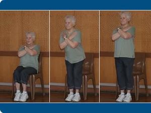
The below video is a great first in the series of exercises for myositis from the myositis society. It links on to further exercises in course.
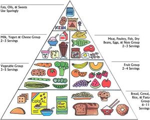
Education is also important for the client and caregivers/family. This can include education on the following aspects
- Diet and nutrition
- Sun protection
- Mind and body practices
- Self-care practices
References[edit | edit source]
- ↑ Gazeley DJ, Cronin ME. Diagnosis and treatment of the idiopathic inflammatory myopathies. Therapeutic advances in musculoskeletal disease. 2011 Dec;3(6):315-24. Available from:https://www.ncbi.nlm.nih.gov/pmc/articles/PMC3383495/ (last accessed 9.12.2019)
- ↑ 2.0 2.1 2.2 Malik A, Hayat G, Kalia JS, Guzman MA. Idiopathic inflammatory myopathies: clinical approach and management. Frontiers in neurology. 2016 May 20;7:64. Available from: https://www.ncbi.nlm.nih.gov/pmc/articles/PMC4873503/ (last accessed 9.12.2019)
- ↑ Mayo clinic Living well with IM Available from: https://www.youtube.com/watch?v=0nMfHDrQn1c&app=desktop (last accessed 9.12.2019)
- ↑ Cheeti A, Brent LH, Panginikkod S. Autoimmune Myopathies.Available from: https://www.ncbi.nlm.nih.gov/books/NBK532860/ (last accessed 21.11.2020)
- ↑ 5.0 5.1 5.2 NIH Inflammatory Myopathies Available from:https://www.ninds.nih.gov/Disorders/Patient-Caregiver-Education/Fact-Sheets/Inflammatory-Myopathies-Fact-Sheet (last accessed 21.11.2020)
- ↑ 6.0 6.1 The myositis association Myositis Available from: https://www.myositis.org/ (last accessed 9.12.2019)
- ↑ Souza FH, Araújo DB, Vilela VS, Bezerra MC, Simões RS, Bernardo WM, Miossi R, Cunha BM, Shinjo SK. Guidelines of the Brazilian Society of Rheumatology for the treatment of systemic autoimmune myopathies. Advances in Rheumatology. 2019;59. Available from:http://www.scielo.br/scielo.php?script=sci_arttext&pid=S2523-31062019000100301 (last accessed 9.12,2019)
- ↑ Myositis association Myositis exercises Available from: https://www.youtube.com/watch?v=In35yxxmiOY&feature=youtu.be (last accessed 9.12.2019)


