Tibialis Posterior: Difference between revisions
Michelle Lee (talk | contribs) No edit summary |
Michelle Lee (talk | contribs) No edit summary |
||
| Line 25: | Line 25: | ||
|} | |} | ||
<ref>Drake RL, Vogl W, Mitchell AWM. Gray's Anatomy for Students. 2nd Ed. Philadelphia: Churchill Livingstone Elsevier, 2010.</ref> | |||
<br> | <ref>Drake RL, Vogl W, Mitchell AWM. Gray's Anatomy for Students. 2nd Ed. Philadelphia: Churchill Livingstone Elsevier, 2010.</ref> <br> | ||
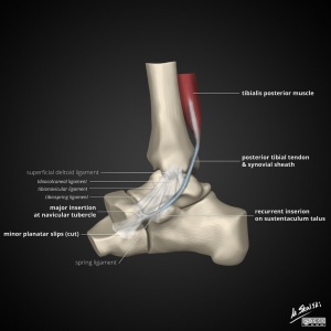
[[Image:Tibialis-posterior-tendon-anatomy.jpg|thumb|left|300x300px]] | |||
Revision as of 17:17, 30 September 2015
Anatomy[edit | edit source]
| Origion |
Proximal posterolateral aspect of the tibia. Proxmial posteromedial aspect of the fibula and the interosseous membrane. |
| Mid portion |
Situated in the deep posterior compartment of the lower leg and runs proximal to the medial malleoli where it is secured by the flexor retinaculum. |
| Insertion |
The major insertion is onto the navicula and the plantar slip attatches to the medial cuniform |
| Innervation |
Tibial Nerve (L4-S3) |
References[edit | edit source]
- ↑ Drake RL, Vogl W, Mitchell AWM. Gray's Anatomy for Students. 2nd Ed. Philadelphia: Churchill Livingstone Elsevier, 2010.


