Thenar and Hypothenar Muscles Of The Hand
Original Editor - User Name
Top Contributors - Saumya Srivastava, Kim Jackson, Leana Louw, Amanda Ager and Ahmed M Diab
Introduction[edit | edit source]
The complex integrity of the human hand is one of the factors that set our spices apart from the primates. The movement of opposition played an important role in the human evolution and this was made possible as more muscles go to the thumb in modern humans than our ancestral primates[1]. Although the muscles of the human hand and forearm are found in one or more extant non –human primates, it is the combination of such what makes up for the uniqueness[1].
In this article, Thenar and Hypothenar muscles of the hands will be discussed.
Description[edit | edit source]
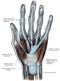
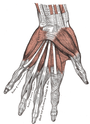
When the human hand is viewed from the palmar side, 2 'fleshy' mounds can be noted[2].
- Thenar eminence - fleshy part at the base of the thumb, made of 3 muscles which control the thumb movements.
- Hypothenar eminence - fleshy part at the base of the fifth digit (little finger), made of 4 muscles which contract to manifest motion through the little finger.
The muscles of the thenar and the hypothenar eminence along with the addcutor compartment make up the intrinsic muscles of the hands as their origin and insertion is within the carpal and metacarpal bones, ligaments, and fascia of the hand. They help with fine motor movements of the hands.[3]
The extrinsic muscles of the hand originate outside the hand, commonly the forearm, and insert into hand structures.
Thenar Eminence (TE) Muscles[edit | edit source]
The muscles of the TE are -[3]
- Opponens Pollicis the largest of the 3 muscles
Origin - at the tubercle of the trapezium
Insertion - lateral margin of the metacarpal of the thumb
Nerve - recurrent branch of the median nerve.
Artery - Superficial palmar arch from the radial artery
Function - perform opposition by flexing and medially rotating the metacarpal on the axis of the trapezium.
- Abductor Pollicis Brevis positioned anteriorly to the opponens pollicis
Origin - tubercles of the scaphoid and trapezium
Insertion - lateral aspect of the proximal phalanx of the thumb.
Nerve - recurrent branch of the median nerve.
Artery - Superficial palmar arch from the radial artery
Function - primary muscle providing opposition. Also leads to abduction of the thumb which is drawing the thumb away from the midline
- Flexor Pollicis Brevis
Origin - tubercle of the trapezium via the deep head, and the associated flexor retinaculum via the superficial head
Insertion - lateral aspect of the proximal phalanx of the thumb.
Nerve - dual innervation with fibers from both the median ( superficial head) and ulnar ( deep head) nerves
Artery - branches of the radial artery; superficial palmar artery, branches of the princeps pollicis artery and radialis indicis artery.
Function - flexion at the metacarpophalangeal and carpometacarpal joints leading to opposition of the thumb and, if continued, produces the medial rotation of thumb.
Hypothenar Eminence (HE) Muscles[edit | edit source]
The HE muscles are[3] -
- Opponens Digiti Minimi
Origin - the hook of hamate and associated transverse carpal ligament
Insertion - lateral aspect of the proximal phalanx of the thumb.
Nerve - Deep branch of ulnar nerve (C8, T1)
Artery - Ulnar artery
Function - flex and laterally rotate the 5th metacarpal about the 5th carpometacarpal joint.
- Abductor digiti minimi
Origin - pisiform bone and the tendon of flexor carpi ulnaris
Insertion - the ulnar base of the proximal phalanx of the small finger
Nerve - Deep branch of ulnar nerve (C8, T1)
Artery - Ulnar artery
Function - abduction of the 5th finger, as well as flexion of its proximal phalanx
- Flexor digiti minimi brevis
Origin - the hook of hamate and associated transverse carpal ligament
Insertion - the base of the proximal phalanx of the small finger
Nerve - Deep branch of ulnar nerve (C8, T1)
Artery - Ulnar artery
Function - flexes the little finger at the metacarpophalangeal joint
- Palmaris brevis
Origin - the transverse carpal ligament
Insertion - on the skin of the medial palm
Nerve - Deep branch of ulnar nerve (C8, T1)
Artery - Ulnar artery
Function - tenses the skin of the palm on the ulnar side during a grip action and deepens the hollow of the palm.
Clinical relevance[edit | edit source]
Fine motor movements are provided by the intrinsic muscles, which include the 3 thenar muscles and 4 hypothenar muscles. The extrinsic muscles provide strength to the hand[3]. The largest of the thenar muscles, opponens pollicis, along with hypothenar muscle, opponens digiti minimi,leads to opposition between little finger and thumb[3]. Palmaris brevis permits the wrinkling of the skin on the palmar surface of the hand[3]. It also deepens the hollow of the palm and protects the neurovasculature of the ulnar canal, which contains the ulnar nerve[4]
Although majority of the injuries resulting in hand deformity and malfunction are due to the involvement of the extrinsic muscles, intrinsic muscle injuries are yet relevant. Median nerve compression at carpal's tunnel will affect the muscles of the thenar compartment. Blunt trauma lesion of the insertion of opponens pollicis can cause the action of thumb opposition and supination to be significantly altered despite intact innervation[3].
As the ulnar nerve serves the hypothenar muscles, damage or compromise to the nerve leads to atrophy of the HT eminence and can be used as a identifying marker for proximal hand ulnar nerve compromise[2]. Another consequence of the ulnar nerve passing through the hook of the hamate and pisiform bone (the Guyon's canal), compression at this level leads to atrophy, numbness, tingling, and pain in the hypothenar eminence along with fourth and fifth digits[2]. This is much like the 'carpel tunnel syndrome' but in this case the ulnar nerve being involved. Cyclist are prone to this kind compression.
Arterial injury to the ulnar artery can also affect the HT eminence due to insufficient collateral blood flow, leading to a condition called 'hypothenar hammer syndrome'. On the degree of blow flow compromise, necrosis of the HT eminence can also occur. Individuals that work with tools needing tight gripping and repetitive pounding can suffer with such injury as the action of gripping and pounding causes recoil impact on the vascular blood supply in the hypothenar eminence[2]
Proximal compromise of the nerve and/or the artery will show symptomatic changes in the hypothenar eminence in both the resting and active presentation of the hand. A loss or decrease of sensation and the feeling of pins and needles (paraesthesia) in the HT eminence can also indicate a C8 spinal root lesion on the ipsilateral side due to the dermatomal distribution for C8[2]. .
Assessment[edit | edit source]
When ulnar nerve compromise occurs at the Guyon's canal it causes weakness with abduction and adduction of the fingers, flexion at the MCP and extension at the PIP, and adduction and flexion at the MCP of the thumb. Hypothenar muscles are also not spared due to their innervation by the ulnar nerve.
In Carpel Tunnel Syndrome (CTS), where median nerve compromise occurs, thenar muscles can show wasting as they are innervated by the median nerve. If noted, other CTS tests like the Phalen's sign and the Tinel's sign should be conducted. Phalen's sign is positive when numbness and tingling occurs when the patient raises the hands out in front of himself, flexes the wrist and places the dorsal wrists together, while flexed, for about 60 seconds.
Treatment[edit | edit source]
Resources[edit | edit source]
- ↑ 1.0 1.1 Diogo R, Richmond BG, Wood B. Evolution and homologies of primate and modern human hand and forearm muscles, with notes on thumb movements and tool use. J Hum Evol. 2012;63(1):64-78. doi:10.1016/j.jhevol.2012.04.001
- ↑ 2.0 2.1 2.2 2.3 2.4 Nguyen J, Duong H. Anatomy, Shoulder and Upper Limb, Hand Hypothenar Eminence. In: StatPearls. Treasure Island (FL): StatPearls Publishing; August 13, 2020.
- ↑ 3.0 3.1 3.2 3.3 3.4 3.5 3.6 Okwumabua E, Sinkler MA, Bordoni B. Anatomy, Shoulder and Upper Limb, Hand Muscles. In: StatPearls. Treasure Island (FL): StatPearls Publishing; July 31, 2020.
- ↑ Moore CW, Rice CL. Structural and functional anatomy of the palmaris brevis: grasping for answers. J Anat. 2017;231(6):939-946. doi:10.1111/joa.12675


