Structure and Function of the Cervical Spine
- Introduction:
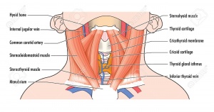
Cervical spine spine starts at base of skull superiorly,ends above thoracic spine inferiorly and it could be called (neck).Neck joins head with trunk and limbs and it works as a major conduit for structures between them. Flexibility of neck movement allows and maximise necessary positions to head functions and its sensory organs. There are many important structures in neck area like nerves, muscles,arteries,veins, vertebrates, lymphatics,glands, oesophagus and trachea. Due to these important structures and lack of bony protection in neck area,it is considered as a region of vulnerability. The main arterial blood supply for head and neck is carotid arteries and principal venous drainage is jugular veins.Carotid and Jugular are commonly injured in penetrating wound of neck. Brachial plexues of nerves originates in neck and travels inferiorly to upper limb. In anterior aspect of neck,there is thyroid cartilage (largest cartilage of thyroid and trachea)[1].
Bony Structure of Neck:[edit | edit source]
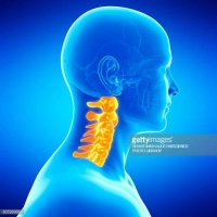
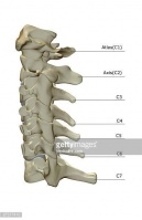
It consists of 7 cervical vertebrates from C1 to C7, hyoid bone,manubrium of sternum and clavicles[1]. Cervical spine has a lordotic curve ( C shaped curve )[2]. According to peculiarities of cervical vertebrates,it could be divided in to two groups:[3]
- Superior cervical group: made up by C1 and C2.
- Inferior cervical group: made up by C3 to C7.
Superior cervical Group:[edit | edit source]
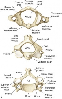
C1 called atlas and C2 called axis and they have some differences than other vertebrae, Atlas C1 is ring like shape , it has no body and no spinous process,
C2 looks like other cervical vertebrae but most distinctive feature is odontoid process or tooth which placed vertically on superior surface of vertebral body[4] with two articular facets( anterior and posterior) which articulate with atlas bone and atlas transverse ligament. C2 has smaller and triangular vertebral foramen.
Inferior Cervical Group:[edit | edit source]
It is made up of 5 vertebrea from C3 TO C7 with similar characteristics like smaller vertebral body, spinous process
References:
- ↑ 1.0 1.1 Lang, J., 1993. Clinical anatomy of the cervical spine. Stuttgart; New York: Thieme.
- ↑ Cervical Spine Anatomy. University of Maryland Clinical Center. http://www.umm.edu/programs/spine/health/guides/cervical-spine-anatomy. accessed Apirl 2018.
- ↑ Spine, S.C., 2015. Anatomy of the Cervical Spine. Cervical Spine: Minimally Invasive and Open Surgery, p.1.
- ↑ Spine, S.C., 2015. Anatomy of the Cervical Spine. Cervical Spine: Minimally Invasive and Open Surgery, p.1.


