Sternum
Original Editor - Grace Barla
Top Contributors - {{Special:Contributors/Template:Sternum}}
Description[edit | edit source]
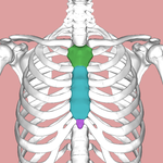
The sternum (Colored part of figure shown below) is a Sword like flat bone that forms the thoracic skeleton's anterior median section. (BD CHAURASIA:BD_Chaurasia’s_Human_Anatomy, Volume 1 - Upper Limb Thorax, 6th Edition.pdf (archive.org))
Length of the bone: 17 cm {Size of sternum in males is longer than in females}
Structure[edit | edit source]
The sternum has 3 anatomical parts:
- Manubrium
- Body of the sternum
- Xiphoid process
1) Manubrium:
- Quadrilateral in shape.
- Thickest and Strongest part
- Surfaces:
- Anterior surface:Convex from side to side; concave from above downwards.
- Posterior Surface:Concave; it forms the anterior boundary of the superior mediastrernum.
- Borders:
- Superior border: Thick,rounded and concave
- marked by the suprasternalnotch or jugular notch or interclavicular notch in the median part
- The clavicular notch on each side
- Inferior border: It forms the secondary cartilaginous joint with the body of the sternum.
- The manubrium makes a slight angle with the body, convex forwards, called the sternal angle of Louis.
- Two lateral borders: It forms the primary cartilaginous joint with the first costal cartilage.
- Superior border: Thick,rounded and concave
2) Body of the sternum:
- The body of the sternum is longer, narrower and thinner than the manubrium
- It is the widest close to its lower end opposite the articulation with the fifth costal cartilage.
- Surfaces:
- Anterior surface: It is flat, directed forward and slightly upwards.
- Posterior surface: slightly concave.
- 2 lateral borders, upper end and lower end.
3) Xiphoid process:
- The smallest part of the sternum
- It is at first cartilaginous, but later in the adult, it becomes ossified near its upper end.
- It varies in shape: It May be bifid or perforated.
- Surfaces:
- Anterior surface
- Posterior surface
- Lateral border


