Sternum: Difference between revisions
Kim Jackson (talk | contribs) (The edits were not acceptable! This page had a banner displayed and was created (and being edited) by another user) Tag: Undo |
Kim Jackson (talk | contribs) (Undo revision 286769 by Vaibhav Panchal (talk)) Tag: Undo |
||
| Line 60: | Line 60: | ||
* Anterior surface:It gives origin on either side to: | * Anterior surface:It gives origin on either side to: | ||
** The | ** The pectoralis major | ||
** The sternal head of the | ** The sternal head of the sternocleidomastoid | ||
* Posterior Surface: It gives origin to: | * Posterior Surface: It gives origin to: | ||
** The sternohyoid in the upper part | ** The sternohyoid in the upper part | ||
** The sternothyroid in the lower part | ** The sternothyroid in the lower part | ||
** The lower part of this surface is related to the arch of the | ** The lower part of this surface is related to the arch of the aorta. | ||
** The upper half is related to the left brachiocephalic vein, the brachiocephalic artery, the left common carotid artery and the left subclavian artery. | ** The upper half is related to the left brachiocephalic vein, the brachiocephalic artery, the left common carotid artery and the left subclavian artery. | ||
** The lateral portion of this surface is related to the corresponding | ** The lateral portion of this surface is related to the corresponding lung and pleura. | ||
* suprasternal notch: | * suprasternal notch: | ||
| Line 75: | Line 75: | ||
* Clavicular notch: | * Clavicular notch: | ||
** The clavicular notch articulates with the medial end of the | ** The clavicular notch articulates with the medial end of the clavicle to form the sternoclavicular joint. | ||
** Margins of clavicular notch give attachment to the capsule of the corresponding sternoclavicular joint. | ** Margins of clavicular notch give attachment to the capsule of the corresponding sternoclavicular joint. | ||
2) '''Body of the sternum''': | 2) '''Body of the sternum''': | ||
* Anterior surface: It gives origin on either side to the | * Anterior surface: It gives origin on either side to the pectoralis major muscles | ||
* Posterior surface: Lower part of the posterior surface gives origin on either side to the sternocostalis muscle. | * Posterior surface: Lower part of the posterior surface gives origin on either side to the sternocostalis muscle. | ||
* Upper end: it forms a secondary cartilaginous joint with the manubrium at the sternal angle | * Upper end: it forms a secondary cartilaginous joint with the manubrium at the sternal angle | ||
* Lower end: It is narrow and forms a primary cartilaginous joint with the xiphisternum. | * Lower end: It is narrow and forms a primary cartilaginous joint with the xiphisternum. | ||
3) '''Xiphoid process:''' | 3) '''Xiphoid process:''' | ||
* Anterior surface: it provides insertion to the medial fibres of the | * Anterior surface: it provides insertion to the medial fibres of the rectus abdminis and the aponeuroses of the abdomen's external and internal oblique muscles. | ||
* Posterior surface: it gives origin to the | * Posterior surface: it gives origin to the diaphragm. It is related to the anterior surface of the liver. | ||
* Lateral border: it gives attachment to the aponeuroses of the internal oblique and transversus abdominis muscles. | * Lateral border: it gives attachment to the aponeuroses of the internal oblique and transversus abdominis muscles. | ||
* Upper end: it forms a primary cartilaginous joint with the body of the sternum | * Upper end: it forms a primary cartilaginous joint with the body of the sternum | ||
| Line 96: | Line 98: | ||
# Bone marrow for examination is usually obtained by manubriosternal puncture. It is done in its upper half to prevent injury to the arch of the aorta, which lies behind its lower half. | # Bone marrow for examination is usually obtained by manubriosternal puncture. It is done in its upper half to prevent injury to the arch of the aorta, which lies behind its lower half. | ||
# The slight movement that takes place at the manubriosternal joint is essential for the direction of the | # The slight movement that takes place at the manubriosternal joint is essential for the direction of the ribs. | ||
# Funnel chest: The sternum is depressed. | # Funnel chest: The sternum is depressed. | ||
# Pigeon chest: Forward projection of the sternum like the keel of the boat and the flattening of the chest wall on either side | # Pigeon chest: Forward projection of the sternum like the keel of the boat and the flattening of the chest wall on either side | ||
| Line 104: | Line 106: | ||
== Resources == | == Resources == | ||
= References = | = References = | ||
Revision as of 10:41, 19 November 2021
Original Editor - Grace Barla
Top Contributors - {{Special:Contributors/Template:Sternum}}
Description[edit | edit source]
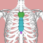
The sternum (Colored part of figure shown below) is a Sword like flat bone that forms the thoracic skeleton's anterior median section. (BD CHAURASIA:BD_Chaurasia’s_Human_Anatomy, Volume 1 - Upper Limb Thorax, 6th Edition.pdf (archive.org))
Length of the bone: 17 cm {Size of sternum in males is longer than in females}
Structure[edit | edit source]
The sternum has 3 anatomical parts:
- Manubrium
- Body of the sternum
- Xiphoid process
1) Manubrium:
- Quadrilateral in shape.
- Thickest and Strongest part
- Surfaces:
- Anterior surface:Convex from side to side; concave from above downwards.
- Posterior Surface:Concave; it forms the anterior boundary of the superior mediastrernum.
- Borders:
- Superior border: Thick,rounded and concave
- marked by the suprasternalnotch or jugular notch or interclavicular notch in the median part
- The clavicular notch on each side
- Inferior border: It forms the secondary cartilaginous joint with the body of the sternum.
- The manubrium makes a slight angle with the body, convex forwards, called the sternal angle of Louis.
- Two lateral borders: It forms the primary cartilaginous joint with the first costal cartilage.
- Superior border: Thick,rounded and concave
2) Body of the sternum:
- The body of the sternum is longer, narrower and thinner than the manubrium
- It is the widest close to its lower end opposite the articulation with the fifth costal cartilage.
- Surfaces:
- Anterior surface: It is flat, directed forward and slightly upwards.
- Posterior surface: slightly concave.
- 2 lateral borders, upper end and lower end.
3) Xiphoid process:
- The smallest part of the sternum
- It is at first cartilaginous, but later in the adult, it becomes ossified near its upper end.
- It varies in shape: It May be bifid or perforated.
- Surfaces:
- Anterior surface
- Posterior surface
- Lateral border
Muscle attachments[edit | edit source]
Manubrium:
- Anterior surface:It gives origin on either side to:
- The pectoralis major
- The sternal head of the sternocleidomastoid
- Posterior Surface: It gives origin to:
- The sternohyoid in the upper part
- The sternothyroid in the lower part
- The lower part of this surface is related to the arch of the aorta.
- The upper half is related to the left brachiocephalic vein, the brachiocephalic artery, the left common carotid artery and the left subclavian artery.
- The lateral portion of this surface is related to the corresponding lung and pleura.
- suprasternal notch:
- It gives attachment to the lower fibres of the interclavicular ligament
- Two subdivisions of the investing layer of cervical fascia.
- Clavicular notch:
- The clavicular notch articulates with the medial end of the clavicle to form the sternoclavicular joint.
- Margins of clavicular notch give attachment to the capsule of the corresponding sternoclavicular joint.
2) Body of the sternum:
- Anterior surface: It gives origin on either side to the pectoralis major muscles
- Posterior surface: Lower part of the posterior surface gives origin on either side to the sternocostalis muscle.
- Upper end: it forms a secondary cartilaginous joint with the manubrium at the sternal angle
- Lower end: It is narrow and forms a primary cartilaginous joint with the xiphisternum.
3) Xiphoid process:
- Anterior surface: it provides insertion to the medial fibres of the rectus abdminis and the aponeuroses of the abdomen's external and internal oblique muscles.
- Posterior surface: it gives origin to the diaphragm. It is related to the anterior surface of the liver.
- Lateral border: it gives attachment to the aponeuroses of the internal oblique and transversus abdominis muscles.
- Upper end: it forms a primary cartilaginous joint with the body of the sternum
- Lower end: It affords attachment to the linea alba.
Clinical relevance[edit | edit source]
- Bone marrow for examination is usually obtained by manubriosternal puncture. It is done in its upper half to prevent injury to the arch of the aorta, which lies behind its lower half.
- The slight movement that takes place at the manubriosternal joint is essential for the direction of the ribs.
- Funnel chest: The sternum is depressed.
- Pigeon chest: Forward projection of the sternum like the keel of the boat and the flattening of the chest wall on either side
- The sternum is protected from injury by attachment of elastic costal cartilages. Indirect violence may lead to fracture of the sternum.
- Ectopia Cordis: Non-fusion of the sternal plates causes this condition, in which the heart lies uncovered on the surface.
- Sternal foramina, bifid xiphoid processes are results of the partial fusion of the sternal plates.


