Search results
Page title matches
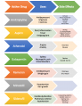
File:Drug use .png (746 × 968 (198 KB)) - 13:22, 11 April 2018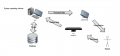
File:Kinect use with PC.png {{fair-use}}(1,264 × 594 (178 KB)) - 17:54, 26 November 2015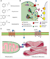
File:Methamphetamine Use and Dopamine.png (629 × 737 (349 KB)) - 01:39, 11 December 2021
File:Cannabis Use Disorder .jpg (1,308 × 1,178 (252 KB)) - 16:41, 6 April 2017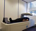
File:DEXA scanner in use ALSPAC.jpeg (1,024 × 881 (107 KB)) - 03:06, 6 August 2022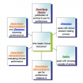
File:Multiple use for clinical outcomes.jpg (732 × 729 (119 KB)) - 16:59, 13 April 2011
File:Wheelchair Use by GMFCS Level.jpeg Rodby-Bousquet E, Hägglund G. Use of Manual and Powered Wheelchair in Children with Cerebral Palsy: A Cross-s {{fair-use}}(885 × 653 (166 KB)) - 00:38, 5 July 2018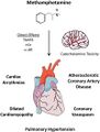
File:Methamphetamine Use and Cardiovascular Disease.jpg (800 × 1,053 (134 KB)) - 01:22, 11 December 2021
File:DVP - How to Use Developmental Chart.pdf {{fair-use}}(0 × 0 (110 KB)) - 01:25, 15 August 2016
File:Video How do I use ES.png (753 × 117 (20 KB)) - 18:00, 27 January 2016
File:CFH 1 How to Use Back.png (516 × 847 (65 KB)) - 18:18, 15 November 2019
File:Key Points How do I use ES.png (769 × 185 (47 KB)) - 18:32, 27 January 2016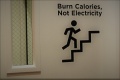
File:Image for sign to use stair 2.jpg (1,024 × 682 (121 KB)) - 23:33, 5 May 2018
File:How do I use ES this section .png (771 × 149 (30 KB)) - 17:38, 27 January 2016
File:Activity One How do I use Es.png (785 × 132 (30 KB)) - 17:44, 27 January 2016
File:Wheelchair Use by Type of Cerebral Palsy.jpeg Rodby-Bousquet E, Hägglund G. Use of Manual and Powered Wheelchair in Children with Cerebral Palsy: A Cross-s {{fair-use}}(878 × 617 (154 KB)) - 00:41, 5 July 2018
File:Activity Two How do I use ES.png (760 × 125 (22 KB)) - 17:52, 27 January 2016
File:Activity 3 How do I use ES.png (706 × 277 (54 KB)) - 18:27, 27 January 2016
File:Activity End of How do you use Es.png (761 × 242 (61 KB)) - 14:30, 28 January 2016
File:Use of Wheelchairs in Cerebral Palsy Related to Age.jpeg Rodby-Bousquet E, Hägglund G. Use of Manual and Powered Wheelchair in Children with Cerebral Palsy: A Cross-s {{fair-use}}(879 × 524 (96 KB)) - 00:42, 5 July 2018


Page text matches
File:Wheelchair Use by GMFCS Level.jpeg Rodby-Bousquet E, Hägglund G. Use of Manual and Powered Wheelchair in Children with Cerebral Palsy: A Cross-s {{fair-use}}(885 × 653 (166 KB)) - 00:38, 5 July 2018File:Wheelchair Use by Type of Cerebral Palsy.jpeg Rodby-Bousquet E, Hägglund G. Use of Manual and Powered Wheelchair in Children with Cerebral Palsy: A Cross-s {{fair-use}}(878 × 617 (154 KB)) - 00:41, 5 July 2018File:Use of Wheelchairs in Cerebral Palsy Related to Age.jpeg Rodby-Bousquet E, Hägglund G. Use of Manual and Powered Wheelchair in Children with Cerebral Palsy: A Cross-s {{fair-use}}(879 × 524 (96 KB)) - 00:42, 5 July 2018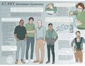
File:Klinefelter Syndrome Image.jpg ...ddress / Organisation]. For more information or to request permission for use, please visit [https://www.webaddress / Organisation] ...d parties. Please seek direct permission from the copyright holder for any use outside Physiopedia.(2,310 × 1,785 (346 KB)) - 10:53, 16 May 2024
File:Flexor Carpi Radialis.png ...ven permission to use this image exclusively in Physiopedia. Please do not use this image outside Physiopedia unless you have prior permission(600 × 600 (116 KB)) - 13:07, 24 February 2022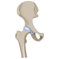
File:HipJoint.png ...ven permission to use this image exclusively in Physiopedia. Please do not use this image outside Physiopedia unless you have prior permission(640 × 640 (51 KB)) - 15:35, 2 March 2022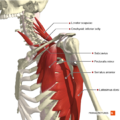
File:Muscles connecting the upper limb to the trunk deep muscles Primal.png ...ven permission to use this image exclusively in Physiopedia. Please do not use this image outside Physiopedia unless you have prior permission(660 × 660 (464 KB)) - 00:59, 8 December 2020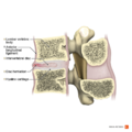
File:Anterior disc hernia sagittal view Primal.png ...ven permission to use this image exclusively in Physiopedia. Please do not use this image outside Physiopedia unless you have prior permission(990 × 990 (660 KB)) - 15:23, 8 December 2020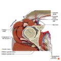
File:Axial section of the hip joint Primal.png ...ven permission to use this image exclusively in Physiopedia. Please do not use this image outside Physiopedia unless you have prior permission(990 × 990 (881 KB)) - 20:57, 7 December 2020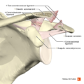
File:Acromioclavicular separation type 3 Primal.png ...ven permission to use this image exclusively in Physiopedia. Please do not use this image outside Physiopedia unless you have prior permission(660 × 660 (300 KB)) - 01:10, 8 December 2020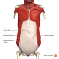
File:Anterior abdominal wall superficial muscles Primal.png ...ven permission to use this image exclusively in Physiopedia. Please do not use this image outside Physiopedia unless you have prior permission(660 × 660 (350 KB)) - 21:49, 8 December 2020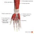
File:Muscles of the hand anterior aspect Primal.png ...ven permission to use this image exclusively in Physiopedia. Please do not use this image outside Physiopedia unless you have prior permission(660 × 660 (265 KB)) - 00:26, 7 December 2020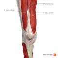
File:Muscles of the knee anterior aspect Primal.png ...ven permission to use this image exclusively in Physiopedia. Please do not use this image outside Physiopedia unless you have prior permission(660 × 660 (289 KB)) - 21:41, 7 December 2020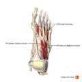
File:Muscles of the foot dorsal aspect Primal.png ...ven permission to use this image exclusively in Physiopedia. Please do not use this image outside Physiopedia unless you have prior permission(660 × 660 (223 KB)) - 00:22, 8 December 2020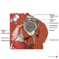
File:Sagittal section of the rotator cuff muscles Primal.png ...ven permission to use this image exclusively in Physiopedia. Please do not use this image outside Physiopedia unless you have prior permission(990 × 990 (905 KB)) - 01:17, 8 December 2020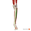
File:Tibialis anterior Primal.png ...ven permission to use this image exclusively in Physiopedia. Please do not use this image outside Physiopedia unless you have prior permission(660 × 660 (118 KB)) - 15:16, 10 January 2021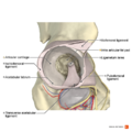
File:Acetabulum Primal.png ...ven permission to use this image exclusively in Physiopedia. Please do not use this image outside Physiopedia unless you have prior permission(990 × 990 (588 KB)) - 20:42, 7 December 2020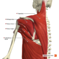
File:Muscles connecting the upper limb to the trunk posterior aspect Primal.png ...ven permission to use this image exclusively in Physiopedia. Please do not use this image outside Physiopedia unless you have prior permission(660 × 660 (487 KB)) - 00:59, 8 December 2020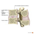
File:Intervertebral disc hernia into adjacent bodies sagittal view Primal.png ...ven permission to use this image exclusively in Physiopedia. Please do not use this image outside Physiopedia unless you have prior permission(990 × 990 (603 KB)) - 15:24, 8 December 2020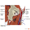
File:Sagittal section of the structures of the hip 1 Primal.png ...ven permission to use this image exclusively in Physiopedia. Please do not use this image outside Physiopedia unless you have prior permission(990 × 990 (1 MB)) - 20:58, 7 December 2020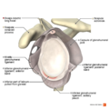
File:Illustration of Bankart lesion Primal.png ...ven permission to use this image exclusively in Physiopedia. Please do not use this image outside Physiopedia unless you have prior permission(990 × 990 (500 KB)) - 01:10, 8 December 2020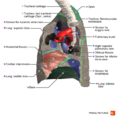
File:Right lung medial surface Primal.png ...ven permission to use this image exclusively in Physiopedia. Please do not use this image outside Physiopedia unless you have prior permission(660 × 660 (324 KB)) - 21:54, 8 December 2020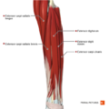
File:Superficial and intermediate extensor muscles of the forearm Primal.png ...ven permission to use this image exclusively in Physiopedia. Please do not use this image outside Physiopedia unless you have prior permission(660 × 660 (236 KB)) - 00:27, 7 December 2020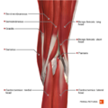
File:Superficial muscles of the knee posterior aspect Primal.png ...ven permission to use this image exclusively in Physiopedia. Please do not use this image outside Physiopedia unless you have prior permission(660 × 660 (372 KB)) - 21:42, 7 December 2020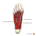
File:Plantar muscles of the foot first layer Primal.png ...ven permission to use this image exclusively in Physiopedia. Please do not use this image outside Physiopedia unless you have prior permission(660 × 660 (239 KB)) - 00:23, 8 December 2020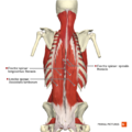
File:Muscles of the back erector spinae group Primal.png ...ven permission to use this image exclusively in Physiopedia. Please do not use this image outside Physiopedia unless you have prior permission(660 × 660 (354 KB)) - 15:12, 8 December 2020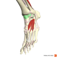
File:Capsule of ankle joint Primal.png ...ven permission to use this image exclusively in Physiopedia. Please do not use this image outside Physiopedia unless you have prior permission(660 × 660 (206 KB)) - 15:18, 10 January 2021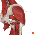
File:Intermediate muscles of the gluteal region Primal.png ...ven permission to use this image exclusively in Physiopedia. Please do not use this image outside Physiopedia unless you have prior permission(660 × 660 (507 KB)) - 20:44, 7 December 2020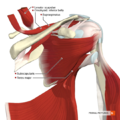
File:Muscles of the scapular region anterior aspect Primal.png ...ven permission to use this image exclusively in Physiopedia. Please do not use this image outside Physiopedia unless you have prior permission(660 × 660 (485 KB)) - 01:00, 8 December 2020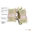
File:Intervertebral disc hernia into anterior body sagittal view Primal.png ...ven permission to use this image exclusively in Physiopedia. Please do not use this image outside Physiopedia unless you have prior permission(990 × 990 (623 KB)) - 15:26, 8 December 2020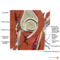
File:Sagittal section of the structures of the hip 2 Primal.png ...ven permission to use this image exclusively in Physiopedia. Please do not use this image outside Physiopedia unless you have prior permission(990 × 990 (1.04 MB)) - 21:02, 7 December 2020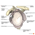
File:Illustration of Buford complex Primal.png ...ven permission to use this image exclusively in Physiopedia. Please do not use this image outside Physiopedia unless you have prior permission(990 × 990 (504 KB)) - 01:11, 8 December 2020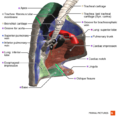
File:Left lung medial surface Primal.png ...ven permission to use this image exclusively in Physiopedia. Please do not use this image outside Physiopedia unless you have prior permission(660 × 660 (340 KB)) - 21:55, 8 December 2020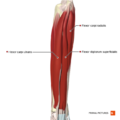
File:Superficial flexor muscles of the forearm Primal.png ...ven permission to use this image exclusively in Physiopedia. Please do not use this image outside Physiopedia unless you have prior permission(660 × 660 (194 KB)) - 00:28, 7 December 2020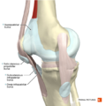
File:Patella bursae Primal.png ...ven permission to use this image exclusively in Physiopedia. Please do not use this image outside Physiopedia unless you have prior permission(660 × 660 (306 KB)) - 21:43, 7 December 2020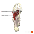
File:Plantar muscles of the foot fourth layer Primal.png ...ven permission to use this image exclusively in Physiopedia. Please do not use this image outside Physiopedia unless you have prior permission(660 × 660 (205 KB)) - 00:24, 8 December 2020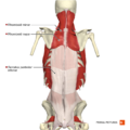
File:Muscles of the back intermediate layer Primal.png ...ven permission to use this image exclusively in Physiopedia. Please do not use this image outside Physiopedia unless you have prior permission(660 × 660 (300 KB)) - 15:14, 8 December 2020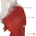
File:Superficial muscles of the gluteal region Primal.png ...ven permission to use this image exclusively in Physiopedia. Please do not use this image outside Physiopedia unless you have prior permission(660 × 660 (474 KB)) - 20:46, 7 December 2020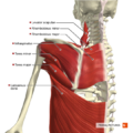
File:Muscles of the scapular region posterior aspect Primal.png ...ven permission to use this image exclusively in Physiopedia. Please do not use this image outside Physiopedia unless you have prior permission(660 × 660 (480 KB)) - 01:01, 8 December 2020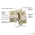
File:Cross-section of a functional spinal unit Primal.png ...ven permission to use this image exclusively in Physiopedia. Please do not use this image outside Physiopedia unless you have prior permission(990 × 990 (535 KB)) - 15:37, 8 December 2020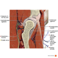
File:Sagittal section of the structures of the hip 3 Primal.png ...ven permission to use this image exclusively in Physiopedia. Please do not use this image outside Physiopedia unless you have prior permission(990 × 990 (1.01 MB)) - 21:03, 7 December 2020
File:Illustration of SLAP II lesion Primal.png ...ven permission to use this image exclusively in Physiopedia. Please do not use this image outside Physiopedia unless you have prior permission(990 × 990 (516 KB)) - 01:11, 8 December 2020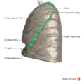
File:Left lung lateral surface Primal.png ...ven permission to use this image exclusively in Physiopedia. Please do not use this image outside Physiopedia unless you have prior permission(660 × 660 (310 KB)) - 21:56, 8 December 2020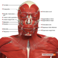
File:Superficial muscles of the head and neck anterior aspect Primal.png ...ven permission to use this image exclusively in Physiopedia. Please do not use this image outside Physiopedia unless you have prior permission(660 × 660 (582 KB)) - 20:00, 7 December 2020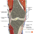
File:Coronal section of the knee joint 1 Primal.png ...ven permission to use this image exclusively in Physiopedia. Please do not use this image outside Physiopedia unless you have prior permission(660 × 660 (658 KB)) - 21:44, 7 December 2020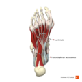
File:Plantar muscles of the foot second layer Primal.png ...ven permission to use this image exclusively in Physiopedia. Please do not use this image outside Physiopedia unless you have prior permission(660 × 660 (223 KB)) - 00:24, 8 December 2020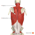
File:Muscles of the back superficial layer Primal.png ...ven permission to use this image exclusively in Physiopedia. Please do not use this image outside Physiopedia unless you have prior permission(660 × 660 (317 KB)) - 15:15, 8 December 2020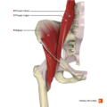
File:Muscles of the iliac region Primal.png ...ven permission to use this image exclusively in Physiopedia. Please do not use this image outside Physiopedia unless you have prior permission(660 × 660 (281 KB)) - 20:47, 7 December 2020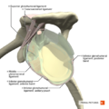
File:Ligaments of the shoulder joint sagittal section Primal.png ...ven permission to use this image exclusively in Physiopedia. Please do not use this image outside Physiopedia unless you have prior permission(660 × 660 (275 KB)) - 01:05, 8 December 2020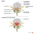
File:Cauda equina syndrome Primal.png ...ven permission to use this image exclusively in Physiopedia. Please do not use this image outside Physiopedia unless you have prior permission(990 × 990 (470 KB)) - 15:37, 8 December 2020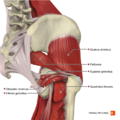
File:Deep muscles of the gluteal region Primal.png ...ven permission to use this image exclusively in Physiopedia. Please do not use this image outside Physiopedia unless you have prior permission(660 × 660 (445 KB)) - 21:20, 7 December 2020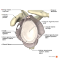
File:Illustration of glenohumeral ligaments lateral view Primal.png ...ven permission to use this image exclusively in Physiopedia. Please do not use this image outside Physiopedia unless you have prior permission(990 × 990 (494 KB)) - 01:12, 8 December 2020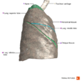
File:Right lung lateral surface Primal.png ...ven permission to use this image exclusively in Physiopedia. Please do not use this image outside Physiopedia unless you have prior permission(660 × 660 (280 KB)) - 21:57, 8 December 2020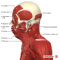
File:Superficial muscles of the head and neck lateral aspect Primal.png ...ven permission to use this image exclusively in Physiopedia. Please do not use this image outside Physiopedia unless you have prior permission(660 × 660 (472 KB)) - 20:01, 7 December 2020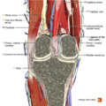
File:Coronal section of the knee joint 2 Primal.png ...ven permission to use this image exclusively in Physiopedia. Please do not use this image outside Physiopedia unless you have prior permission(660 × 660 (629 KB)) - 21:45, 7 December 2020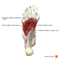
File:Plantar muscles of the foot third layer Primal.png ...ven permission to use this image exclusively in Physiopedia. Please do not use this image outside Physiopedia unless you have prior permission(660 × 660 (218 KB)) - 00:25, 8 December 2020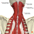
File:Muscles of the cervical region intermediate muscles Primal.png ...ven permission to use this image exclusively in Physiopedia. Please do not use this image outside Physiopedia unless you have prior permission(660 × 660 (619 KB)) - 15:16, 8 December 2020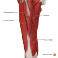
File:Muscles of the thigh anterior compartment Primal.png ...ven permission to use this image exclusively in Physiopedia. Please do not use this image outside Physiopedia unless you have prior permission(660 × 660 (326 KB)) - 20:48, 7 December 2020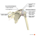
File:Ligaments of the shoulder anterior aspect Primal.png ...ven permission to use this image exclusively in Physiopedia. Please do not use this image outside Physiopedia unless you have prior permission(990 × 990 (340 KB)) - 01:05, 8 December 2020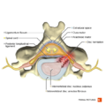
File:Posterolateral disc hernia axial view Primal.png ...ven permission to use this image exclusively in Physiopedia. Please do not use this image outside Physiopedia unless you have prior permission(660 × 660 (287 KB)) - 15:38, 8 December 2020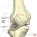
File:Knee joint anterior aspect Primal.png ...ven permission to use this image exclusively in Physiopedia. Please do not use this image outside Physiopedia unless you have prior permission(660 × 660 (279 KB)) - 21:36, 7 December 2020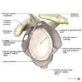
File:Illustration of sublabral foramen Primal.png ...ven permission to use this image exclusively in Physiopedia. Please do not use this image outside Physiopedia unless you have prior permission(990 × 990 (510 KB)) - 01:13, 8 December 2020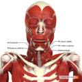
File:Intermediate muscles of the head and neck anterior aspect Primal.png ...ven permission to use this image exclusively in Physiopedia. Please do not use this image outside Physiopedia unless you have prior permission(660 × 660 (557 KB)) - 20:05, 7 December 2020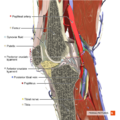
File:Sagittal section of the knee joint Primal.png ...ven permission to use this image exclusively in Physiopedia. Please do not use this image outside Physiopedia unless you have prior permission(660 × 660 (607 KB)) - 21:46, 7 December 2020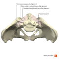
File:Sacro-iliac joint Primal.png ...ven permission to use this image exclusively in Physiopedia. Please do not use this image outside Physiopedia unless you have prior permission(660 × 660 (239 KB)) - 00:49, 8 December 2020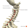
File:Muscles of the cervical region multifidus deep layer Primal.png ...ven permission to use this image exclusively in Physiopedia. Please do not use this image outside Physiopedia unless you have prior permission(660 × 660 (290 KB)) - 15:17, 8 December 2020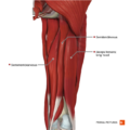
File:Muscles of the thigh posterior compartment Primal.png ...ven permission to use this image exclusively in Physiopedia. Please do not use this image outside Physiopedia unless you have prior permission(660 × 660 (315 KB)) - 20:49, 7 December 2020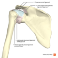
File:Ligaments of the shoulder posterior aspect Primal.png ...ven permission to use this image exclusively in Physiopedia. Please do not use this image outside Physiopedia unless you have prior permission(660 × 660 (256 KB)) - 01:06, 8 December 2020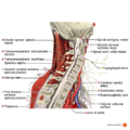
File:Sagittal section of the cervical spine Primal.png ...ven permission to use this image exclusively in Physiopedia. Please do not use this image outside Physiopedia unless you have prior permission(990 × 990 (792 KB)) - 15:39, 8 December 2020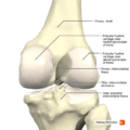
File:Knee joint posterior aspect Primal.png ...ven permission to use this image exclusively in Physiopedia. Please do not use this image outside Physiopedia unless you have prior permission(660 × 660 (289 KB)) - 21:37, 7 December 2020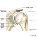
File:Illustration of type I biceps labral complex Primal.png ...ven permission to use this image exclusively in Physiopedia. Please do not use this image outside Physiopedia unless you have prior permission(990 × 990 (498 KB)) - 01:13, 8 December 2020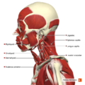
File:Intermediate muscles of the head and neck lateral aspect Primal.png ...ven permission to use this image exclusively in Physiopedia. Please do not use this image outside Physiopedia unless you have prior permission(660 × 660 (437 KB)) - 20:06, 7 December 2020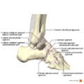
File:Ligaments of the ankle lateral aspect Primal.png ...ven permission to use this image exclusively in Physiopedia. Please do not use this image outside Physiopedia unless you have prior permission(990 × 990 (433 KB)) - 00:16, 8 December 2020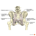
File:Ligaments of the pelvis posterior aspect Primal.png ...ven permission to use this image exclusively in Physiopedia. Please do not use this image outside Physiopedia unless you have prior permission(990 × 990 (537 KB)) - 00:50, 8 December 2020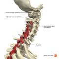
File:Muscles of the cervical region multifidus intermediate layer Primal.png ...ven permission to use this image exclusively in Physiopedia. Please do not use this image outside Physiopedia unless you have prior permission(660 × 660 (307 KB)) - 15:18, 8 December 2020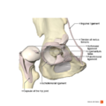
File:Hip joint Primal.png ...ven permission to use this image exclusively in Physiopedia. Please do not use this image outside Physiopedia unless you have prior permission(990 × 990 (443 KB)) - 20:52, 7 December 2020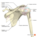
File:Shoulder anterior aspect Primal.png ...ven permission to use this image exclusively in Physiopedia. Please do not use this image outside Physiopedia unless you have prior permission(660 × 660 (266 KB)) - 01:07, 8 December 2020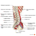
File:Sagittal section of the lumbar spine Primal.png ...ven permission to use this image exclusively in Physiopedia. Please do not use this image outside Physiopedia unless you have prior permission(990 × 990 (683 KB)) - 15:41, 8 December 2020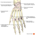
File:Ligaments of the hand dorsal aspect Primal.png ...ven permission to use this image exclusively in Physiopedia. Please do not use this image outside Physiopedia unless you have prior permission(660 × 660 (297 KB)) - 00:20, 7 December 2020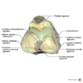
File:Ligaments of the knee joint superior aspect Primal.png ...ven permission to use this image exclusively in Physiopedia. Please do not use this image outside Physiopedia unless you have prior permission(990 × 990 (409 KB)) - 21:37, 7 December 2020
File:Illustration of type II biceps labral complex Primal.png ...ven permission to use this image exclusively in Physiopedia. Please do not use this image outside Physiopedia unless you have prior permission(990 × 990 (497 KB)) - 01:14, 8 December 2020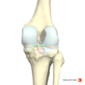
File:Posterior menisco-meniscal ligament Primal.png ...ven permission to use this image exclusively in Physiopedia. Please do not use this image outside Physiopedia unless you have prior permission(660 × 660 (173 KB)) - 15:13, 10 January 2021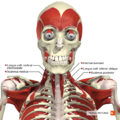
File:Deep muscles of the head and neck anterior aspect Primal.png ...ven permission to use this image exclusively in Physiopedia. Please do not use this image outside Physiopedia unless you have prior permission(660 × 660 (524 KB)) - 20:07, 7 December 2020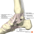
File:Ligaments of the ankle medial aspect Primal.png ...ven permission to use this image exclusively in Physiopedia. Please do not use this image outside Physiopedia unless you have prior permission(660 × 660 (335 KB)) - 00:18, 8 December 2020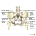
File:Ligaments of the pelvis anterior aspect Primal.png ...ven permission to use this image exclusively in Physiopedia. Please do not use this image outside Physiopedia unless you have prior permission(990 × 990 (557 KB)) - 00:51, 8 December 2020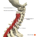
File:Muscles of the cervical region multifidus superficial layer Primal.png ...ven permission to use this image exclusively in Physiopedia. Please do not use this image outside Physiopedia unless you have prior permission(660 × 660 (314 KB)) - 15:19, 8 December 2020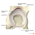
File:Peripheral longitudinal labral tear Primal.png ...ven permission to use this image exclusively in Physiopedia. Please do not use this image outside Physiopedia unless you have prior permission(990 × 990 (552 KB)) - 20:53, 7 December 2020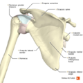
File:Shoulder posterior aspect Primal.png ...ven permission to use this image exclusively in Physiopedia. Please do not use this image outside Physiopedia unless you have prior permission(660 × 660 (280 KB)) - 01:07, 8 December 2020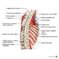
File:Sagittal section of the thoracic spine Primal.png ...ven permission to use this image exclusively in Physiopedia. Please do not use this image outside Physiopedia unless you have prior permission(990 × 990 (709 KB)) - 15:41, 8 December 2020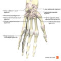
File:Ligaments of the hand palmar aspect Primal.png ...ven permission to use this image exclusively in Physiopedia. Please do not use this image outside Physiopedia unless you have prior permission(660 × 660 (288 KB)) - 00:22, 7 December 2020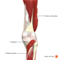
File:Deep muscles of the knee posterior aspect Primal.png ...ven permission to use this image exclusively in Physiopedia. Please do not use this image outside Physiopedia unless you have prior permission(660 × 660 (250 KB)) - 21:38, 7 December 2020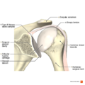
File:Illustration of type III biceps labral complex Primal.png ...ven permission to use this image exclusively in Physiopedia. Please do not use this image outside Physiopedia unless you have prior permission(990 × 990 (510 KB)) - 01:15, 8 December 2020
File:Extensor retinaculum of hand Primal.png ...ven permission to use this image exclusively in Physiopedia. Please do not use this image outside Physiopedia unless you have prior permission(660 × 660 (193 KB)) - 15:14, 10 January 2021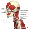
File:Deep muscles of the head and neck lateral aspect Primal.png ...ven permission to use this image exclusively in Physiopedia. Please do not use this image outside Physiopedia unless you have prior permission(660 × 660 (418 KB)) - 20:09, 7 December 2020
File:Ligaments of the ankle posterior aspect Primal.png ...ven permission to use this image exclusively in Physiopedia. Please do not use this image outside Physiopedia unless you have prior permission(660 × 660 (286 KB)) - 00:18, 8 December 2020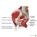
File:Muscles of the pelvic diaphragm Primal.png ...ven permission to use this image exclusively in Physiopedia. Please do not use this image outside Physiopedia unless you have prior permission(990 × 990 (574 KB)) - 00:51, 8 December 2020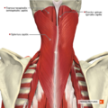
File:Muscles of the cervical region superficial muscles Primal.png ...ven permission to use this image exclusively in Physiopedia. Please do not use this image outside Physiopedia unless you have prior permission(660 × 660 (604 KB)) - 15:20, 8 December 2020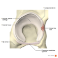
File:Radial flap labral tear Primal.png ...ven permission to use this image exclusively in Physiopedia. Please do not use this image outside Physiopedia unless you have prior permission(990 × 990 (542 KB)) - 20:54, 7 December 2020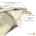
File:Acromioclavicular separation type 1 Primal.png ...ven permission to use this image exclusively in Physiopedia. Please do not use this image outside Physiopedia unless you have prior permission(660 × 660 (274 KB)) - 01:08, 8 December 2020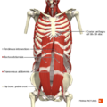
File:Anterior abdominal wall deep muscles Primal.png ...ven permission to use this image exclusively in Physiopedia. Please do not use this image outside Physiopedia unless you have prior permission(660 × 660 (412 KB)) - 21:47, 8 December 2020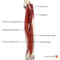
File:Deep extensor muscles of the forearm Primal.png ...ven permission to use this image exclusively in Physiopedia. Please do not use this image outside Physiopedia unless you have prior permission(660 × 660 (208 KB)) - 00:24, 7 December 2020
File:Intermediate muscles of the knee posterolateral aspect Primal.png ...ven permission to use this image exclusively in Physiopedia. Please do not use this image outside Physiopedia unless you have prior permission(660 × 660 (342 KB)) - 21:39, 7 December 2020
File:Axial section of the shoulder joint Primal.png ...ven permission to use this image exclusively in Physiopedia. Please do not use this image outside Physiopedia unless you have prior permission(990 × 990 (707 KB)) - 01:15, 8 December 2020
File:Peroneus brevis Primal.png ...ven permission to use this image exclusively in Physiopedia. Please do not use this image outside Physiopedia unless you have prior permission(660 × 660 (113 KB)) - 15:15, 10 January 2021
File:Ligaments of the hip joint anterior aspect Primal.png ...ven permission to use this image exclusively in Physiopedia. Please do not use this image outside Physiopedia unless you have prior permission(660 × 660 (361 KB)) - 20:29, 7 December 2020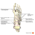
File:Ligaments of the foot dorsal aspect Primal.png ...ven permission to use this image exclusively in Physiopedia. Please do not use this image outside Physiopedia unless you have prior permission(660 × 660 (230 KB)) - 00:20, 8 December 2020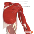
File:Muscles connecting the upper limb to the trunk anterior aspect Primal.png ...ven permission to use this image exclusively in Physiopedia. Please do not use this image outside Physiopedia unless you have prior permission(660 × 660 (485 KB)) - 00:58, 8 December 2020
File:Posterior disc hernia sagittal view Primal.png ...ven permission to use this image exclusively in Physiopedia. Please do not use this image outside Physiopedia unless you have prior permission(990 × 990 (660 KB)) - 15:21, 8 December 2020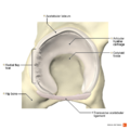
File:Radial fibrillated labral tear Primal.png ...ven permission to use this image exclusively in Physiopedia. Please do not use this image outside Physiopedia unless you have prior permission(990 × 990 (539 KB)) - 20:56, 7 December 2020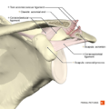
File:Acromioclavicular separation type 2 Primal.png ...ven permission to use this image exclusively in Physiopedia. Please do not use this image outside Physiopedia unless you have prior permission(660 × 660 (287 KB)) - 01:09, 8 December 2020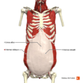
File:Anterior abdominal wall intermediate muscles Primal.png ...ven permission to use this image exclusively in Physiopedia. Please do not use this image outside Physiopedia unless you have prior permission(660 × 660 (345 KB)) - 21:48, 8 December 2020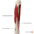
File:Deep flexor muscles of the forearm Primal.png ...ven permission to use this image exclusively in Physiopedia. Please do not use this image outside Physiopedia unless you have prior permission(660 × 660 (189 KB)) - 00:25, 7 December 2020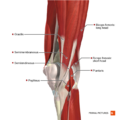
File:Intermediate muscles of the knee posteromedial aspect Primal.png ...ven permission to use this image exclusively in Physiopedia. Please do not use this image outside Physiopedia unless you have prior permission(660 × 660 (355 KB)) - 21:40, 7 December 2020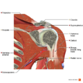
File:Coronal section of the tendon of long head of biceps Primal.png ...ven permission to use this image exclusively in Physiopedia. Please do not use this image outside Physiopedia unless you have prior permission(990 × 990 (900 KB)) - 01:17, 8 December 2020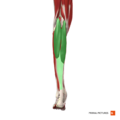
File:Gastrocnemius Primal.png ...ven permission to use this image exclusively in Physiopedia. Please do not use this image outside Physiopedia unless you have prior permission(660 × 660 (112 KB)) - 15:16, 10 January 2021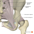
File:Ligaments of the hip joint posterior aspect Primal.png ...ven permission to use this image exclusively in Physiopedia. Please do not use this image outside Physiopedia unless you have prior permission(660 × 660 (395 KB)) - 20:40, 7 December 2020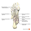
File:Ligaments of the foot plantar aspect Primal.png ...ven permission to use this image exclusively in Physiopedia. Please do not use this image outside Physiopedia unless you have prior permission(660 × 660 (234 KB)) - 00:22, 8 December 2020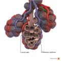
File:Healthy alveoli Primal.png ...ven permission to use this image exclusively in Physiopedia. Please do not use this image outside Physiopedia unless you have prior permission(990 × 990 (1.04 MB)) - 23:58, 6 December 2020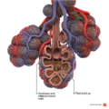
File:Pneumonia Primal.png ...ven permission to use this image exclusively in Physiopedia. Please do not use this image outside Physiopedia unless you have prior permission(990 × 990 (1.05 MB)) - 00:02, 7 December 2020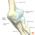
File:Elbow joint Primal.png ...ven permission to use this image exclusively in Physiopedia. Please do not use this image outside Physiopedia unless you have prior permission(660 × 660 (235 KB)) - 00:11, 7 December 2020
File:Cervical plexus phrenic nerve Primal.png ...ven permission to use this image exclusively in Physiopedia. Please do not use this image outside Physiopedia unless you have prior permission(660 × 660 (358 KB)) - 18:43, 14 June 2021
File:3D check socket.jpg The copyright holder has given special permission to use this work in Physiopedia.</div> ...ission does not extend to any other person or organisation. Please do not use this image without permission from the copyright holder [https://www.ascen(1,512 × 2,016 (1.12 MB)) - 12:58, 27 October 2022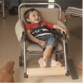
File:Incorrect wc posture.jpeg ...mission does not extend to any other person or organisation. Please do not use this image without permission.(1,024 × 1,024 (68 KB)) - 18:32, 29 February 2024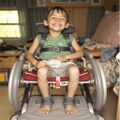
File:Correct wc position.jpeg ...mission does not extend to any other person or organisation. Please do not use this image without permission.(1,024 × 1,024 (78 KB)) - 18:27, 29 February 2024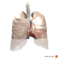
File:Lung tissue.png ...ven permission to use this image exclusively in Physiopedia. Please do not use this image outside Physiopedia unless you have prior permission(660 × 660 (276 KB)) - 17:59, 21 June 2020
File:What is pneumonia.png ...ven permission to use this image exclusively in Physiopedia. Please do not use this image outside Physiopedia unless you have prior permission(637 × 378 (141 KB)) - 18:09, 21 June 2020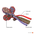
File:Healthy alveoli and bronchiole.png ...ven permission to use this image exclusively in Physiopedia. Please do not use this image outside Physiopedia unless you have prior permission(990 × 990 (610 KB)) - 19:00, 21 June 2020
File:Respiratory membrane.png ...ven permission to use this image exclusively in Physiopedia. Please do not use this image outside Physiopedia unless you have prior permission(660 × 660 (659 KB)) - 19:20, 21 June 2020
File:Causes of pneumonia.png ...ven permission to use this image exclusively in Physiopedia. Please do not use this image outside Physiopedia unless you have prior permission(454 × 259 (80 KB)) - 19:30, 21 June 2020
File:Symptoms of pneumonia.png ...ven permission to use this image exclusively in Physiopedia. Please do not use this image outside Physiopedia unless you have prior permission(708 × 412 (176 KB)) - 19:35, 21 June 2020
File:Treatment of pneumonia.png ...ven permission to use this image exclusively in Physiopedia. Please do not use this image outside Physiopedia unless you have prior permission(708 × 412 (171 KB)) - 20:02, 21 June 2020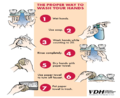
File:Effective hand washing - Virginia Dep(t of Health.png 2. Use soap 6. Use paper towel to turn off faucet/tap(873 × 713 (539 KB)) - 12:13, 15 March 2020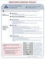
File:RExI Muscle Regeneration.jpg This image is part of the RExI. For permission to use this image visit [https://physicaltherapy.med.ubc.ca/physical-therapy-knowl The copyright holder has given special permission to use this work in Physiopedia.</div>(1,784 × 2,322 (997 KB)) - 09:56, 19 April 2022File:RExExercisesLyingSitting.pdf This image is part of the RExI. For permission to use this image visit [https://physicaltherapy.med.ubc.ca/physical-therapy-knowl The copyright holder has given special permission to use this work in Physiopedia.</div>(0 × 0 (1.83 MB)) - 08:12, 21 April 2022File:RExExercisesStanding.pdf This image is part of the RExI. For permission to use this image visit [https://physicaltherapy.med.ubc.ca/physical-therapy-knowl The copyright holder has given special permission to use this work in Physiopedia.</div>(0 × 0 (1.23 MB)) - 08:21, 21 April 2022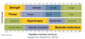
File:RExi Appropriate Exercise Intensity.png This image is part of the RExI. For permission to use this image visit [https://physicaltherapy.med.ubc.ca/physical-therapy-knowl The copyright holder has given special permission to use this work in Physiopedia.</div>(411 × 187 (698 KB)) - 09:58, 19 April 2022
File:Calcification of rotator cuff.png ...ven permission to use this image exclusively in Physiopedia. Please do not use this image outside Physiopedia unless you have prior permission(704 × 575 (450 KB)) - 02:28, 21 June 2020
File:Muscles scapular region anterior aspect.png ...ven permission to use this image exclusively in Physiopedia. Please do not use this image outside Physiopedia unless you have prior permission.(660 × 660 (485 KB)) - 00:54, 21 June 2020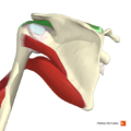
File:Supraspinatus muscle.png ...ven permission to use this image exclusively in Physiopedia. Please do not use this image outside Physiopedia unless you have prior permission(660 × 660 (257 KB)) - 01:04, 21 June 2020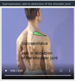
File:Supraspinatus aids in abduction.png ...ven permission to use this image exclusively in Physiopedia. Please do not use this image outside Physiopedia unless you have prior permission(261 × 286 (85 KB)) - 01:35, 21 June 2020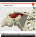
File:Rotator cuff tear.png ...ven permission to use this image exclusively in Physiopedia. Please do not use this image outside Physiopedia unless you have prior permission(561 × 587 (190 KB)) - 01:50, 21 June 2020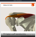
File:Rotator cuff tendonitis.png ...ven permission to use this image exclusively in Physiopedia. Please do not use this image outside Physiopedia unless you have prior permission(570 × 589 (168 KB)) - 01:57, 21 June 2020
File:Illustration of SLAP II lesion.png ...ven permission to use this image exclusively in Physiopedia. Please do not use this image outside Physiopedia unless you have prior permission(990 × 990 (516 KB)) - 02:13, 21 June 2020
File:Illustration of sublabral foramen.png ...ven permission to use this image exclusively in Physiopedia. Please do not use this image outside Physiopedia unless you have prior permission(990 × 990 (510 KB)) - 02:20, 21 June 2020
File:Tear of supraspinatus tendon.png ...ven permission to use this image exclusively in Physiopedia. Please do not use this image outside Physiopedia unless you have prior permission(868 × 568 (432 KB)) - 02:24, 21 June 2020
File:Buffalo hump.jpg You don't need to ask permission to use these images provided: -the use is non-commercial(512 × 512 (92 KB)) - 21:59, 12 October 2023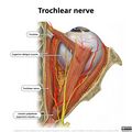
File:Trochlear-nerve-illustration.jpg ...mission does not extend to any other person or organisation. Please do not use this image without permission from the copyright holder.(2,400 × 2,400 (496 KB)) - 18:18, 18 April 2022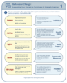
File:Exercise behaviour change.png This image is part of the RExI. For permission to use this image visit [https://physicaltherapy.med.ubc.ca/physical-therapy-knowl The copyright holder has given special permission to use this work in Physiopedia.</div>(665 × 826 (364 KB)) - 10:08, 19 April 2022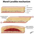
File:Morel-lavallee-illustrations.jpg ...mission does not extend to any other person or organisation. Please do not use this image without permission from the copyright holder.</div>(1,600 × 1,600 (929 KB)) - 18:44, 25 November 2022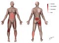
File:Morel-lavallee sites.jpg ...mission does not extend to any other person or organisation. Please do not use this image without permission from the copyright holder.</div>(1,691 × 1,240 (104 KB)) - 18:52, 25 November 2022
File:Teamwork MDT.png ✓ Use Content for free ✓ Use Content without having to attribute the author (although giving credit is a(640 × 480 (345 KB)) - 14:29, 23 April 2023
File:HIV medication.jpg ✓ Use Content for free ✓ Use Content without having to attribute the author (although giving credit is a(1,920 × 1,277 (886 KB)) - 11:16, 22 April 2023
File:Hiv-ga1d0b4868 1280.jpg ✓ Use Content for free ✓ Use Content without having to attribute the author (although giving credit is a(1,280 × 800 (338 KB)) - 11:09, 22 April 2023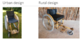
File:Bamboo Wheelchair.png ...osornu Y. Local Bamboo as a Cost-Effective Construction Material for Daily Use Wheelchairs and Sports Chairs: Increasing Mobility Accessibility for Person {{fair-use}}(1,330 × 641 (1.35 MB)) - 06:23, 23 November 2023
File:Physiotitle.jpg {{fair-use}}(524 × 215 (54 KB)) - 18:02, 13 November 2013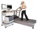
File:VO2max.jpeg {{fair-use}}(740 × 600 (60 KB)) - 23:55, 16 January 2014File:PDF2.pdf {{fair-use}}(0 × 0 (50 KB)) - 20:12, 25 March 2014
File:PTSD image 2.jpg {{fair-use}}(300 × 300 (16 KB)) - 01:36, 3 April 2017
File:Hormone sides.png {{fair-use}}(481 × 394 (18 KB)) - 15:45, 4 December 2014
File:Thor 1.png {{fair-use}}(198 × 351 (61 KB)) - 13:11, 18 June 2016
File:Causes.png {{fair-use}}(300 × 300 (9 KB)) - 14:21, 31 May 2015
File:Cervical dystonia ppt.PNG {{fair-use}}(466 × 272 (63 KB)) - 15:57, 23 October 2013
File:NICEPathways.PNG {{fair-use}}(583 × 285 (14 KB)) - 16:37, 25 November 2015
File:Scapulohumeral rhythm-1-.png {{fair-use}}(720 × 929 (331 KB)) - 18:29, 25 February 2019
File:Lumbar traction title page.png {{fair-use}}(336 × 180 (66 KB)) - 02:37, 25 January 2014
File:Balance.gif {{fair-use}}(350 × 263 (27 KB)) - 16:00, 7 March 2017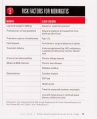
File:Meningitis Risk Factors.jpg {{fair-use}}(390 × 480 (60 KB)) - 22:51, 5 April 2017File:Group 1 WIKI PROJECT.pdf {{fair-use}}(0 × 0 (200 KB)) - 15:28, 4 December 2014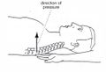
File:Vertebral pressure.jpg {{fair-use}}(1,080 × 739 (82 KB)) - 17:58, 8 May 2020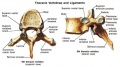
File:Vertebra.jpg {{fair-use}}(539 × 300 (43 KB)) - 17:54, 12 June 2016
File:2 clam hep2go.JPG {{fair-use}}(598 × 600 (43 KB)) - 22:33, 30 March 2015
File:CP-riskfactors1-2.png {{fair-use}}(1,014 × 787 (130 KB)) - 12:20, 13 August 2016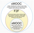
File:Wcpt-xmooc-in-cmooc.png {{fair-use}}(485 × 465 (143 KB)) - 22:54, 27 April 2015File:Jones - 1995 - Clinical reasoning and pain.pdf {{fair-use}}(0 × 0 (5.51 MB)) - 16:05, 6 August 2013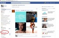
File:FB1.jpg {{fair-use}}(592 × 384 (76 KB)) - 17:12, 29 October 2013
File:Active Living.png {{fair-use}}(339 × 265 (89 KB)) - 15:05, 3 November 2013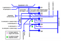
File:Cervical plexus diagram.PNG {{fair-use}}(800 × 541 (71 KB)) - 19:08, 24 February 2014
File:A&E NHS.png {{fair-use}}(180 × 264 (104 KB)) - 13:46, 11 January 2016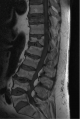
File:Vertebral Fracture.png {{fair-use}}(343 × 511 (179 KB)) - 22:58, 17 February 2015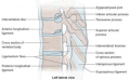
File:Baastrup 2.png {{fair-use}}(619 × 380 (238 KB)) - 12:27, 18 June 2016
File:4 extensionplank.JPG {{fair-use}}(599 × 593 (34 KB)) - 22:47, 30 March 2015
File:Screenshot (laptop) for media page.PNG {{fair-use}}(1,304 × 831 (95 KB)) - 21:31, 6 November 2017File:Rehabilitation Problem Solving Form.pdf {{fair-use}}(0 × 0 (32 KB)) - 20:44, 20 August 2016
File:PP WHO image.png {{fair-use}}(630 × 440 (69 KB)) - 22:35, 17 May 2015
File:Fig 1 for nobles test.png {{fair-use}}(410 × 266 (53 KB)) - 00:35, 30 August 2013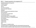
File:Therapeutic Approaches CP.jpeg {{fair-use}}(658 × 504 (92 KB)) - 01:19, 17 September 2016
File:PhysioPEP.png {{fair-use}}(1,313 × 729 (1,023 KB)) - 18:24, 13 November 2013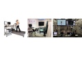
File:Triplegoldstandard.jpeg {{fair-use}}(720 × 540 (51 KB)) - 00:08, 17 January 2014File:PDF3.pdf {{fair-use}}(0 × 0 (364 KB)) - 20:15, 25 March 2014
File:PTSD image 3.jpg {{fair-use}}(700 × 477 (36 KB)) - 01:37, 3 April 2017File:Fitts & Posner Stages.pdf {{fair-use}}(0 × 0 (121 KB)) - 11:49, 11 March 2016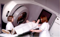
File:Radiotherapy photo.png {{fair-use}}(350 × 219 (116 KB)) - 20:22, 1 December 2014
File:Thor 2.png {{fair-use}}(320 × 356 (31 KB)) - 13:11, 18 June 2016
File:Bruce cpet protocol.jpg {{fair-use}}(1,080 × 1,444 (229 KB)) - 08:58, 14 October 2020
File:Chris physio pedia.jpg {{fair-use}}(150 × 150 (4 KB)) - 00:40, 17 June 2013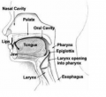
File:Swallow 1.png {{fair-use}}(492 × 448 (110 KB)) - 12:24, 2 September 2016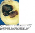
File:Thrombosis.jpg {{fair-use}}(355 × 335 (34 KB)) - 18:57, 3 June 2015
File:APchestwall pain ppt.PNG {{fair-use}}(617 × 371 (57 KB)) - 16:19, 23 October 2013
File:Table 1 Vocation Training.png {{fair-use}}(1,629 × 525 (236 KB)) - 20:24, 30 September 2016
File:SMARTERGoals.PNG {{fair-use}}(657 × 372 (23 KB)) - 16:44, 25 November 2015
File:Parkinsons Flow Chart.png {{fair-use}}(459 × 500 (127 KB)) - 00:34, 9 February 2014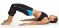
File:Bridge.jpg {{fair-use}}(577 × 278 (23 KB)) - 15:53, 7 March 2017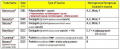
File:Meningococcal Vacccine.png {{fair-use}}(801 × 324 (301 KB)) - 23:12, 5 April 2017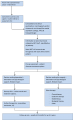
File:Care pathway.png {{fair-use}}(1,872 × 3,070 (141 KB)) - 15:30, 4 December 2014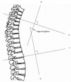
File:Cobbs angle.jpg {{fair-use}}(901 × 1,050 (86 KB)) - 18:03, 12 June 2016
File:2 hip abduction hep2go.JPG {{fair-use}}(1,196 × 724 (57 KB)) - 22:36, 30 March 2015
File:Typical Development.png {{fair-use}}(865 × 370 (59 KB)) - 22:05, 13 August 2016
File:Wcpt-revenue.png {{fair-use}}(770 × 264 (34 KB)) - 23:05, 27 April 2015
File:Icfinteractions.gif {{fair-use}}(491 × 303 (5 KB)) - 14:03, 17 August 2013
File:Quad 1.jpeg {{fair-use}}(566 × 237 (124 KB)) - 13:48, 4 September 2016
File:FB2.jpg {{fair-use}}(706 × 484 (67 KB)) - 17:13, 29 October 2013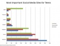
File:Socialnetworkteen.jpg {{fair-use}}(634 × 489 (37 KB)) - 15:23, 3 November 2013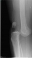
File:Knee dislocation.jpg {{fair-use}}(343 × 608 (15 KB)) - 19:03, 1 January 2014
File:Pain diary figure 1.png {{fair-use}}(377 × 198 (30 KB)) - 17:47, 14 January 2017
File:HYDROLYSIS-570x235.jpg {{fair-use}}(570 × 235 (41 KB)) - 20:13, 2 December 2015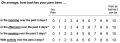
File:P4 Questionnaire.png {{fair-use}}(793 × 283 (18 KB)) - 03:22, 7 March 2014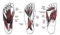
File:Intrinsic foot muscles.png {{fair-use}}(668 × 395 (249 KB)) - 10:49, 19 May 2016
File:PP Parkinsons Outcome Measures.jpg {{fair-use}}(628 × 532 (124 KB)) - 00:04, 2 March 2015
File:Fall on out-stretched hand.png {{fair-use}}(267 × 219 (129 KB)) - 15:59, 21 June 2020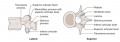
File:Baastrup 1.png {{fair-use}}(910 × 318 (151 KB)) - 12:28, 18 June 2016
File:4 GobletSquat.JPG {{fair-use}}(569 × 390 (29 KB)) - 22:48, 30 March 2015
File:Screenshot (smartphone) for media page half size.png {{fair-use}}(349 × 600 (40 KB)) - 21:35, 6 November 2017File:Rehabilitation Problem Solving Assessment Form.pdf {{fair-use}}(0 × 0 (59 KB)) - 23:50, 20 August 2016
File:Cervicogenic Headache ppt.PNG {{fair-use}}(546 × 317 (344 KB)) - 13:36, 10 October 2013
File:Therapeutic CP 1.jpeg {{fair-use}}(390 × 516 (57 KB)) - 01:30, 17 September 2016
File:Rhabdomyolysis lab values.jpg {{fair-use}}(620 × 373 (66 KB)) - 21:00, 19 November 2015
File:Blahblah.png {{fair-use}}(1,003 × 399 (532 KB)) - 18:43, 13 November 2013
File:ECG wave on ECG paper.png free to use(225 × 225 (7 KB)) - 11:41, 5 December 2018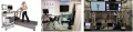
File:TriageGoldStandard.jpg {{fair-use}}(695 × 168 (50 KB)) - 00:23, 17 January 2014
File:Group 3 Keys to life.png {{fair-use}}(307 × 273 (101 KB)) - 13:07, 26 January 2017File:PDF4.pdf {{fair-use}}(0 × 0 (135 KB)) - 20:18, 25 March 2014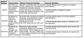
File:Fitts & Posner Stages.jpg {{fair-use}}(1,526 × 705 (1.06 MB)) - 11:53, 11 March 2016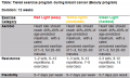
File:Beauty programme.png {{fair-use}}(540 × 323 (21 KB)) - 14:41, 4 December 2014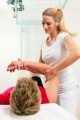
File:PT photo.JPG {{fair-use}}(175 × 262 (26 KB)) - 03:30, 30 March 2015
File:Thor 3.png {{fair-use}}(376 × 547 (62 KB)) - 13:12, 18 June 2016
File:CPET treadmill protocol.jpg {{fair-use}}(1,080 × 1,365 (228 KB)) - 09:00, 14 October 2020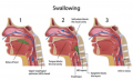
File:Swallow 2.png {{fair-use}}(633 × 380 (267 KB)) - 12:26, 2 September 2016
File:Thrust techniques in LBP ppt.PNG {{fair-use}}(531 × 340 (84 KB)) - 11:53, 27 October 2013
File:SMART-goals.png {{fair-use}}(792 × 341 (91 KB)) - 12:02, 26 November 2015
File:Parkinsons Outcome Measures Chart.png {{fair-use}}(745 × 448 (67 KB)) - 00:41, 19 February 2014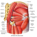
File:Muscles.png {{fair-use}}(350 × 350 (259 KB)) - 17:13, 7 March 2017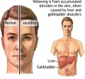
File:Gilbertjaundice.jpg {{fair-use}}(462 × 432 (49 KB)) - 00:02, 6 April 2017
File:Exercise risks ca.png {{fair-use}}(422 × 406 (21 KB)) - 15:48, 4 December 2014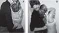
File:Manual mobilisations.jpg {{fair-use}}(1,338 × 752 (107 KB)) - 18:25, 12 June 2016
File:3 hiphinge.JPG {{fair-use}}(479 × 484 (37 KB)) - 22:40, 30 March 2015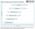
File:Six-gross-motor-milestones.jpg {{fair-use}}(846 × 708 (88 KB)) - 22:12, 13 August 2016
File:Wcpt-21-century.jpg {{fair-use}}(470 × 470 (55 KB)) - 17:36, 30 April 2015
File:EF.gif {{fair-use}}(400 × 385 (12 KB)) - 14:03, 17 August 2013
File:Quad 2.jepg.jpg {{fair-use}}(475 × 245 (107 KB)) - 13:50, 4 September 2016
File:FB3.jpg {{fair-use}}(696 × 467 (67 KB)) - 17:13, 29 October 2013


_for_media_page.PNG)


_for_media_page_half_size.png)


