Soleus
Introduction
[edit | edit source]
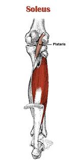
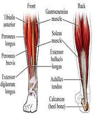
Soleus is a powerfull lower limb muscle which along with gastronemius and plantaris forms the CALF muscle or TRICEPS SURAE.It runs from back of knee to the ankle. Its name is derived from the Latin word, "solea," meaning "sandal."It is located in superficial posterior compartment of the leg.
Soleus muscle is not present in all mammal and it differ in shape in various mammals.In humans the soleus muscle is multipennate but in some mammals it is unipennate.[1][2] Soleus is absent in dogs and vestiginal in horses.[3]
.
Anatomy[4][5][edit | edit source]
ORIGIN
- posterior surface of the head and upper 1/3 of the shaft of the fibula;
- middle 1/3 of the medial border of the tibia, tendinous arch between tibia and fibula.
INSERTION
Into calcaneus with gastrocnemius by way of achilles tendon
ACTION
- Plantar flexion of the foot at the ankle;
- Reversed origin insertion action: when standing, the calcanius becomes the fixed origin of the muscle;
- Soleus muslce stabilizes the tibia on the calcaneus limiting forward sway.
NERVE SUPPLY
Tibial nerve, L4, L5, S1 , S2
SYNERGISTS
Gastrocnemius, Plantaris, Tibialis posterior, Peroneus longus and Brevis, FHL and FDL.
ANTAGONIST
Tibialis anterior
BLOOD SUPPLY
- Blood supply of the soleus muscle is from peroneal artery proximally and the posterior tibial artery distally;
- Muscle has a mixed blood supply;
- Vascular supply of the soleus is from popliteal, posterior tibial, & peroneal vascular pedicles to the proximal muscle, peroneal pedicles to distal lateral belly, and segmental posterior tibial pedicles to distal medial belly;
- w/ distal pedicles from the posterior tibial artery ligated & based on proximal pedicles from the posterior tibial and peroneal arteries, muscle can be transposed medially or laterally to cover defects in middle third of the leg;
- Proximal vasculature arises directly from the popliteal vessels and can reliably carry all but the distal 4 to 5 cm of the muscle;
- Intramuscularly, vasculature of the soleus divides into a bipenniform segmental pattern;
- w/ this vascular pattern, either half of the soleus muscle can be used, leaving a functional hemisoleus muscle intact
Soleus muscle palpation[edit | edit source]
Accessory soleus muscle (ASM)
[edit | edit source]
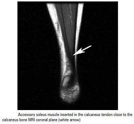
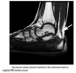
It is present in 0.7 to 5.5% of humans.[6]It is usually observed during the second or third decade of life and is more commonly seen in females than males at a ratio of 2:1.[7][8][9][10][11][12]. This supernumerary muscle is located under the gastrocnemius muscle, in the posterior upper third of the fibula, in the oblique soleus line, between the fibular head and the posterior part of the tibia. From its origin, the ASM runs anteriorly and medially until it reaches the Achilles tendon.[13]
TYPES:
Depending upon its insertion it is of 5 types, or in other words it can origninate from 5 sites
- Achilles tendon
- Upper calcaneus region
- Insertion in the upper calcaneus,
- Medial calcaneus region,
- Medial part of the calcaneus
Sometimes it is impossible to precisely identify the ASM origin and insertion, since the MRI fails to show details, depending on the slices.[14]
References[edit | edit source]
- ↑ Botta G, Piccinetti A, Giontella M, Mancini S (2001). "Il potenziamento dell’attività di pompa venosa del tricipite surale in ortopedia e traumatologia mediante l’utilizzo di una nuova apparecchiatura di ginnastica vascolare" [Strengthening of venous pump activity of the sural tricipital in orthopaedics and traumatology by means of a new equipment for vascular exercise]. Giornale Italiano di Ortopedia e Traumatologia (in Italian) 27: 84–8.
- ↑ Ariano MA, Armstrong RB, Edgerton VR (January 1973). "Hindlimb muscle fiber populations of five mammals". The Journal of Histochemistry and Cytochemistry 21 (1): 51–5.
- ↑ Meyers, Ron A.; Hermanson, John W. (2006). "Horse Soleus Muscle: Postural Sensor or Vestigial Structure?". The Anatomical Record Part a 288A 288 (10): 1068–1076.
- ↑ Gray's anatomy.Student edition.
- ↑ Duke's orthopaedics.Wheeless' Textbook of Orthopaedics
- ↑ Sookur PA, Naraghi AM, Bleakney RR, Jalan R, Chan O, White LM. Accessorymuscles: anatomy, symptoms and radiology evaluation. Radiographics. 2008;28(2):481-99.
- ↑ Crespo E, Minguez MF, Gascó J, Silvestre A, Jolín T, et al. Músculo sóleo accesorio como diagnóstico diferencial de un tumor de partes blandas del tobillo. Acta Ortop Castellano-Manch. 2004;(5):37-41.
- ↑ Romanus B, Lindahl S, Sterner B. Accessory soleus muscle. A clinical and radiographic presentation of eleven cases. J Bone Joint Surg Am. 1986; 68(5):731-4.
- ↑ Salomão O, Carvalho Junior AE, Fernandes TD, Romano D, Adachi PP, Sampaio Neto R. Músculo solear acessório: aspectos clínicos e achados cirúrgicos. Rev Bras Ortop. 1994;29(4):251-5.
- ↑ Leswick DA, Chow V, Stoneham GW. Resident's corner. Can Assoc Radiol J. 2003;54(5):313-5.
- ↑ Featherstone T. MRI diagnosis of accessory soleus muscle strain. Br J Sports Med. 1995;29(4):277-8.
- ↑ Doda N, Peh WC, Chawla A. Symptomatic accessory soleus muscle: diagnosis and follow-up on magnetic resonance imaging. Br J Radiol. 2006;79(946):e129-32.
- ↑ Flavio Belmont Del Nero; Cristiane Regina Ruiz; Roberto Aliaga Junior.The presence of accessory soleous muscle in humans.Einstein (São Paulo) vol.10 no.1 São Paulo Jan./Mar. 2012.
- ↑ Flavio Belmont Del Nero; Cristiane Regina Ruiz; Roberto Aliaga Junior.The presence of accessory soleous muscle in humans.Einstein (São Paulo) vol.10 no.1 São Paulo Jan./Mar. 2012.


