Sinding Larsen Johansson Syndrome: Difference between revisions
Michelle Lee (talk | contribs) No edit summary |
Michelle Lee (talk | contribs) No edit summary |
||
| Line 40: | Line 40: | ||
== Physical Therapy Treatment == | == Physical Therapy Treatment == | ||
Referral to | Referral to a doctor for anti-inflammatories should be initiated. First and foremost, physical therapists must educate the patient on activity modification. Kneeling, jumping, squatting, stair climbing, and running on the affected knee should be avoided at least for the short term. Lower extremity strength needs to be tested, especially at the ankle and the hip to find any muscle weaknesses that may be contributing to the overuse syndrome. Core strengthening should be initiated al well as exercise addressing flexibility or strength issues. The goal in patients with SLJ is to avoid muscle atrophy. An adjunct can include electrotherapy. <ref name="Physioadvisor" /><ref name="Wilson">Wilson, J.K., et al., 'Comparison of rehabilitation methods in the treatment of patellar tendinitis', Journal of Sports Rehabilitation, 2000, 9(4), p. 304-314. (Level of Evidence 1B)</ref> | ||
A safe progression back to sports or high-level activities may happen when each of the following happens in this specific order: | A safe progression back to sports or high-level activities may happen when each of the following happens in this specific order: | ||
| Line 57: | Line 55: | ||
*Ability to jump on both legs without pain and hop on the injured leg without pain. | *Ability to jump on both legs without pain and hop on the injured leg without pain. | ||
Stretching and proprioceptive muscle strengthening exercises in patients with anterior knee pain, have been proven beneficial. <ref name="Clark">Clark, D.I., et al., ‘Physiotherapy for Anterior Knee Pain. A Randomised Controlled Trial’, Ann Rheum Dis, 2000, 59, p. 700-704. (Level of Evidence 1B)</ref> An improvement of strength and functionality can be accomplished by both open and closed kinetic chain exercises.<ref name="Witvrouw">Witvrouw, E., et al., ‘Open Versus Closed Kinetic Chain Exercises for Patellofemoral Pain. A Prospective Randomised Study’, The American Journal of Sports Medicine, 2000, vol. 28, no. 5, p. 687-694. (Level of Evidence 1B)</ref> | Stretching, load management and proprioceptive muscle strengthening exercises in patients with anterior knee pain, have been proven beneficial. <ref name="Clark">Clark, D.I., et al., ‘Physiotherapy for Anterior Knee Pain. A Randomised Controlled Trial’, Ann Rheum Dis, 2000, 59, p. 700-704. (Level of Evidence 1B)</ref> An improvement of strength and functionality can be accomplished by both open and closed kinetic chain exercises.<ref name="Witvrouw">Witvrouw, E., et al., ‘Open Versus Closed Kinetic Chain Exercises for Patellofemoral Pain. A Prospective Randomised Study’, The American Journal of Sports Medicine, 2000, vol. 28, no. 5, p. 687-694. (Level of Evidence 1B)</ref> | ||
Patients who followed a 30 to 60 minute therapy, once a week during six weeks showed a decrease in patellofemoral pain. Table one shows the content of the therapy.<ref name="Crossley">Crossley, K., et al., ‘Physical Therapy for Patellofemoral Pain. A Randomised, Double-Blinded, Placebo-Controlled Trial’, The American Journal of Sports Medicine, 2002, vol. 30, no. 6, p. 856-865. (Level of Evidence 1B)</ref> | Patients who followed a 30 to 60 minute therapy, once a week during six weeks showed a decrease in patellofemoral pain. Table one shows the content of the therapy.<ref name="Crossley">Crossley, K., et al., ‘Physical Therapy for Patellofemoral Pain. A Randomised, Double-Blinded, Placebo-Controlled Trial’, The American Journal of Sports Medicine, 2002, vol. 30, no. 6, p. 856-865. (Level of Evidence 1B)</ref> | ||
Revision as of 16:21, 14 November 2016
Original Editor - Andrew Klaehn, Yelena Gesthuizen as part of the Vrije Universiteit Brussel Evidence-based Practice Project
Top Contributors - Admin, Yelena Gesthuizen, Laure VanderDonckt, Andrew Klaehn, Michelle Lee, Adam Vallely Farrell, Lucinda hampton, Wanda van Niekerk, Kim Jackson, Charlotte Moreau, Candace Goh, Claire Knott, Rucha Gadgil, Jess Bell, 127.0.0.1, Naomi O'Reilly and Gitte Verdoodt
Definition/Description[edit | edit source]
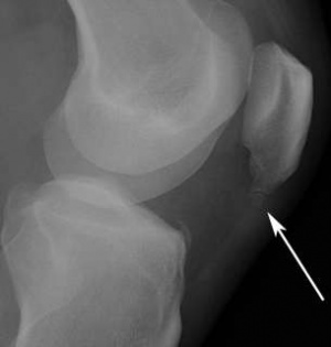
The Sinding Larsen Johansson Syndrome (SLJ) is an osteochondroses and traction epiphysitis affecting the extensor mechanism of the knee. SLJ occurs at the inferior pole of the patella, at the superior attachment of the patella tendon. The tenderness of the inferior pole of the patella is usually accompanied by roentgen graphic evidence of splintering of that pole. Most patients with SLJ also show a calcification at the inferior pole of the patella.[1]
The syndrome usually appears in adolescence, during the growth spurt. It’s associated with localized pain which is worsened by exercise. Usually we observe a localized tenderness and soft tissue swelling. There’s also a tightness of the surrounding muscles, the quadriceps, hamstrings and gastrocnemius in particular. This tightness usually results in inflexibilities of the kneejoint, altering the stress through the patellofemoral joint.[2]
Clinically Relevant Anatomy[edit | edit source]
The Sinding Larsen Johansson Syndrome occurs in the knee joint. It is an inflammation of the growth plate at the inferior pole of the patella. At the superior pole of the patella, the quadriceps tendon inserts. The patella tendon inserts at the inferior pole of the patella.
The growth plate of the patella, or apophysis, is consists of cartilage cells. These cells are softer and more vulnerable to injury. Increased tension and pressure on the growth centre causes SLJ. [3]
Epidemiology/Etiology[edit | edit source]
SLJ is caused by microtraumata to this area and can be followed by calcification and ossification if the condition becomes chronic. It typically affects children and adolescents between the ages of 10 and 15 years old. However, it can also affect active adults who run for moderate to long distances or are involved in sports that require much jumping or squatting. It is similiar to Osgood-Schlatter's disease of the distal patellar tendon.
Characteristics/Clinical Presentation[edit | edit source]
US or MRI imaging may show osseus fragmentation of the distal patellar pole, or it may be irregular, with chondral changes and thickening at the insertion of the patellar tendon. Any activity, from normal walking to climbing stairs, may increase the person's pain depending upon the severity of the condition. In less severe cases, a person may not begin to feel pain until after extended activity, such as running for several miles. The pain occurs when straightening the knee against force: deep knee bends, kneeling, jumping, climbing stairs, squatting, running, weightlifting. [4] Tenderness to touch, limping and a tender bump in the infrapatellar area are all common signs. Lower extremity neurovascular signs or crepitus in the knee are rare and may be indicative of another pathology.
Differential Diagnosis[edit | edit source]
Other diseases with similar symptoms to the Sinding Larsen Johansson Syndrome are injury to the infrapatellar fat pad, Hoffa disease, patellofemoral joint dysfunction, mucoid degeneration of the infrapatellar tendon. [5]
Diagnostic Procedures[edit | edit source]
The physiotherapist performs a physical examination of the knee and reviews the patient’s symptoms. [3]
In case of anterior knee pain there are three important tests to perform (in all tests the patient is in supine position) [6]
Physical Therapy Treatment[edit | edit source]
Referral to a doctor for anti-inflammatories should be initiated. First and foremost, physical therapists must educate the patient on activity modification. Kneeling, jumping, squatting, stair climbing, and running on the affected knee should be avoided at least for the short term. Lower extremity strength needs to be tested, especially at the ankle and the hip to find any muscle weaknesses that may be contributing to the overuse syndrome. Core strengthening should be initiated al well as exercise addressing flexibility or strength issues. The goal in patients with SLJ is to avoid muscle atrophy. An adjunct can include electrotherapy. [4][7]
A safe progression back to sports or high-level activities may happen when each of the following happens in this specific order:
- The lower kneecap is no longer tender and there is no swelling.
- The injured knee can be fully straightened and bent without pain.
- The knee and leg have regained normal strength compared to the uninjured knee and leg
- Ability to jog straight ahead without limping.
- Ability to sprint straight ahead without limping.
- Ability to do 45-degree cuts.
- Ability to do 90-degree cuts.
- Ability to do 20-yard figure-of-eight runs.
- Ability to do 10-yard figure-of-eight runs.
- Ability to jump on both legs without pain and hop on the injured leg without pain.
Stretching, load management and proprioceptive muscle strengthening exercises in patients with anterior knee pain, have been proven beneficial. [8] An improvement of strength and functionality can be accomplished by both open and closed kinetic chain exercises.[9]
Patients who followed a 30 to 60 minute therapy, once a week during six weeks showed a decrease in patellofemoral pain. Table one shows the content of the therapy.[10]
Image:Used_therapy_and_home_exercises.jpg
Resources[edit | edit source]
Recent Related Research (from Pubmed)[edit | edit source]
Failed to load RSS feed from http://www.ncbi.nlm.nih.gov/entrez/eutils/erss.cgi?rss_guid=1P_FVbJaBNrJLV8hGBvqJDFdcq_9bsMgNlHMz-gAbS6EL_eEVv|charset=UTF-8|short|max=10: Error parsing XML for RSS
References[edit | edit source]
- ↑ Medlar, R. C., et al., ‘Sinding-Larsen-Johansson Disease. Its Etiology and Natural History’, Journal of Bone &amp;amp;amp;amp;amp;amp;amp;amp;amp;amp;amp;amp;amp; Joint Surgery, December 1978, vol. 60, no. 8, p. 1113-1116. (Level of Evidence 1B)
- ↑ Houghton, K. M., ‘Review for the generalist: evaluation of anterior knee pain’, Paediatric Rheumatology, (2007), vol. 5, p. 4-10. (Level of Evidence 2B)
- ↑ 3.0 3.1 Sinding-Larsen- Johansson Syndrome (distal patella apophysitis)’, internet, (19 november 2010), (http://www.childrensmemorial.org/depts/sportsmedicine/slj.aspx). (Level of Evidence 5)
- ↑ 4.0 4.1 Demetrious, T. and B., Harrop (red.), Sinding-Larsen-Johansson Disease, internet, 2008, (http://www.physioadvisor.com.au/10246650/sindinglarsenjohansson-disease-physioadvisor.htm). (Level of Evidence 5)
- ↑ Klucinec, B., ‘Recalcitrant Infrapatellar Tendinitis and Surgical Outcome in a Collegiate Basketball Player: A Case Report’, Journal of Athletic Training, June 2001, vol. 36, no. 2, p. 174-181. (Level of Evidence 1C)
- ↑ Hagen, K., ‘Anterieure Kniepijn’, Afstudeeropdracht fysiotherapie HvU, 2005, p. 1-8. (Level of Evidence 2A)
- ↑ Wilson, J.K., et al., 'Comparison of rehabilitation methods in the treatment of patellar tendinitis', Journal of Sports Rehabilitation, 2000, 9(4), p. 304-314. (Level of Evidence 1B)
- ↑ Clark, D.I., et al., ‘Physiotherapy for Anterior Knee Pain. A Randomised Controlled Trial’, Ann Rheum Dis, 2000, 59, p. 700-704. (Level of Evidence 1B)
- ↑ Witvrouw, E., et al., ‘Open Versus Closed Kinetic Chain Exercises for Patellofemoral Pain. A Prospective Randomised Study’, The American Journal of Sports Medicine, 2000, vol. 28, no. 5, p. 687-694. (Level of Evidence 1B)
- ↑ Crossley, K., et al., ‘Physical Therapy for Patellofemoral Pain. A Randomised, Double-Blinded, Placebo-Controlled Trial’, The American Journal of Sports Medicine, 2002, vol. 30, no. 6, p. 856-865. (Level of Evidence 1B)


